
A 44 year old woman came to Gynaecology OP for routine gynaecological examination. This is her first visit for routine gynaecological screening. She had a history of three pregnancies resulting in three full term normal vaginal delivery of healthy children. She was leading a sexually active life. Her examination both general and local was normal. Pap smear performed as a part of the examination. The Pap smear report showed the presence of cervical intra epithelial neoplasia –CIN. a) What is the most likely infectious cause of this lesion? b) Enumerate the serotypes of the causative agent most likely associated with this condition. c) Describe briefly the pathogenesis and laboratory diagnosis of this infectiou
Reading File
Now let me search the medical library for HPV, CIN, and cervical cancer pathogenesis simultaneously.
Finding Sources
Finding Sources
Good - found relevant sections in Berek & Novak's Gynecology. Let me read the key blocks in parallel.
Reading File
Finding Sources
Finding Sources
Excellent content found. Let me read further into the pathogenesis section and also search for HPV virology and diagnosis sections.
Reading File
Finding Sources
Finding Sources
Good, rich content in the file. Let me read further to get the pathogenesis/virology section and also search for HPV microbiology content.
Reading File
Finding Sources
Excellent - this has the key information. Let me read further to get the laboratory diagnosis section.
Reading File
I now have comprehensive information. Let me also fetch the CIN diagram image for illustration.
Loading Image
Loading Image
I now have all the information needed for a comprehensive answer. Let me compile the complete response.
Case Analysis: CIN on Pap Smear - HPV Infection
a) Most Likely Infectious Cause
The most likely infectious cause of Cervical Intraepithelial Neoplasia (CIN) is Human Papillomavirus (HPV).
HPV is the primary cause of cancer of the cervix and its precursor lesions. Persistent high-risk oncogenic HPV infection is the principal risk factor for the development of CIN. Detection of HPV is associated with a 250-fold increase in the risk of high-grade CIN, and the percentage of intraepithelial neoplasia attributed to HPV infection approaches 90%.
This 44-year-old sexually active woman with multiple full-term deliveries fits the classic epidemiological profile - sexual activity is the main route of transmission for genital HPV.
b) HPV Serotypes Associated with CIN
There are more than 120 types of HPV identified, with about 30 primarily infecting the squamous epithelium of the lower anogenital tract. They are classified into:
High-Risk (Oncogenic) Types - Cause CIN and Cervical Cancer
These account for approximately 90% of high-grade intraepithelial lesions and invasive cancers:
| Type | Significance |
|---|---|
| HPV-16 | Most common in invasive cancer and CIN 2/3; most common type in women with normal cytology |
| HPV-18 | Second most common; associated with adenocarcinoma |
| HPV-31 | High-risk |
| HPV-33 | High-risk |
| HPV-35 | High-risk |
| HPV-39 | High-risk |
| HPV-45 | High-risk |
| HPV-51 | High-risk |
| HPV-52 | High-risk |
| HPV-56 | High-risk |
| HPV-58 | High-risk |
| HPV-59 | High-risk |
| HPV-68 | High-risk |
Low-Risk Types - Cause Benign Warts (Condylomata Acuminata), Not CIN
- HPV-6 and HPV-11 - cause anogenital warts
HPV-16 and HPV-18 together account for ~70% of cervical cancers and are the primary targets of available vaccines.
c) Pathogenesis and Laboratory Diagnosis
Pathogenesis
Step 1: Infection and Site of Entry
HPV infects the transformation zone of the cervix - the area between the squamocolumnar junction (SCJ) and the external os. CIN characteristically originates here, at the advancing SCJ where squamous metaplasia is actively occurring. Microabrasions during sexual intercourse allow HPV access to the basal layer of the epithelium.
Step 2: Productive Infection (Low-Grade CIN / CIN 1)
- In early infection (CIN 1), HPV replicates productively within differentiating keratinocytes
- The virus exists as an episomal (extrachromosomal) element - it does not integrate into the host genome at this stage
- High copy numbers of HPV DNA and capsid antigens are present
- The hallmark cytological change is koilocytosis - cells in the upper layers of the epithelium develop perinuclear halos, well-defined cell borders, and nuclear hyperchromasia/enlargement
- In the vast majority of cases (~70-90%), HPV infection clears spontaneously within 9-15 months
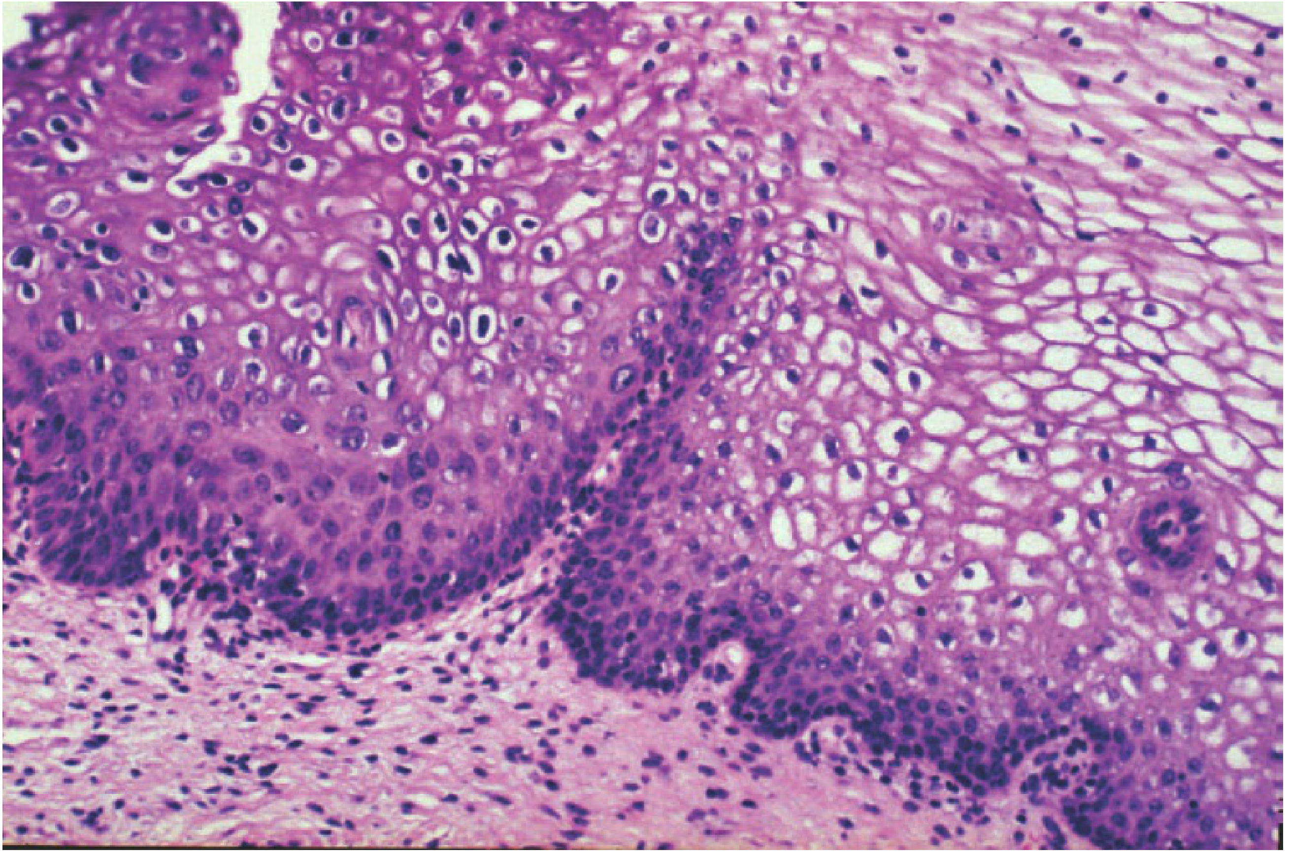
Step 3: Progression to High-Grade CIN (CIN 2 and CIN 3)
- As lesions become more severe, koilocytes disappear, HPV copy numbers decrease, and capsid antigen disappears
- The virus is no longer capable of productive replication in less differentiated cells
- Instead, portions of HPV DNA integrate into the host cell genome - this integration event is essential for malignant transformation
- Integration disrupts the E2 regulatory gene of HPV, leading to uncontrolled overexpression of the E6 and E7 oncoproteins
Step 4: Oncogenic Transformation via E6 and E7
Malignant transformation requires expression of the two key oncoproteins:
| Oncoprotein | Target | Effect |
|---|---|---|
| E6 | p53 tumor suppressor | Binds and degrades p53 via the ubiquitin-proteasome pathway → loss of apoptosis and cell cycle checkpoint control |
| E7 | Retinoblastoma protein (pRb) | Binds and inactivates pRb → releases transcription factor E2F → uncontrolled cell proliferation |
These two events together disable both major tumor suppressor pathways and drive progressive genomic instability, resulting in CIN 2 → CIN 3 → invasive carcinoma.
CIN Grading (Histological):
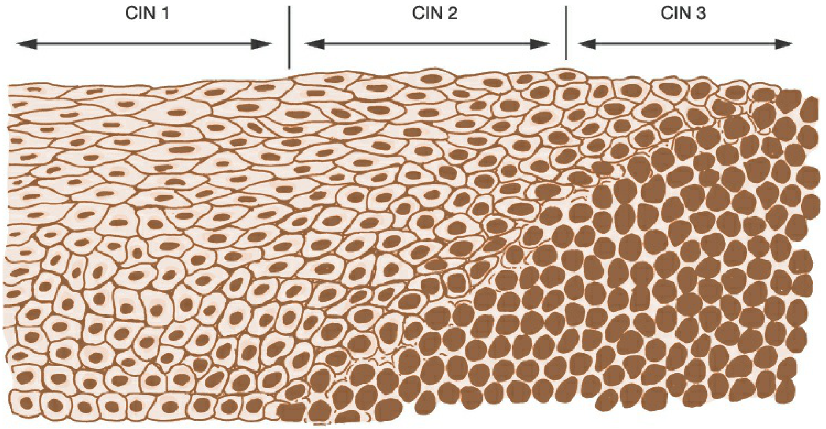
- CIN 1: Mitoses and immature cells limited to the lower 1/3 of epithelium; koilocytes in upper layers
- CIN 2: Abnormal cells in lower 2/3; moderate dysplasia
- CIN 3: Abnormal cells throughout the full thickness; severe dysplasia / carcinoma in situ
Laboratory Diagnosis
1. Cytological Screening - Pap Smear
- Cells scraped from the cervical transformation zone are examined microscopically
- Liquid-based cytology (LBC) is now preferred over conventional slides - better cell preservation and allows reflex HPV testing on the same sample
- Results are reported using the Bethesda System:
- NILM (Negative for Intraepithelial Lesion or Malignancy)
- ASC-US (Atypical Squamous Cells of Undetermined Significance)
- LSIL (Low-Grade Squamous Intraepithelial Lesion)
- HSIL (High-Grade Squamous Intraepithelial Lesion)
- ASC-H, AGC (Atypical Glandular Cells)
- Cytological hallmark of HPV infection: koilocytes (perinuclear clearing, nuclear atypia)
2. High-Risk HPV Testing
Because HPV cannot be grown in conventional cell culture, molecular methods are used for direct detection. FDA-approved HPV assays include:
| Assay | Method | Product Examples |
|---|---|---|
| DNA hybridization | Hybrid Capture 2 (HC2) | Detects a pool of 13 high-risk types |
| DNA PCR | Cobas HPV Test | Genotypes HPV-16/18 separately |
| E6/E7 mRNA detection | APTIMA | More specific for clinically active infection |
| Invader technology | Cervista | Detects 14 high-risk types |
Clinical indications for HPV testing:
- ASC-US triage: HPV reflex testing identifies 90% of women with CIN 2/3 - those who test HPV-negative can return to routine screening
- Co-testing: HPV + cytology every 5 years for women aged 30-65
- Primary HPV screening: Stand-alone primary screening modality (Cobas approved)
3. Colposcopy with Directed Biopsy
- Indicated when cytology shows LSIL or higher, or HPV-positive ASC-US
- The cervix is examined under magnification after application of acetic acid (acetowhitening of abnormal areas) and Lugol's iodine (Schiller's test)
- Directed punch biopsies are taken from the most abnormal colposcopic areas
- Endocervical curettage (ECC) is performed to sample the endocervical canal
4. Histopathological Examination
- Gold standard for definitive diagnosis and grading
- Confirms CIN grade based on the degree of epithelial involvement by immature, dysplastic cells
- Immunohistochemistry for p16: Surrogate marker for high-risk HPV activity; strong diffuse positivity supports CIN 2/3 diagnosis
5. Colposcopic Biopsy Features to Note
- Acetowhite areas, mosaicism, punctation, irregular vascular patterns
- HSIL lesions often show dense acetowhitening with atypical vessels
Key Takeaway for this Case:
This 44-year-old woman's CIN was almost certainly caused by persistent infection with a high-risk HPV type, most likely HPV-16 or HPV-18, transmitted through sexual contact. The recommended next step after a CIN result on Pap smear is colposcopy with directed biopsy for histological confirmation and grading.
Sources: Berek & Novak's Gynecology (9781496380357), Chapter 16 - Cervical Intraepithelial Neoplasia; Goldman-Cecil Medicine
This is a shared conversation. Sign in to Orris to start your own chat.