
Amylodosis for 15 mark for pathology
Reading File
Now let me search the medical library for comprehensive amyloidosis content.
Finding Sources
Finding Sources
Robbins is the best source for a pathology exam. Let me read the amyloidosis section from Robbins and the dermatology book for comprehensive content.
Reading File
Finding Sources
Reading File
Reading File
Excellent - I have comprehensive content. Let me also fetch the key images and read the pathogenesis section.
Reading File
Loading Image
I now have comprehensive material for a 15-mark pathology answer on amyloidosis. Let me compose the full answer.
Amyloidosis - Pathology (15 Marks)
Source: Robbins, Cotran & Kumar - Pathologic Basis of Disease
Definition
Amyloidosis is a group of disorders characterized by the extracellular deposition of misfolded proteins that aggregate into insoluble fibrils, causing progressive tissue damage and functional impairment. The name "amyloid" was a misnomer based on its starch-like staining (resembling amylose), but the deposits are entirely proteinaceous.
Physical Nature of Amyloid
- By electron microscopy: continuous, non-branching fibrils, ~8-10 nm in diameter
- Each fibril consists of stacked protofilaments arranged in a beta-pleated sheet conformation
- This beta-pleated sheet structure is responsible for the characteristic Congo red staining
- All amyloid types share this identical physical structure regardless of their protein composition
Chemical Nature of Amyloid
Amyloid is composed of:
- ~95% fibril proteins (the disease-specific protein)
- ~5% serum amyloid P-component (SAP) and glycoproteins
The three major fibril proteins are:
| Type | Precursor | Associated Condition |
|---|---|---|
| AL (amyloid light chain) | Immunoglobulin light chains (mainly λ) | Multiple myeloma, plasma cell dyscrasias |
| AA (amyloid-associated) | SAA (serum amyloid-associated protein from liver) | Chronic inflammatory states |
| ATTR (transthyretin) | Transthyretin (binds thyroxine + retinol) | Senile cardiac (wild-type, males >70 yrs); familial neuropathies (variant) |
Other minor forms:
- Aβ (beta-amyloid): from APP cleavage - Alzheimer disease
- Aβ2m: β2-microglobulin - long-term hemodialysis (carpal tunnel syndrome)
- ACal: calcitonin precursor - medullary carcinoma of thyroid
- AIAPP: islet amyloid peptide - Type 2 diabetes mellitus
- Prion proteins: a minority of CJD/prion disease cases
Pathogenesis
Amyloidosis results from failure of the normal quality-control mechanisms (proteasomal degradation intracellularly; macrophage clearance extracellularly) that handle misfolded proteins.
Two broad mechanisms lead to amyloid formation:
- Normal protein overproduced - e.g., in chronic inflammation, SAA is overproduced as an acute-phase reactant. The liver synthesizes excess SAA; proteolysis yields AA protein which aggregates.
- Variant (mutant) protein - inherently prone to misfolding regardless of quantity (e.g., variant transthyretin in familial amyloidosis).
Once formed, amyloid fibrils bind proteoglycans (heparan sulfate, dermatan sulfate), glycosaminoglycans, and serum amyloid P-component, stabilizing the deposits and resisting clearance.
Organ dysfunction occurs through direct mechanical disruption (compression of parenchymal cells) and interference with cell signaling.
Classification
I. Systemic (Generalized) Amyloidosis
A. AL (Primary) Amyloidosis
- Most common systemic form
- Associated with multiple myeloma and other B-cell/plasma cell neoplasms
- Bence-Jones proteins (free κ or λ light chains) are amyloidogenic
- λ chains more amyloidogenic than κ chains
- Not all myeloma patients develop amyloidosis - specific amino acid sequence determines amyloidogenic potential
B. AA (Secondary/Reactive) Amyloidosis
- Complicates chronic inflammatory conditions:
- Rheumatoid arthritis, ankylosing spondylitis, IBD (Crohn disease)
- Chronic infections: tuberculosis, osteomyelitis, bronchiectasis
- Familial Mediterranean fever (hereditary AA amyloid)
- SAA is produced by the liver as an acute-phase reactant - chronically elevated levels lead to deposition
C. ATTRwt (Senile Systemic Amyloidosis)
- Wild-type transthyretin aggregates in the heart, predominantly in males >70 years
- Increasingly prevalent with aging populations
II. Hereditary (Familial) Amyloidosis
- Familial amyloidotic neuropathies: variant TTR mutations - peripheral nerve + cardiac involvement
- Familial Mediterranean fever: AA amyloid, autosomal recessive
- ATTRv: variant transthyretin
III. Localized Amyloidosis
- Deposits confined to a single organ/tissue
- Senile cerebral (Alzheimer disease) - Aβ
- Endocrine organs: islets of Langerhans (Type 2 DM), medullary carcinoma thyroid
- Isolated atrial amyloidosis (ANF)
Morphology (Gross and Microscopic)
Gross Appearance
- Affected organs enlarge, firm, waxy, pale
- Liver/spleen: cut surface has "lardaceous" (bacon-like) waxy appearance
- "Sago spleen": early deposits around follicles giving grey, translucent granules resembling sago grains
- "Lardaceous spleen": diffuse deposits replacing red pulp
Microscopic Appearance
H&E stain:
- Amyloid appears as amorphous, eosinophilic, hyaline, extracellular substance
- Progressively obliterates the parenchymal architecture
Congo red stain (Gold Standard):
- Under ordinary light: pink-red deposits
- Under polarized light: characteristic apple-green birefringence - this is pathognomonic
Other stains:
- Crystal violet / methyl violet: metachromasia (purple-red color)
- Thioflavin T and S: fluorescence under UV light (yellow-green)
- PAS positive, diastase resistant
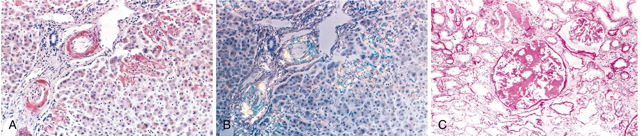
Fig. 6.46 - Robbins: (A) Congo red stain - pink-red amyloid deposits in liver vessel walls and sinusoids. (B) Apple-green birefringence under polarized light. (C) Glomerular architecture almost totally obliterated by amyloid deposition.
Organ-Specific Morphology
Kidney (most common and clinically important):
- Amyloid deposits first in mesangium and glomerular capillary walls
- Progressive glomerular obliteration
- Vascular wall deposits
- Results in nephrotic syndrome → renal failure
Spleen:
- Sago spleen (periarterial/follicular deposits) vs. lardaceous spleen (diffuse red pulp)
Liver:
- Deposits in space of Disse (between sinusoids and hepatocytes)
- Hepatomegaly; hepatic function remarkably well preserved early on
Heart:
- Subendocardial deposits and within myocardium
- Restrictive cardiomyopathy pattern
- Conduction disturbances and arrhythmias (serious, potentially fatal)
- "Rubber-like" rigid heart on gross exam
Other organs:
- Macroglossia (tongue involvement in AL amyloidosis)
- Respiratory tract - larynx to bronchioles
- Peripheral + autonomic nerves in familial forms
- Joints (hemodialysis-related Aβ2m - carpal tunnel syndrome)
Clinical Features
| System | Manifestation |
|---|---|
| Renal | Proteinuria → nephrotic syndrome → renal failure (leading cause of death) |
| Cardiac | Restrictive cardiomyopathy, CHF, arrhythmias, conduction blocks |
| GI | Macroglossia, dysphagia, malabsorption, diarrhea |
| Vascular | Vascular fragility → spontaneous bleeding; AL amyloid binds Factor X → bleeding disorder |
| General | Weakness, weight loss, lightheadedness, syncope |
Diagnosis
-
Tissue biopsy + Congo red staining with polarized microscopy - gold standard
- Sites: kidney (if renal features present), rectal mucosa, gingival tissue, abdominal fat aspirate
- Abdominal fat aspirate: high specificity but low sensitivity for systemic amyloidosis
-
Serum/urine protein electrophoresis and immunoelectrophoresis - for AL type (detect M protein)
-
Bone marrow biopsy - often shows increased plasma cells in AL type
-
Imaging - echocardiography for cardiac amyloidosis (granular sparkling appearance)
-
Potassium permanganate pretreatment (useful to distinguish types):
- AA amyloid loses Congo red affinity after permanganate treatment
- AL amyloid retains Congo red affinity
-
Immunohistochemistry on biopsy - to definitively type the amyloid protein
Prognosis and Treatment
- Prognosis depends on the organs involved and degree of functional compromise
- Renal failure and cardiac arrhythmias are the most common causes of death
- AL amyloidosis: treat the underlying plasma cell dyscrasia (chemotherapy, stem cell transplant)
- AA amyloidosis: treat the underlying chronic inflammatory disease; colchicine useful in FMF
- ATTR amyloidosis: tafamidis (TTR stabilizer), small interfering RNA agents (patisiran, vutrisiran)
- No treatment reverses established amyloid deposits effectively
Summary Box for Exam:
| Feature | Detail |
|---|---|
| Structure | Beta-pleated sheet, non-branching fibrils, 8-10 nm |
| Stain (routine) | H&E: eosinophilic hyaline |
| Gold standard stain | Congo red + polarized light → apple-green birefringence |
| Most common systemic type | AL (associated with myeloma) |
| Reactive type | AA (chronic inflammation) |
| Key organ complication | Kidney (nephrotic syndrome) |
| Cardiac finding | Restrictive cardiomyopathy |
| Diagnosis | Biopsy + Congo red |
| KMnO4 test | Abolishes Congo red in AA, not in AL |
(Robbins, Cotran & Kumar - Pathologic Basis of Disease, Chapter 6)
This is a shared conversation. Sign in to Orris to start your own chat.