
ecg showing sinus arrest, what are next step as a junior doctor
Reading File
Finding Sources
Finding Sources
Reading File
Reading File
Finding Sources
Finding Sources
Reading File
Reading File
Reading File
Now let me get the ECG image and check for causes/workup from Harrison's:
Loading Image
Finding Sources
I now have comprehensive information from multiple authoritative sources. Here is a structured clinical response:
ECG Showing Sinus Arrest — Next Steps as a Junior Doctor
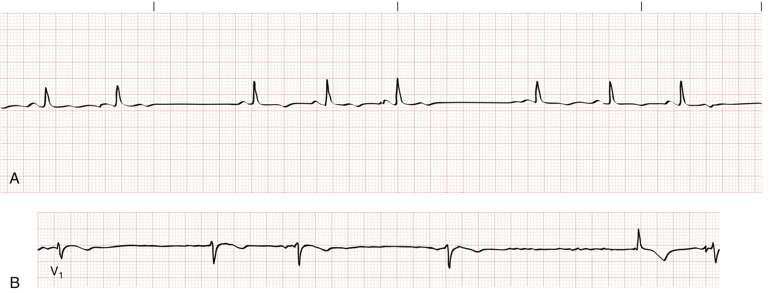
What Is Sinus Arrest?
Sinus arrest = failure of the SA node to generate an impulse, producing a pause on ECG where there are no P waves and the P-P interval delimiting the pause is not a multiple of the basic P-P interval (this distinguishes it from SA exit block, where the pause is a multiple).
If no escape pacemaker fires, the result is ventricular asystole. — Braunwald's Heart Disease
Step 1: Assess the Patient Immediately (ABCDE)
Is the patient symptomatic? This is the key decision point.
| Symptomatic | Features |
|---|---|
| Haemodynamically unstable | Hypotension, syncope/pre-syncope, chest pain, acute pulmonary oedema, altered consciousness |
| Haemodynamically stable | Asymptomatic, normal BP, well-perfused |
Emergent treatment is required if HR <50–60 bpm with hypotension or hypoperfusion. A patient with a "low-normal" HR who is simultaneously in shock may also need rhythm-directed therapy. — Tintinalli's Emergency Medicine
Step 2: Call for Help
- Senior doctor/registrar immediately if patient is symptomatic or unstable
- Alert the resuscitation team if there is haemodynamic compromise or prolonged asystole
Step 3: Urgent Investigations
Run these in parallel while monitoring:
| Investigation | Rationale |
|---|---|
| 12-lead ECG (if not done) | Confirm rhythm, look for signs of inferior MI (ST elevation in II, III, aVF), AV block, ischaemia |
| Continuous cardiac monitoring | Track rate, pauses, escape rhythms |
| IV access + bloods: U&E, Mg, Ca, glucose, TFTs | Electrolyte abnormalities (hyperkalaemia), hypothyroidism |
| ABG / SpO2 | Hypoxia as a reversible cause |
| Drug history review | β-blockers, calcium-channel blockers, digoxin, amiodarone, antiarrhythmics (class I agents) are common culprits |
| Troponin / cardiac enzymes | Rule out acute MI — inferior/posterior MI can cause SA node ischaemia |
| Echo (if available) | Structural/cardiomyopathy assessment |
Step 4: Identify and Treat the Underlying Cause
Common reversible causes (the "4 H's and 4 T's" plus drug causes):
- Drugs: β-blockers, CCBs, digoxin toxicity, membrane-active antiarrhythmics, cardiac glycosides — Fuster & Hurst's The Heart
- Inferior/posterior MI: SA node ischaemia ± Bezold-Jarisch reflex — Harrison's 22E
- Excessive vagal tone: vasovagal, carotid sinus hypersensitivity, sleep apnoea
- Metabolic: hypoxia, hypothermia, hyperkalaemia, hypothyroidism
- Intrinsic disease: sick sinus syndrome (SSS), fibrosis, cardiomyopathy, myocarditis
Step 5: Acute Treatment (Symptomatic Patient)
If haemodynamically unstable:
-
Atropine 0.5–1 mg IV — repeat every 3–5 minutes, max 3 mg total
- Effective for sinus arrest because it is vagolytic, enhancing SA node automaticity — Rosen's / Braunwald's
- Not effective in post-transplant hearts (use isoprenaline 2–10 mcg/min instead)
-
If atropine fails:
- Transcutaneous pacing — most appropriate method for the acutely symptomatic patient; confirm electrical capture (paced spike + QRS) then mechanical capture (palpable pulse)
- Isoprenaline/dopamine/adrenaline infusion as a bridge to pacing
-
Transvenous pacing — if transcutaneous fails or for more prolonged support; requires specialist expertise
Drug treatment is generally a temporary bridge to pacing unless the cause is fully reversible. — Tintinalli's
Step 6: Asymptomatic / Incidental Sinus Arrest
- Transient sinus arrest (especially during sleep) may have no clinical significance if escape rhythms prevent asystole — Braunwald's
- Still requires close monitoring, investigation for reversible causes, and senior review
- Withhold/reduce offending medications if a drug cause is identified
Step 7: Disposition and Long-Term Planning
| Scenario | Management |
|---|---|
| Reversible cause found and corrected | Monitor, outpatient follow-up |
| Recurrent/symptomatic, no reversible cause | Permanent pacemaker — indicated for symptomatic sinus node disease (SSS) — Braunwald's / Rosen's |
| Tachy-Brady syndrome | Permanent pacemaker + rate-controlling therapy for tachycardia |
| Digoxin toxicity with serious features (HR <40, sinus arrest, ventricular arrhythmia) | Digoxin-specific Fab antibody fragments — Rosen's |
Quick Summary
Sinus Arrest on ECG
│
├── ASSESS: Symptomatic? (BP, GCS, perfusion)
│
├── CALL FOR HELP (senior + resus if unstable)
│
├── 12-lead ECG + monitoring + bloods + drug history
│
├── Reversible cause? → Treat it
│
├── Symptomatic/unstable?
│   ├── YES → Atropine IV → Transcutaneous pacing → Senior/ICU
│   └── NO  → Monitor closely, investigate, senior review
│
└── Long-term → Permanent pacemaker if symptomatic & no reversible cause
Key sources: Rosen's Emergency Medicine; Braunwald's Heart Disease; Tintinalli's Emergency Medicine; Harrison's Principles of Internal Medicine 22E; Fuster & Hurst's The Heart
This is a shared conversation. Sign in to Orris to start your own chat.