
Hey
Hey! How can I help you today?
Define joints
A joint (also called an articulation) is a point where two or more bones meet. Joints provide the skeleton with both stability and mobility, depending on their type.
Searching sources...
Reading File
Finding Sources
Reading File
Reading File
Here is a complete definition and classification of joints, drawn from Firestein & Kelley's Textbook of Rheumatology:
Definition of Joints
A joint (articulation) is the point of connection between two or more adjacent bones. Joints are essential for skeletal motion, function, and quality of life. They display a broad spectrum of shapes, sizes, tissue composition, and overall organization - each combination uniquely fitted to its anatomic location and the specific type and range of motion required.
Classification of Joints
Joints are most commonly classified by their main histologic (tissue) component and by their degree of movement. These two systems correlate with and inform each other.
1. Fibrous Joints (Synarthroses)
- Tissue: Dense connective tissue bridges the adjacent bones
- Movement: Minimal to none (immovable)
- No joint cavity
- Subtypes & Examples:
- Sutures - skull bones (flexible at birth, eventually ossify)
- Syndesmoses - ligament-like tissue bridging two bones, e.g., ulna and radius (slightly movable)
- Gomphoses - periodontal tissue connecting a tooth to its bone socket
2. Cartilaginous Joints (Amphiarthroses)
- Tissue: Hyaline cartilage or fibrocartilage
- Movement: Slightly movable
- Subtypes & Examples:
- Primary cartilaginous (Synchondroses) - first rib to sternum, epiphyseal growth plates
- Secondary cartilaginous - pubic symphysis, intervertebral disks (fibrocartilaginous)
3. Synovial Joints (Diarthroses)
- Tissue: Synovial cavity lined by synovial membrane, filled with synovial fluid (water, plasma filtrate, hyaluronic acid, lubricin, phospholipids)
- Movement: Freely movable - the major functional joints of the skeleton
- Features: Enclosed by a fibrous capsule; opposing bone ends covered by articular cartilage for nearly frictionless motion
- Further classified by axes of motion:
- Uniaxial - e.g., elbow (hinge joint)
- Multiaxial - e.g., shoulder (ball-and-socket)
- Classified by shape/architecture:
- Ball and socket - hip, shoulder
- Hinge - elbow
- Saddle - first carpometacarpal joint
- Plane - patellofemoral joint
- Movements permitted: Flexion, extension, abduction, adduction, rotation
Summary Table
| Type | Tissue | Movement | Examples | Location |
|---|---|---|---|---|
| Fibrous (Synarthroses) | Connective tissue | Minimally mobile | Sutures, Syndesmoses, Gomphoses | Skull, between long bones, teeth |
| Cartilaginous (Amphiarthroses) | Cartilage/fibrocartilage | Slightly mobile | Pubic symphysis, intervertebral disk | Pelvis, spine |
| Synovial (Diarthroses) | Cavity, fluid, articular cartilage | Freely moveable | Elbow, shoulder, knee, hip, wrist | Limbs, jaw |
Below is a diagram of synovial joint structure (frontal section of knee, stained to show cartilage and tissues):
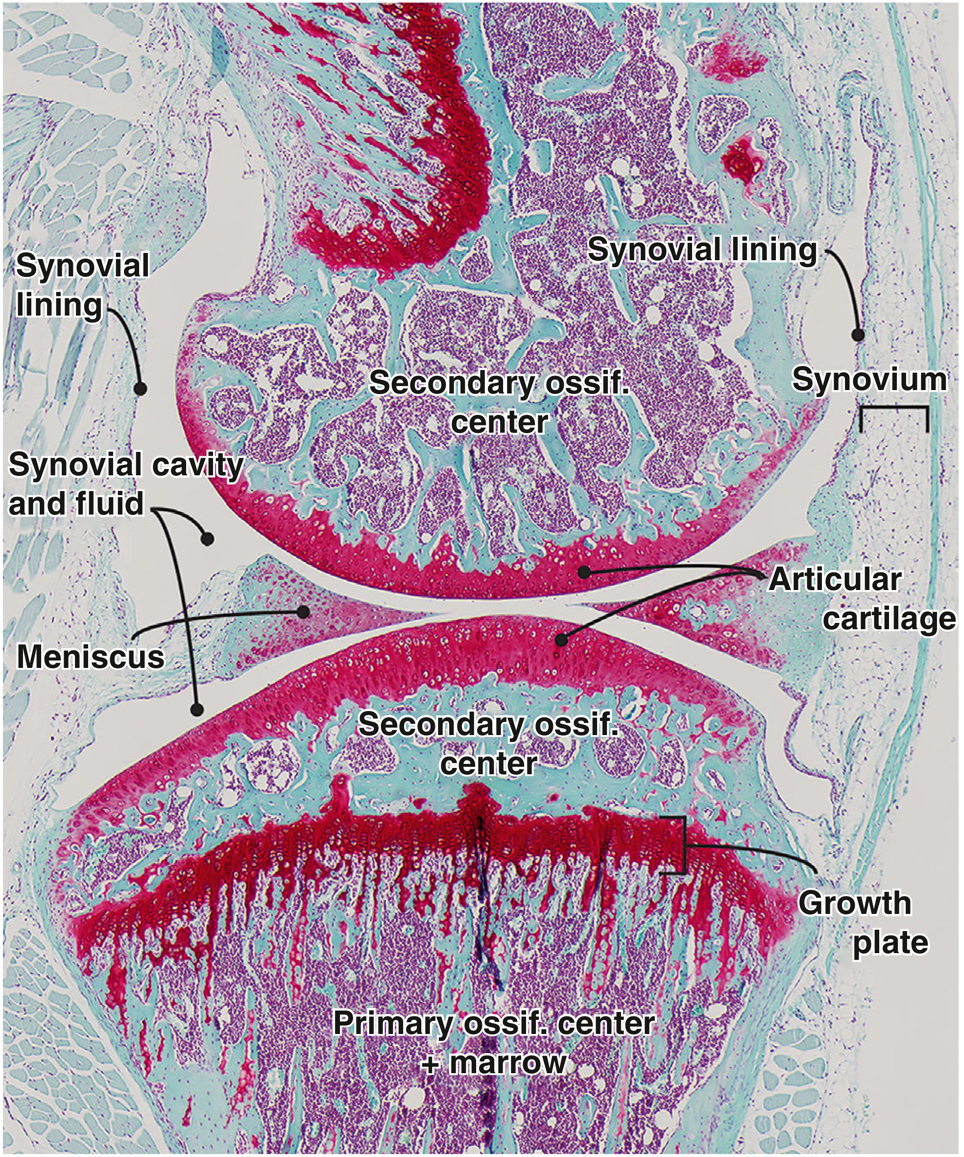
- Firestein & Kelley's Textbook of Rheumatology
This is a shared conversation. Sign in to Orris to start your own chat.