Can you explain how umbilicus is the watershed or like it acts as a watershed and why is it so that lymph and venous blood flow upwards above the spleen and downwards below the spleen? Because venous blood has to flow towards the heart. So, how is the above situation possible?

Reading File
Finding Sources
Finding Sources
Finding Sources
Reading File
Reading File
Reading File
Reading File
Loading Image
Loading Image
I now have all the textbook material and diagrams needed for a thorough answer. Let me put it all together clearly.

The Umbilicus as a Watershed - A Full Explanation

This is a brilliant question and the confusion is completely understandable. Let me break it into two separate phenomena - they are related but follow different anatomical logic.

First: What Does "Watershed" Mean Here?
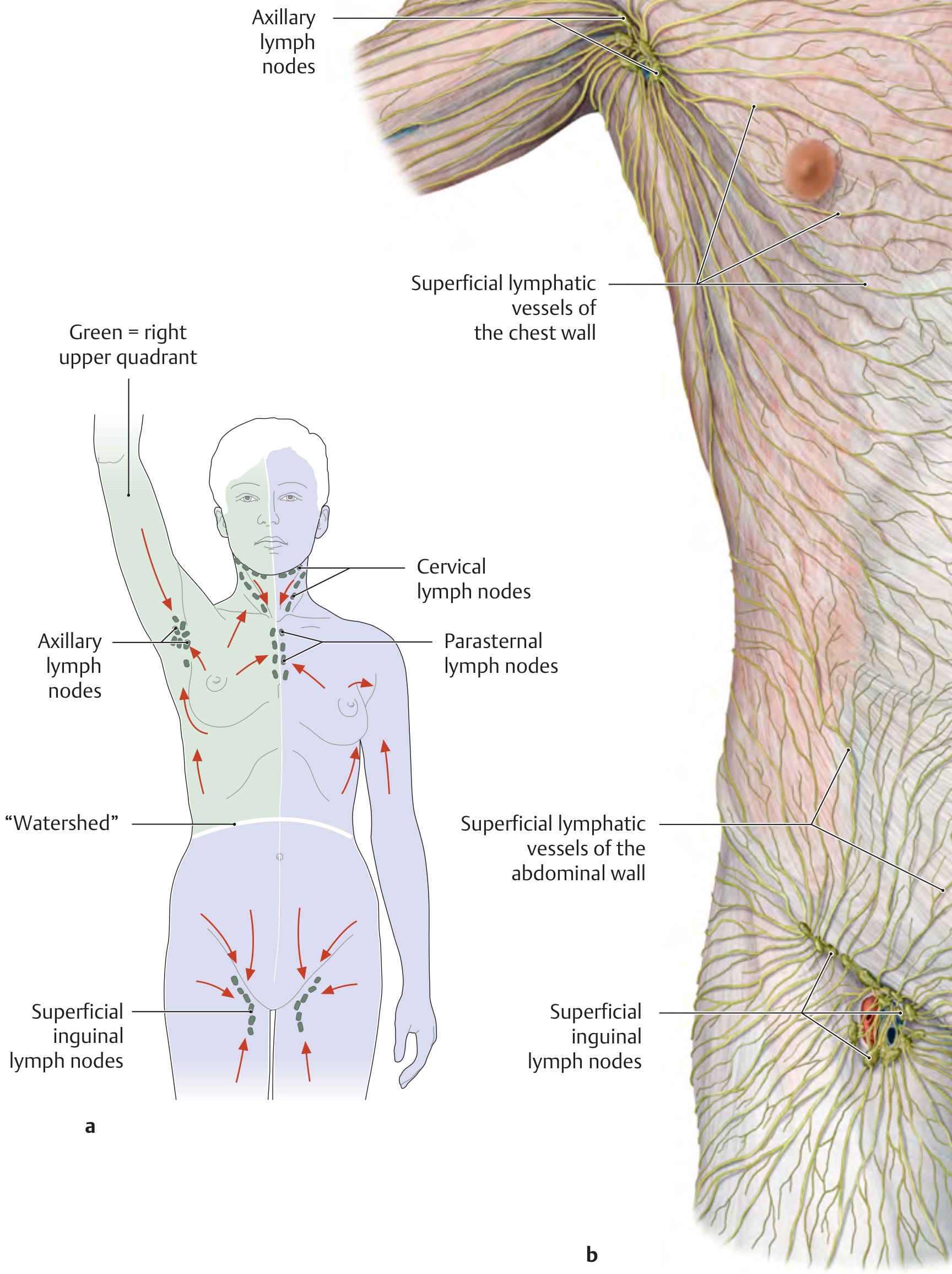
A watershed is a dividing ridge where flow splits in opposite directions depending on which side of the ridge something sits. The umbilicus acts exactly like this ridge - not for gravity, but because of where the draining vessels happen to connect into the body's larger venous and lymphatic trunks.
Watershed lymphatic drainage of trunk wall
Thieme Atlas of Anatomy - The "watershed" zone is clearly labeled at the umbilical level, with arrows showing upward flow above it (to axillary nodes) and downward flow below it (to inguinal nodes)

The Lymphatic Watershed

Above the umbilicus: Superficial lymphatics of the skin and anterior abdominal wall drain upward to the axillary lymph nodes, following the same pattern as the superficial venous drainage.
Below the umbilicus: Superficial lymphatics drain downward to the superficial inguinal lymph nodes.
The textbook defines it precisely:
"The 'watershed' zone between the two drainage regions is defined by a curved line above the umbilicus and below the costal arch. Lymph from the regional axillary and inguinal lymph nodes is finally collected by two lymphatic trunks, each of which drains into the jugulosubclavian venous junction on the corresponding side."
  • General Anatomy and Musculoskeletal System, THIEME Atlas
The key insight: lymphatics do not drain by gravity. They drain based on where the collecting ducts connect. The skin above the umbilicus developed embryologically in the same territory as the chest wall and upper limb, so its lymphatics connect to axillary nodes. The skin below developed with the lower limb territory, so it connects to inguinal nodes. Both eventually return to the venous system at the subclavian angles (one way or another), just via different routes.

The Venous Watershed - Addressing Your Core Question
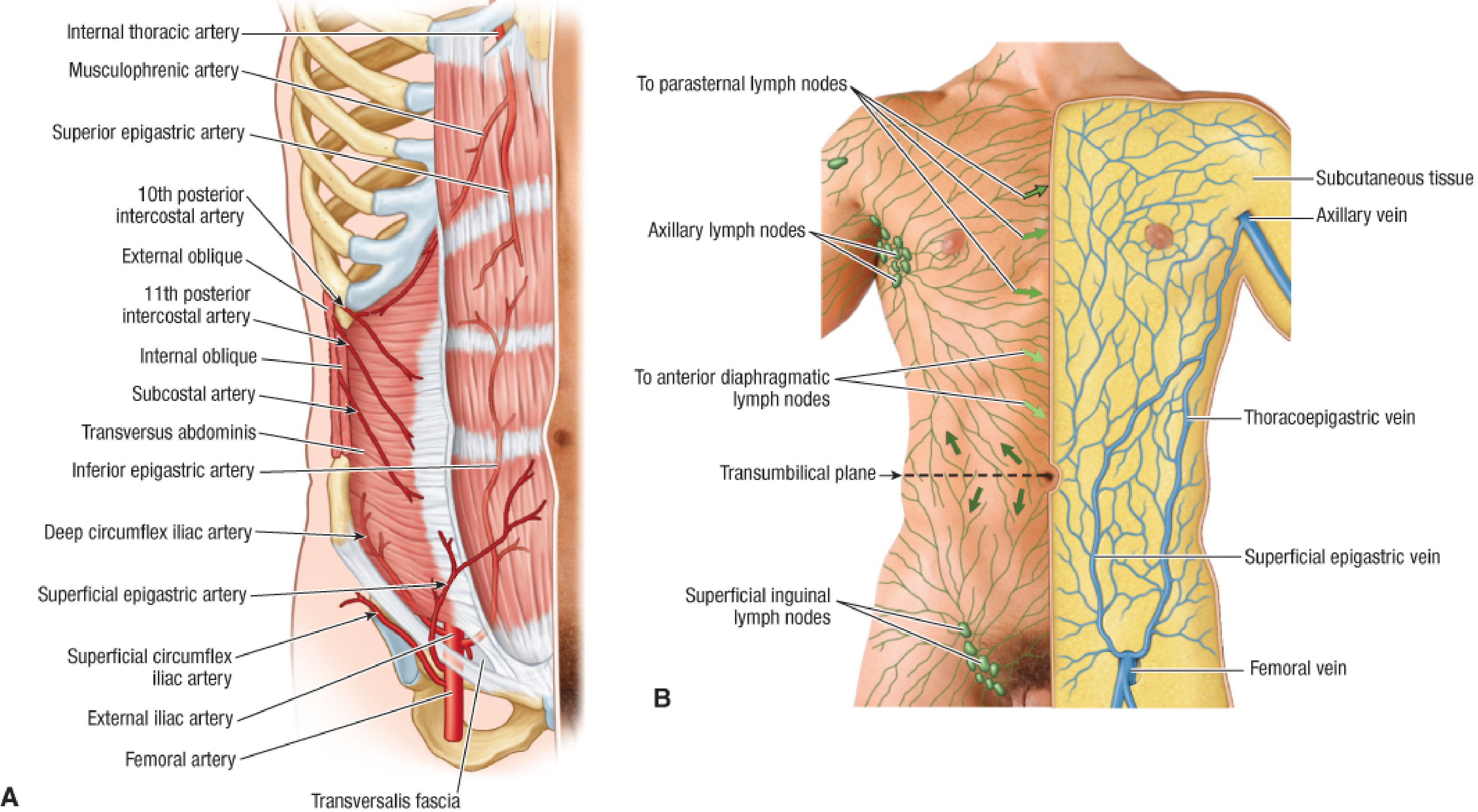
Venous and lymphatic drainage of abdominal wall
Fischer's Mastery of Surgery - Panel B shows the transumbilical plane as the watershed, with the thoracoepigastric vein bridging the two systems, and veins diverging above and below
Here is where your logic is exactly right, but the anatomy accounts for it beautifully:
Supraumbilical wall: The superficial veins drain into tributaries of the internal thoracic (mammary) vein, intercostal veins, and lateral thoracic vein, which all flow into the superior vena cava (SVC). Since the heart is superior to this region, venous blood does indeed flow upward toward the SVC - which is exactly what you'd expect.
Infraumbilical wall: The superficial epigastric, superficial circumflex iliac, and superficial external pudendal veins drain into the great saphenous vein, which drains into the femoral vein, and then into the inferior vena cava (IVC). Blood here flows downward into the saphenous/femoral system before turning and heading back up to the heart via the IVC. So it is not flowing away from the heart forever - it is first flowing down into the lower limb venous system, then returning to the heart via the IVC.
"Supraumbilical part of abdominal wall: tributaries of internal thoracic (mammary), intercostal and lateral thoracic veins that indirectly drain into the superior caval system. Infraumbilical part: superficial epigastric, superficial circumflex iliac, and superficial external pudendal veins... drain into the great saphenous vein."
  • Fischer's Mastery of Surgery, 8th ed.
So the "downward" flow below the umbilicus is not going away from the heart permanently - it is draining into the lower limb venous system, which loops back to the heart via the IVC.

The Thoracoepigastric Vein - The Bridge

There is one more elegant structure tying this together: the thoracoepigastric vein. This runs vertically along the lateral trunk wall and connects the supra- and infraumbilical venous systems, forming a collateral channel between the SVC and IVC systems. This becomes clinically important in portal hypertension and SVC/IVC obstruction.

The Portosystemic Anastomosis at the Umbilicus

The umbilicus is also a site where the portal system meets the systemic venous system - the paraumbilical veins (portal tributaries) anastomose with the periumbilical superficial epigastric veins (systemic). In portal hypertension, blood backs up through this connection and dilates the periumbilical veins, producing the visible "caput medusae" sign.

Summary Table

FeatureAbove UmbilicusBelow Umbilicus
Lymph drains toAxillary lymph nodesSuperficial inguinal lymph nodes
Superficial veins drain intoInternal thoracic / intercostal / lateral thoracic veinsGreat saphenous → femoral vein
Ultimately empties intoSuperior vena cava (SVC)Inferior vena cava (IVC)
Route back to heartDirectly upward via SVCDown into saphenous/femoral, then up via IVC
The reason veins "flow downward" below the umbilicus is not that they defy the need to return to the heart - they simply take a detour into the lower limb venous network (which is anatomically "below" them) and then return via the IVC. The umbilicus is where the two great venous territories - the SVC drainage field above and the IVC/femoral drainage field below - happen to meet, making it the watershed.

But venous blood from the superior vena cava drains into the heart from the above. So, how is the flow maintained?

Finding Sources
Finding Sources
Finding Sources
Finding Sources
Reading File
Reading File
Reading File
Reading File
Loading Image
Loading Image
This is a perfect question that gets to the heart of venous physiology (pun intended). The short answer: venous blood does NOT flow against gravity by itself - gravity is actually a problem the body has to actively overcome, and it does so through four distinct mechanisms.

Why SVC Blood Can Flow "Upward" Into the Heart
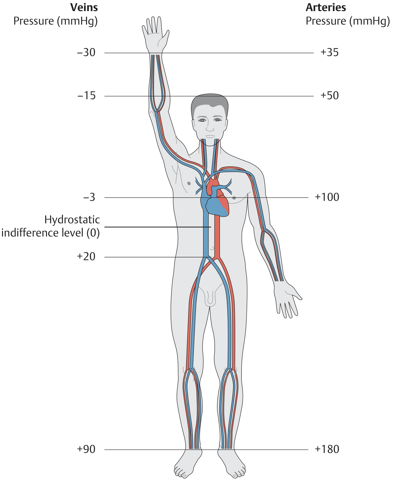
First, let's dissolve a common misconception. Look at this pressure diagram from a person standing upright:
Arterial and venous pressures in the standing position
THIEME Atlas of General Anatomy - Venous pressures in the standing position
Notice something striking on the venous side: the pressure in the upper body veins (head, neck, upper limb) is actually negative - around -15 to -30 mmHg in the raised arm! The pressure at heart level is -3 mmHg. The pressure in the foot veins is +90 mmHg.
This is the key insight: venous blood in the upper body does not need to be pushed upward toward the heart - it is actually being sucked there, because the pressure at the heart is lower (more negative) than the pressure in the neck and chest veins above it.
Gravity is pulling the blood column downward, which creates a negative pressure (a partial vacuum) in the upper body veins. Since pressure is lower at the heart than in the peripheral upper body veins, blood flows DOWN the pressure gradient - which also happens to be physically downward toward the chest. This is not fighting gravity; it is flowing WITH gravity from the upper body into the chest.

The Four Mechanisms of Venous Return

For the lower body (where blood genuinely must fight gravity to reach the heart), and to support overall venous return, the body uses four mechanisms:
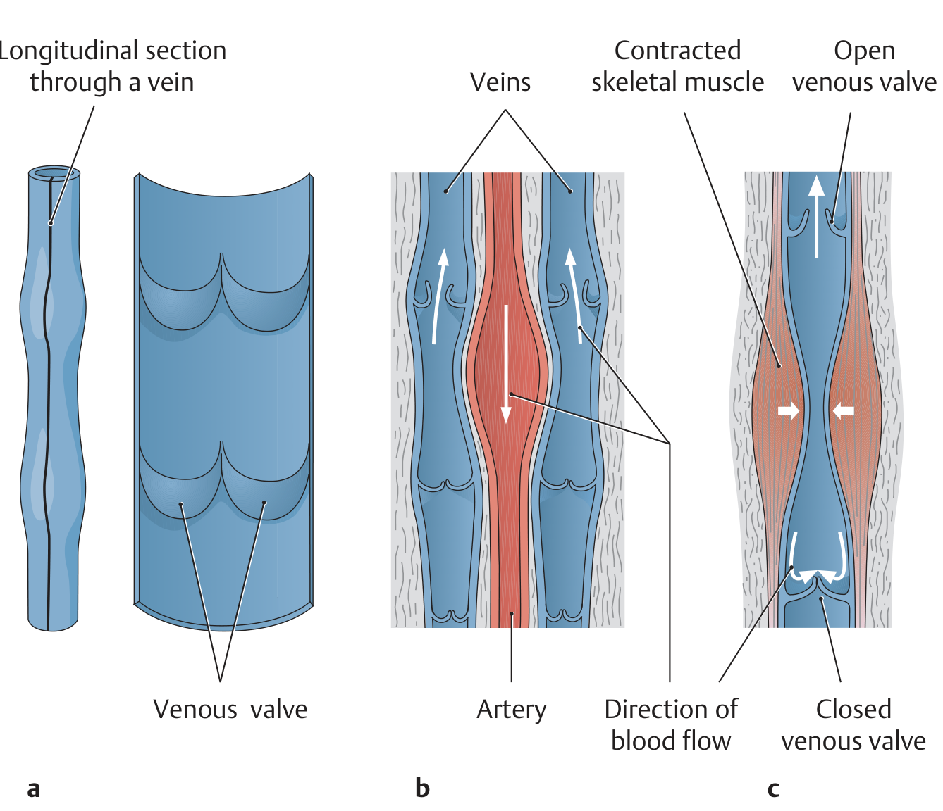
Venous valves and muscle pump mechanism
THIEME Atlas - (a) venous valves, (b) arteriovenous coupling, (c) muscle pump with valve closure preventing backflow

1. The Residual Arterial Pressure ("Vis a Tergo")

Blood leaving the capillaries still has some residual pressure (~15-20 mmHg) pushing it into the venules and veins. This is the primary driving force.

2. The Muscle Pump + Venous Valves

When skeletal muscles contract, they compress the adjacent veins and squeeze blood forward. The valves, oriented to allow flow only toward the heart, prevent backflow when the muscle relaxes. This is particularly critical in the lower limbs. As the textbook states:
"The contraction of skeletal muscle can also affect pressure and flow in veins. Large veins in the lower limbs are equipped with valves..."
  • Medical Physiology (Boron & Boulpaep)
This is why prolonged standing without movement causes venous pooling, edema, and eventually varicose veins - the pump stops working.

3. The Respiratory Pump

During inspiration, the diaphragm descends and intrathoracic pressure becomes more negative (falls below atmospheric). This expands the great veins and right atrium inside the thorax, lowering their pressure and sucking blood in from the periphery. Every breath you take helps pull venous blood toward the heart.

4. The Cardiac Suction Effect ("Vis a Fronte")

The textbook directly describes this:
"Venous return is also aided by the 'suction effect' of the heart, i.e., the negative pressure produced when the valve plane moves toward the cardiac apex during systole."
  • THIEME Atlas of General Anatomy
During ventricular systole, the AV valve plane (mitral + tricuspid rings) is pulled downward toward the apex, which transiently enlarges the atria and creates a brief suction that pulls blood into them.

Putting It All Together

RegionSituationWhy blood flows toward heart
Upper body (SVC territory)Blood is above the heartGravity + negative intrathoracic pressure create a pressure gradient pulling blood downward into the chest
Lower body (IVC territory)Blood must fight gravityResidual arterial pressure + muscle pump + respiratory pump + cardiac suction
So your intuition was pointing at the right problem. The SVC actually has it easier than the IVC - upper body blood essentially falls down into the chest. It is the lower body that needs all the mechanical assistance to push blood upward against gravity, which is why lower limb venous disease (varicose veins, DVT, chronic venous insufficiency) is far more common than upper limb venous disease.

In the earlier answer, you stated that the superficial veins drain into tributaries of internal thoracic, intercostal, and lateral thoracic vein, which all flow into the superior vena cava. And since the heart is superior to this region, venous blood does indeed flow upward toward the SVC. But superior vena cava blood, I mean, the venous blood from the superior vena cava, it drains into the heart from up. From the above, so how can you say that the blood is is flowing upwards

This is a really sharp observation, and you've caught an imprecise statement in my earlier answer. Let me correct it properly.

You Are Right - I Was Imprecise

When I said "venous blood flows upward toward the SVC," that was a loose and misleading way of putting it. Let me be precise.

The Actual Anatomy of Where Things Sit

The heart sits in the mediastinum at roughly the level of T4-T8. The SVC enters the right atrium from above, yes - but look at where the supraumbilical abdominal wall actually is relative to the heart:
  • The supraumbilical abdominal wall is at roughly T10 downward - it is below the heart, not above it.
  • The internal thoracic vein runs upward along the posterior surface of the sternum and drains into the brachiocephalic vein, which then drains into the SVC.
  • The SVC itself descends from above and enters the right atrium from the superior aspect.
So the flow path for supraumbilical abdominal wall blood is:
Supraumbilical skin veins → internal thoracic / intercostal veins → brachiocephalic vein → SVC → right atrium
The blood does travel upward from the abdominal wall to reach the brachiocephalic vein and SVC. Then the SVC carries it a short distance downward into the right atrium. There is no contradiction - the SVC is a collecting channel that sits above the heart and delivers blood downward into it.

The Key Point You Were Questioning

You were essentially asking: "If the SVC empties into the heart from above, then blood in the SVC must be flowing downward - so how was it ever flowing upward?"
The answer is - you are absolutely right that the final segment (SVC into right atrium) is a downward flow. The word "upward" I used earlier only referred to the journey of blood from the lower abdominal wall up to the level of the brachiocephalic veins and SVC. Once it reaches the SVC, gravity actually assists it downward into the heart. This is consistent with what we discussed in the last answer - upper body blood falls toward the heart, it does not have to be pushed.
So to re-state the full journey cleanly:
  1. Blood from the supraumbilical abdominal wall travels upward through intercostal and internal thoracic tributaries to reach the brachiocephalic veins (which are at the level of the clavicles, above the heart).
  2. From the brachiocephalic veins, it enters the SVC.
  3. The SVC then carries it downward (with gravity assisting) into the right atrium.
The SVC is not a vessel that goes against gravity - it runs from the level of the first rib downward to the right atrium. Blood in it is helped by gravity, not fighting it.

Why "SVC territory = upward flow" is a simplification

The phrase "drains into the SVC system" just means it ends up in the SVC before the heart - it does not mean all that blood travels upward the entire way. Blood from the chest wall travels upward to reach the brachiocephalic level, but once collected into the SVC, it falls the last short distance down into the right atrium. The net pressure gradient at every point along that path is toward the heart, whether the vessel is ascending or descending at any given segment.
So to directly answer your challenge: the SVC drains into the heart from above by flowing downward - and that is perfectly consistent with the physiology, because gravity assists that final step rather than opposing it.
This is a shared conversation. Sign in to Orris to start your own chat.