
Risk factor for varicose vein
varicose veins risk factors venous insufficiency
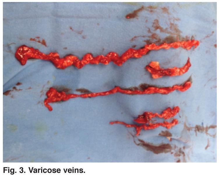
This clinical photograph displays gross surgical specimens of human varicose veins post-phlebectomy. The specimens consist of several segments of diseased venous tissue laid out on a blue surgical drape. The veins exhibit classic gross pathological features of chronic venous insufficiency, including extreme tortuosity, irregular dilation (varicosity), and a twisted, rope-like appearance. The texture is nodular and uneven, with visible focal distensions and constrictions along the length of the vessels. The specimens appear bright red and congested, likely due to intraluminal blood and the inflammatory nature of the diseased vessels. This image serves as a pathological reference for the morphological changes occurring in the great saphenous vein system or its branches during advanced venous disease, highlighting the structural degradation that necessitates surgical intervention such as ambulatory phlebectomy or radiofrequency ablation.
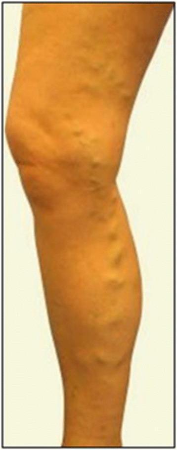
This clinical photograph displays a lateral view of a right lower limb exhibiting varicose syndrome. The image demonstrates significant venous insufficiency characterized by prominent, dilated, and tortuous superficial veins. These enlarged vessels are particularly visible along the posterior and medial aspects of the calf and around the knee joint. The veins appear as irregular, bulging, rope-like elevations beneath the skin surface. The skin over the affected areas shows mild distention due to the underlying venous pressure, but there are no overt signs of chronic venous stasis such as hyperpigmentation, stasis dermatitis, or ulceration in this specific view. The anatomical distribution is consistent with involvement of the saphenous system. This visual represents a classic presentation of severe varicose veins, which clinically correlates with symptoms like heaviness, pain, or edema, and often serves as a preoperative baseline for surgical interventions such as saphenectomy or vein stripping.
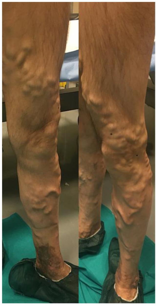
A preoperative clinical photograph showing the posterior and medial views of a lower left limb with severe chronic venous insufficiency. The image demonstrates extensive varicose veins involving the great saphenous vein distribution and its collateral branches. The veins appear markedly enlarged, tortuous, and protuberant, extending from the distal thigh through the popliteal region and down to the mid-calf. In the lower calf and ankle region, there is significant hyperpigmentation and skin discoloration consistent with venous stasis dermatitis or lipodermatosclerosis. The morphology of the veins is characteristic of advanced primary or secondary varicosities, indicating venous hypertension. This visual serves as an educational example of advanced peripheral vascular disease, highlighting the physical examination findings of varicosity, tortuosity, and secondary cutaneous changes associated with chronic venous stasis.
Risk Factors for Varicose Veins
1. Gender (Female Sex)
2. Pregnancy
- Direct mechanical compression of the IVC by the uterus, raising lower limb venous pressure
- Hormonal effects (progesterone) causing venous wall relaxation and dilation
3. Age
- 11.5% in age 18-24 years
- 14.6% in age 25-34 years
- 28.8% in age 35-44 years
- 41.9% in age 45-54 years
- 55.7% in age 55-64 years
4. Obesity (High BMI)
5. Family History / Hereditary Predisposition
6. Prolonged Standing (Occupational)
7. Inactivity / Sedentary Lifestyle
8. Height
9. Ethnicity
10. Smoking and Constipation
Pathophysiological Summary
Quick Reference Table
| Risk Factor | Strength of Evidence |
|---|---|
| Female sex | Strong |
| Pregnancy | Strong |
| Increasing age | Strong |
| Obesity (high BMI) | Moderate-Strong |
| Family history | Moderate-Strong |
| Prolonged standing | Moderate |
| Physical inactivity | Moderate |
| Tall stature | Moderate |
| Ethnicity | Variable |
| Smoking / Constipation | Inconclusive |
Which nutrient deficiency contribute to this
varicose veins nutrient deficiency vitamin
varicose veins vitamin C deficiency venous wall
nutrient deficiency contributing to varicose veins vitamin C fiber magnesium
https://pmc.ncbi.nlm.nih.gov/articles/PMC8751841
varicose veins Mendelian randomization nutrition minerals
Nutrient Deficiencies and Varicose Veins
1. Vitamin C (Ascorbic Acid) - Most Relevant
- Reduced collagen production weakens the venous wall structurally
- Veins lose tensile strength and elasticity, making them prone to dilation under normal venous pressure
- Increased vascular fragility and permeability
- Reduced antioxidant protection against oxidative damage to the endothelium
2. Dietary Fiber
- Increases intra-abdominal pressure with each straining episode
- Chronically elevates pressure transmitted to the inferior vena cava and iliac veins
- Raises venous back-pressure in the lower limbs, promoting valvular incompetence over time
3. Minerals - Evidence from Mendelian Randomization
| Nutrient | Association with Varicose Veins | Direction |
|---|---|---|
| Iron (high circulating) | Strongly associated | Risk increased (OR 1.22 per 1-SD) |
| Calcium | Inversely associated | Protective (low calcium = higher risk) |
| Zinc | Inversely associated | Protective (low zinc = higher risk) |
| Magnesium | Suggestive inverse association | Protective (low magnesium = possibly higher risk) |
| Vitamin B12 (high) | Suggestive positive association | Complex - needs more study |
4. Bioflavonoids (Vitamin P)
- Reduce capillary fragility and permeability
- Have anti-inflammatory and anti-edema effects on veins
- Support venous tone
5. Vitamin E
Summary Table
| Nutrient | Mechanism | Strength of Evidence |
|---|---|---|
| Vitamin C | Collagen synthesis, vascular integrity | Moderate (mechanistic) |
| Dietary fiber | Reduces constipation/intra-abdominal pressure | Low-Moderate (indirect) |
| Magnesium | Vascular tone, smooth muscle relaxation | Low-Moderate (MR study) |
| Zinc | Collagen cross-linking, tissue repair | Low-Moderate (MR study) |
| Calcium | Vascular smooth muscle function | Low-Moderate (MR study) |
| Bioflavonoids | Capillary integrity, venous tone | Low (indirect/therapeutic) |
| Vitamin E | Antioxidant, endothelial protection | Low |