
There are 2 fasciae, which are the parietal and visceral layers The parietal layer covers the wall: while the visceral layer surrounds the pelvic organs and forms 2 septa which pass in the sagittal direction beside the pelvic organs. The visceral fascia separates the fatty spaces located around the organs of the lesser pelvis. The peritoneoperineal fascia (Denonvilliers' fascia) stretches in the transverse direction between 2 septa of the visceral fascia and separates the fatty spaces of the lesser pelvis into 2 compartments: Pirogov-Retzius' and Amusat's capsules Can you generate a transverse section of pelvic cavity for explaining above section, do include 2 septa and Denonvilliers fascia
transverse section male pelvis rectovesical septum Denonvilliers fascia prostate rectum
Labeled anatomical diagram of a transverse (axial) section of the male lesser pelvis at the level of the bladder/prostate and rectum, viewed from above. Show: the parietal pelvic fascia lining the pelvic wall; the visceral pelvic fascia surrounding the pelvic organs and forming two sagittal septa (right and left lateral vesicorectal/sagittal septa) running anterior-posterior beside the organs; the peritoneoperineal fascia (Denonvilliers' fascia) stretching transversely between the two sagittal septa, between the bladder/prostate (anterior) and the rectum (posterior); the resulting anterior fatty space (Pirogov-Retzius' space, prevesical/paravesical) and posterior fatty space (Amusat's space, retrorectal/pararectal). Label bladder or prostate, rectum, pubic symphysis, sacrum/coccyx, obturator internus muscles, pelvic bones. Clean textbook style, clear labels with leader lines.
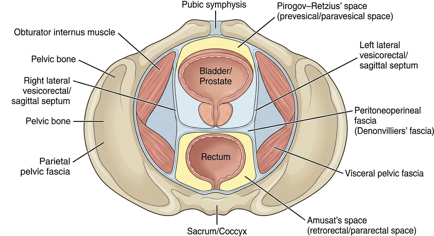
| Structure | Where it is on the section |
|---|---|
| Parietal pelvic fascia | The outer layer lining the pelvic wall, over the obturator internus and the pelvic bones. |
| Visceral pelvic fascia | The layer wrapping the bladder/prostate and the rectum. |
| Two sagittal septa (right and left) | These arise from the visceral fascia. They run anteroposteriorly on each side of the organs, from the pubic symphysis toward the sacrum. |
| Peritoneoperineal (Denonvilliers') fascia | A transverse sheet stretching between the two septa. It lies between the bladder/prostate in front and the rectum behind. |
| Pirogov-Retzius' space | The anterior fat compartment (prevesical/paravesical, shown in yellow), in front of the transverse sheet. |
| Amusat's space | The posterior fat compartment (retrorectal/pararectal, shown in yellow), behind the transverse sheet. |
- The drawing is schematic and AI-generated, so use it as a teaching sketch rather than a dissection-accurate atlas figure. I found no matching textbook image in the library.
- The labels "Pirogov-Retzius' capsule" and "Amusat's capsule" come from the Russian anatomical tradition. Western textbooks mostly call these the retropubic (Retzius) space and the retrorectal/presacral space.
- The library confirms the position of Denonvilliers' fascia as a layer between the rectum and the prostate/seminal vesicles (Bailey and Love's Short Practice of Surgery, 28th ed., Table 79.1, "Anatomical relations of the rectum").
- The library also says it is derived from peritoneum of the rectovesical pouch (Hinman's Atlas of Urologic Surgery, "Development of the Posterior Plane"). That fits the term "peritoneoperineal."
- In females, the equivalent layer is the rectovaginal septum. The section would show the vagina and uterus in place of the prostate and bladder.
Generate answer
"Denonvilliers fascia" AND anatomy
- It lines the inner surface of the pelvic wall and the pelvic muscles: obturator internus, levator ani, coccygeus and piriformis. Fischer's Mastery of Surgery, 8th ed. (section "Perirectal Tissue Planes and Spaces") describes it this way.
- The presacral fascia is part of this layer.
- It envelops the pelvic organs: bladder, prostate or vagina, and rectum. Fischer's Mastery of Surgery, 8th ed. (same section).
- The rectum has its own envelope, the mesorectal ("proper") fascia.
- Gynecology texts describe selective thickenings of this fascia forming ligaments and septa that divide the subperitoneal pelvis into potential spaces. Berek & Novak's Gynecology, section "Retroperitoneum and Retroperitoneal Spaces".
- This corresponds to your description of two sagittal septa on either side of the organs. The library calls them "bladder septa" and "descending rectal septa."
- Together with the organ fasciae, they separate the fatty spaces from one another.
- It is a two-layered sheet of fibrous connective tissue between the anterior rectum and the prostate. It extends up to cover the seminal vesicles and the ampulla of the vas deferens, where it is thickest. It extends down to the urethral sphincter, where it is thinnest. Hinman's Atlas of Urologic Surgery, section "Pelvic Fascia".
- Anteriorly it forms the border of the mesorectum. Fischer's Mastery of Surgery, 8th ed.
- Hinman's Atlas describes it as derived from an extension of the peritoneum of the pouch of Douglas (the rectovesical pouch in men). That explains the name "peritoneoperineal."
- Together with the two septa, it divides the fat of the lesser pelvis into two compartments.
| Compartment (Russian terminology) | Western equivalent | Location relative to Denonvilliers' fascia |
|---|---|---|
| Pirogov-Retzius' space | Prevesical / retropubic (Retzius) and paravesical spaces | Anterior. It holds loose connective tissue and fat. |
| Amusat's space | Retrorectal / pararectal spaces | Posterior |
- The fascial planes define the surgical planes in rectal cancer surgery (total mesorectal excision) and in prostatectomy.
- Denonvilliers' fascia should not be violated during proctectomy, to avoid sexual dysfunction. Mulholland and Greenfield's Surgery, 7e.
- Recent evidence: a 2024 meta-analysis of Denonvilliers' fascia retention versus resection (PMID 38302364) compared urinary and sexual function after laparoscopic rectal cancer surgery in men. It did not contradict the anatomy above. I only saw the title, not the abstract, so I can't say what it found.
Generate more realistic anatomical diagram
Realistic medical-textbook anatomical illustration (Netter / Gray's Anatomy style, detailed watercolor-like shading) of an axial transverse section through the male lesser pelvis at the level of the prostate and rectum, viewed from below, anterior at top. Show realistic anatomy: pubic symphysis and pubic bones, obturator internus muscles, levator ani, ischioanal fossa fat, prostate with urethra, seminal vesicles region, rectum with muscular wall and mucosa, sacrum/coccyx behind, pelvic vessels. Show fascial layers as fine distinct lines: parietal pelvic fascia lining the pelvic wall; visceral pelvic fascia enveloping the bladder/prostate and the rectum; two sagittal septa (right and left) running anteroposteriorly beside the organs; Denonvilliers' fascia (peritoneoperineal fascia) as a thin transverse sheet between the two septa separating prostate from rectum; anterior fatty compartment Pirogov-Retzius space and posterior compartment Amusat space shaded pale yellow fat. Clean leader-line labels for each structure including: parietal fascia, visceral fascia, right and left sagittal septum, Denonvilliers' fascia, Pirogov-Retzius' space, Amusat's space, prostate, rectum, pubic symphysis, obturator internus, levator ani.
axial section male pelvis prostate rectum Denonvilliers fascia levator ani
pelvic fascia transverse section parietal visceral fascia rectum bladder
Realistic textbook anatomical illustration (Netter style) of an axial transverse section through the male lesser pelvis at the level of the prostate and rectum, anterior at top. NO TEXT, NO LABELS, NO LETTERS anywhere in the image. Anatomy from front to back: pubic symphysis at top midline between the pubic bones; behind it a small pale yellow fat space; the prostate with central urethra (round, pinkish-tan) in the midline; directly behind the prostate a thin transverse fibrous membrane (Denonvilliers fascia) drawn as a distinct thin blue-white line spanning side to side; behind it the rectum (round, muscular wall, collapsed mucosal lumen) in the midline; behind the rectum a pale yellow fat space then sacrum/coccyx at bottom midline. On each side, a thin blue-white sagittal fascial septum running front to back alongside the prostate and rectum, joining the transverse membrane in an H shape. Lateral to them levator ani and obturator internus muscles in red along the pelvic walls, the pelvic bones, ischioanal fossa fat in yellow, and a thin parietal fascia line lining the pelvic wall. Clean, cream background, symmetric, high detail.
mkdir -p /tmp/workspace/pelvic-fascia && cd /tmp/workspace/pelvic-fascia && curl -s -o base.png https://cdn.orris.care/image-gen/ce76c5e0d0474403817504949abda6bd.png && python3 -c " from PIL import Image; im=Image.open('base.png'); print(im.size)"; fc-list | grep -i dejavu | head -3
cd /tmp/workspace/pelvic-fascia && cat > label.py <<'EOF' from PIL import Image, ImageDraw, ImageFont import glob base = Image.open('base.png').convert('RGB') W,H = base.size M = 330 canvas = Image.new('RGB',(W+2*M,H+80),(255,252,238)) canvas.paste(base,(M,60)) d = ImageDraw.Draw(canvas) fp = [f for f in glob.glob('/usr/share/fonts/**/DejaVuSans*.ttf',recursive=True)] bold = [f for f in fp if 'Bold' in f and 'Oblique' not in f and 'Mono' not in f and 'Serif' not in f] reg = [f for f in fp if f.endswith('DejaVuSans.ttf')] font = ImageFont.truetype((reg or bold)[0],21) tfont = ImageFont.truetype((bold or reg)[0],26) def pt(x,y): return (x+M,y+60) # (label, text anchor side, text pos (canvas coords), target (base coords)) L = [ ("Pubic symphysis","L",(20,40),(702,95)), ("Pirogov-Retzius' space\n(prevesical fat)","L",(20,150),(690,180)), ("Prostate (with urethra)","L",(20,260),(650,260)), ("Sagittal septum\n(visceral fascia)","L",(20,350),(470,330)), ("Obturator internus","L",(20,445),(400,300)), ("Levator ani","L",(20,520),(400,420)), ("Parietal pelvic fascia","L",(20,610),(330,510)), ("Ischioanal fossa fat","L",(20,690),(400,570)), ("Denonvilliers' fascia\n(peritoneoperineal)","R",(W+M+20,40),(780,353)), ("Sagittal septum\n(visceral fascia)","R",(W+M+20,170),(940,330)), ("Obturator internus","R",(W+M+20,260),(1000,290)), ("Rectum","R",(W+M+20,400),(800,470)), ("Amusat's space\n(retrorectal fat)","R",(W+M+20,500),(820,570)), ("Sacrum / coccyx","R",(W+M+20,610),(700,640)), ("Parietal pelvic fascia","R",(W+M+20,700),(1100,520)), ] for text,side,(tx,ty),(px,py) in L: lines=text.split('\n') d.multiline_text((tx,ty),text,fill=(20,20,20),font=font,spacing=3) w=max(d.textlength(l,font=font) for l in lines) h=len(lines)*26 sx = tx+w+8 if side=="L" else tx-8 sy = ty+h/2 tx2,ty2 = pt(px,py) d.line([(sx,sy),(tx2,ty2)],fill=(30,30,30),width=2) d.ellipse([tx2-4,ty2-4,tx2+4,ty2+4],fill=(30,30,30)) d.text((canvas.width//2,25),"Transverse section of the male lesser pelvis: fascial septa and compartments",fill=(10,10,10),font=tfont,anchor="mm") canvas.save('pelvic_fascia_labeled.png') print(canvas.size) EOF python3 label.py
ls /usr/share/fonts/ 2>&1 | head; python3 -c "import matplotlib, os; print(os.path.join(os.path.dirname(matplotlib.__file__),'mpl-data/fonts/ttf'))"
cd /tmp/workspace/pelvic-fascia && sed -i "s#^fp = .*#fp = glob.glob('/opt/amazon/genesis1p-tools/venv/lib64/python3.12/site-packages/matplotlib/mpl-data/fonts/ttf/DejaVuSans*.ttf')#" label.py && python3 label.py
cd /tmp/workspace/pelvic-fascia && python3 -c " from PIL import Image im=Image.open('pelvic_fascia_labeled.png'); im.save('preview.jpg',quality=80); import os; print(os.path.getsize('preview.jpg'), os.path.getsize('pelvic_fascia_labeled.png'))"
cd /tmp/workspace/pelvic-fascia && python3 - <<'EOF' s=open('label.py').read() s=s.replace('("Sagittal septum\\n(visceral fascia)","L",(20,350),(470,330)),\n ("Obturator internus","L",(20,445),(400,300)),', '("Obturator internus","L",(20,345),(430,290)),\n ("Sagittal septum\\n(visceral fascia)","L",(20,430),(470,330)),') s=s.replace('("Sagittal septum\\n(visceral fascia)","R",(W+M+20,170),(940,330)),\n ("Obturator internus","R",(W+M+20,260),(1000,290)),', '("Obturator internus","R",(W+M+20,130),(1000,290)),\n ("Sagittal septum\\n(visceral fascia)","R",(W+M+20,220),(940,330)),') open('label.py','w').write(s) EOF python3 label.py && python3 -c " from PIL import Image Image.open('pelvic_fascia_labeled.png').save('preview.jpg',quality=80)"
pelvic fascia labeled
Image · PNG
- Two sagittal septa: these are the blue-white bands running front to back on each side of the prostate and rectum, labeled "Sagittal septum (visceral fascia)."
- Denonvilliers' fascia: this is the transverse blue-white band between the two septa, lying between the prostate in front and the rectum behind. Together with the septa it forms an "H."
- Pirogov-Retzius' space: this is the yellow fat in front, behind the pubic symphysis, in the anterior compartment.
- Amusat's space: this is the yellow fat behind the rectum, in front of the sacrum and coccyx, in the posterior compartment.
- Parietal pelvic fascia: this is the thin blue line lining the pelvic wall, over the obturator internus and the muscles beside the sacrum.
- The fascial layers are drawn thicker and more obvious than they appear in real anatomy, so they are easy to see.
- The bones, muscle outlines and the exact level of the section are the AI's interpretation, not a traced dissection or CT. Treat it as a teaching illustration, not an atlas plate.
- The leader lines point to approximate positions. Because the fascia is drawn as a continuous web, a few endpoints (for example "Parietal pelvic fascia" and "Ischioanal fossa fat") may sit a few millimetres off.
- I did not draw a separate visceral fascia envelope around the prostate and rectum. The septa and Denonvilliers' fascia stand in for it.
- I found no real textbook figure of this exact section. My two image-library searches returned no matches. A third database search returned only surgical photographs of the prostate and Denonvilliers' fascia, which don't show this section. For exam or publication use, redraw it from a standard atlas such as Netter or Gray's.