
Unilateral ptosis not corrected with ucepack test RNS negative
ptosis ice pack test myasthenia gravis negative
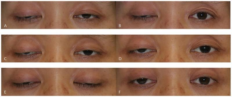
This set of six clinical photographs (A-F) illustrates the variability of the ice test in a patient with myasthenia gravis presenting with bilateral asymmetric ptosis. The images are arranged in pairs representing three different days, showing the patient's eyes before and after a 2-minute ice application. Frames A, C, and E show baseline bilateral blepharoptosis with varying degrees of upper eyelid drooping obscuring the pupil and iris. Frames B, D, and F show the post-intervention state. The series demonstrates three distinct clinical responses in the right eye: (B) a negative result with no significant change, (D) an equivocal result showing subtle 1.0 mm elevation, and (F) a positive result with a marked 2.0 mm increase in the palpebral fissure. This comparison serves as an educational tool for neurology and ophthalmology students to understand the fluctuating nature of neuromuscular junction disorders and the diagnostic utility (and inconsistency) of the ice pack test, which temporarily improves ptosis by inhibiting acetylcholinesterase at the motor endplate.
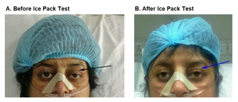
Two side-by-side clinical photographs labeled A and B demonstrate a bedside ice pack test in a patient with suspected myasthenia gravis. Image A (Before Ice Pack Test) shows significant left-sided blepharoptosis, with the upper eyelid drooping to partially cover the pupil (indicated by a black arrow). Image B (After Ice Pack Test) demonstrates a positive result, where the left eyelid shows temporary elevation and resolution of ptosis (indicated by a blue arrow) following local cooling. The patient is shown in a clinical setting wearing a blue surgical cap, a nasal cannula for oxygen delivery, and an adhesive nasal strip for stabilization. This comparative series illustrates the clinical utility of the ice pack test, which inhibits acetylcholinesterase activity at the neuromuscular junction to temporarily improve muscle strength in myasthenic patients. The images highlight key diagnostic physical exam findings in neuro-ophthalmology and neuromuscular medicine.

A clinical photograph focused on the periocular region of a 59-year-old female patient, demonstrating bilateral ptosis (upper eyelid drooping). The presentation is asymmetrical, with the right upper eyelid showing a more pronounced descent, covering a larger superior portion of the iris compared to the left eye. A vertical ruler is positioned on the left side of the frame as a measuring tool to quantify the palpebral fissure height or marginal reflex distance. This image represents the baseline state 'before ice test' in a clinical evaluation for neuromuscular junction disorders, such as ocular myasthenia gravis or Lambert-Eaton myasthenic syndrome (LEMS). The surrounding skin appears normal without visible inflammation or edema. This visual material is used to teach diagnostic bedside maneuvers for evaluating fatiguable ptosis and distinguishing between various causes of blepharoptosis in an ophthalmological or neurological context.
Unilateral Ptosis: Ice Pack Test Negative + RNS Negative
What These Two Negative Tests Mean
"RNS confirms impaired NMT but is frequently normal in mild or purely ocular disease."
- Bradley and Daroff's Neurology in Clinical Practice
Differential Diagnosis: If Not MG, Then What?
| Cause | Key Features |
|---|---|
| Horner Syndrome | Mild unilateral ptosis (2-3 mm), miosis (small pupil), anhidrosis, lower lid elevation (upside-down ptosis); no EOM involvement |
| CN III (Oculomotor) Palsy | Complete ptosis, eye deviated "down and out," dilated pupil (if compressive - e.g. aneurysm), or pupil-sparing (if ischemic - diabetes/HTN) |
| Aponeurotic/Involutional Ptosis | Most common in elderly, elevated superior eyelid crease, good levator function, no neurological signs |
| CPEO (Mitochondrial) | Bilateral (often asymmetric) slowly progressive ptosis + ophthalmoplegia; Kearns-Sayre syndrome if + retinopathy + cardiac conduction defects |
| Oculopharyngeal Muscular Dystrophy | Bilateral ptosis + dysphagia, late-onset (>50 years), autosomal dominant, GCG repeat in PABPN1 gene |
| Myotonic Dystrophy | Bilateral ptosis + facial weakness, myotonia, systemic features |
| Mechanical ptosis | Lid mass, inflammation, infiltration |
| Cerebral (Supranuclear) ptosis | Right hemispheric lesion, gaze deviation, upgaze paresis |
How to Differentiate Further
- Look for miosis (anisocoria greater in dim light)
- Apraclonidine 1% eye drop test (reversal of anisocoria confirms Horner)
- Cocaine 4-10% test (pupil fails to dilate on affected side)
- Hydroxyamphetamine test localizes to preganglionic vs postganglionic
- Is there EOM restriction (adduction, elevation, depression)?
- Is the pupil dilated? - if yes, urgent neuroimaging to rule out posterior communicating artery aneurysm
- Pupil-sparing CN III palsy in a diabetic/hypertensive patient = microvascular
- Even with negative RNS and negative ice pack test, MG is not fully excluded
- Proceed to Single-Fiber EMG (SFEMG) - the most sensitive test for NMJ dysfunction, showing increased jitter in almost all MG patients, even those with purely ocular disease (60% show abnormal jitter even in a limb muscle)
- Check AChR antibodies and anti-MuSK antibodies (AChR Ab negative does not exclude MG)
- Consider a tensilon/edrophonium test if SFEMG not available
Bottom Line
| Test | Result | Implication |
|---|---|---|
| Ice Pack Test | Negative | MG unlikely but not excluded |
| RNS | Negative | Common in ocular MG; does NOT rule it out |
| Next step | SFEMG | Most sensitive - abnormal in ~98% of MG; can confirm or exclude |
| If SFEMG also negative | Consider non-MG causes | Horner, CN III palsy, aponeurotic, myogenic |
- SFEMG (if MG still clinically suspected)
- Pupil examination - miosis = Horner; mydriasis + EOM palsy = CN III
- AChR / anti-MuSK antibodies
- Neuroimaging (MRI brain/orbits, MRA if CN III palsy with pupil involvement)
- Bradley and Daroff's Neurology in Clinical Practice (p. 2776)
- Bradley and Daroff's Neurology in Clinical Practice (p. 2770, Table 17.2)