
How to manage if pneumothorax is on mediastinum side?
Reading File
A pneumothorax on the mediastinum side refers to a tension pneumothorax - air accumulates and pushes the mediastinum away from the affected side, compressing the opposite lung and great vessels. This is a life-threatening emergency.
Searching sources...
Finding Sources
Finding Sources
Searching PubMed
tension pneumothorax management treatment
Good sources found. Now reading the key sections in parallel:
Reading File
Reading File
Reading File
Reading File
Excellent content. Let me also fetch the key image and check the Bailey & Love section for completeness:
Loading Image
Reading File
Reading Article
PMID: 40383767
I now have comprehensive information. Here is the full answer:
Tension Pneumothorax with Mediastinal Shift - Management
When a pneumothorax causes mediastinal shift (structures pushed toward the opposite side), it is by definition a tension pneumothorax - a life-threatening emergency requiring immediate action.
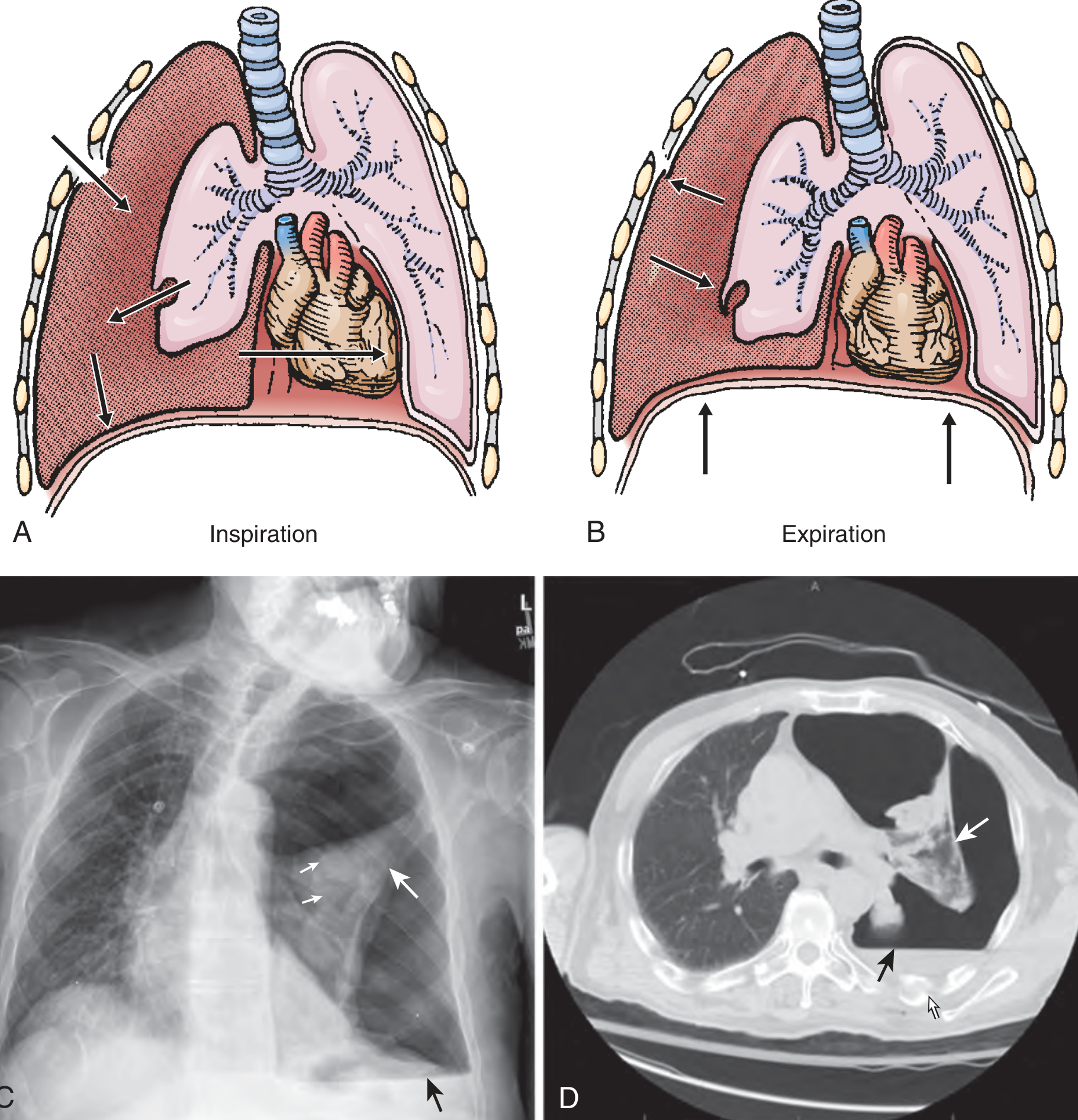
A & B: Mechanism - air enters through one-way valve during inspiration but cannot escape. C: CXR showing total left lung collapse with mediastinal shift to right. D: CT confirming findings.
Pathophysiology
Air accumulates under positive pressure via a one-way valve mechanism (from lung, airway, or chest wall). It:
- Collapses the ipsilateral lung completely
- Pushes the mediastinum toward the opposite side
- Compresses the SVC/IVC, reducing venous return and cardiac preload
- Compresses the contralateral lung, worsening hypoxia
The result is combined respiratory and circulatory failure that is rapidly fatal if untreated.
- Roberts and Hedges' Clinical Procedures in Emergency, p. 264
- Current Surgical Therapy 14e, p. 1310
Clinical Recognition (Diagnose Clinically - Do NOT Wait for CXR)
| Finding | Detail |
|---|---|
| Air hunger / tachypnea | Often the earliest sign |
| Hypoxia | Due to lung collapse |
| Absent breath sounds | On the affected side |
| Hyperresonance | On the affected side |
| Tracheal deviation | Away from the affected side |
| Distended neck veins | May be absent if hypovolemic |
| Hypotension + tachycardia | Due to reduced cardiac output |
| Increased ventilator resistance | In intubated patients - earliest sign |
Differentiate from cardiac tamponade: Both cause JVD, hypotension, and restlessness - but tamponade causes muffled heart sounds with no tracheal deviation and symmetric breath sounds.
- Current Surgical Therapy 14e
- Bailey and Love's Short Practice of Surgery 28th Ed
Management - Step by Step
STEP 1: Immediate Needle Decompression (Emergency)
Do not delay for imaging. This converts tension pneumothorax to a simple pneumothorax.
Sites (choose one):
- 2nd intercostal space, midclavicular line (2nd ICS MCL) - traditional ATLS approach; insert needle just above the 3rd rib to avoid the neurovascular bundle
- 4th-5th intercostal space, anterior axillary line - recommended by Rosen's; preferred in obese patients and may be more reliable
Technique:
- Large-bore needle/angiocatheter (14-gauge), at least 5 cm long (ideally 7 cm)
- A rush of air confirms correct placement
- Immediate improvement in vital signs expected
A 2025 meta-analysis (n = 8,046) found a 32.84% failure rate when the needle did not penetrate the pleural cavity - longer needles (7 cm) significantly reduce failure. For left-sided tension pneumothorax, the 2nd MCL is safer to avoid cardiac injury; for right-sided cases, either the 2nd MCL or 5th midaxillary line is acceptable. - Ahmad et al., World J Emerg Surg, 2025 (PMID 40383767)
STEP 2: Chest Tube (Tube Thoracostomy) - Definitive Treatment
-
Perform immediately after (or instead of, in the ED setting) needle decompression
-
In the ED, it may be just as fast to insert a chest tube directly
-
Alternatively, finger thoracostomy (without inserting the tube yet) can provide immediate relief
-
The chest tube drains residual air and any associated blood (hemopneumothorax)
-
Re-expansion of the lung usually seals the lung defect
-
Rosen's Emergency Medicine
STEP 3: Supportive Measures
- High-flow supplemental oxygen (100% O2 speeds nitrogen absorption)
- IV access, fluid resuscitation for hemodynamic instability
- Continuous monitoring (SpO2, BP, HR, ECG)
- If on positive pressure ventilation (PPV): temporarily disconnect from ventilator during decompression - PPV actively drives more air in and worsens tension
STEP 4: Post-Stabilization
- Confirm chest tube position with CXR
- Ensure lung re-expansion; if not re-expanded consider bronchial plug, contralateral tension, or malpositioned tube
- Treat underlying cause (e.g., close open chest wound with occlusive dressing - three-sided, to allow air escape)
Special Situations
| Situation | Action |
|---|---|
| Open (sucking) chest wound | Apply 3-sided occlusive dressing first; place chest tube; do NOT fully seal without a tube (risks converting to tension) |
| Ventilated patient | Disconnect from ventilator briefly during decompression; has highest risk of developing tension PTX |
| Penetrating chest trauma | Consider prophylactic chest tube before starting mechanical ventilation even if no PTX evident on imaging |
| Bilateral pneumothorax | Mediastinal shift may be absent - do not rely on it; decompress both sides |
| Asthma/emphysema on PPV | High-pressure ventilation is a common cause; have low threshold to decompress |
- Roberts and Hedges', Bailey and Love's, Rosen's EM
Key Principle
"Tension pneumothorax is diagnosed clinically, constituting a life-threatening emergency. Chest x-rays are not necessary to confirm the diagnosis, and delays to definitive treatment significantly increase the risk of circulatory collapse and cardiorespiratory arrest." - Current Surgical Therapy 14e
The sequence is: Recognize clinically → Needle decompress immediately → Follow with chest tube → Supportive care → Identify and treat the underlying cause.
This is a shared conversation. Sign in to Orris to start your own chat.