
Approach to diagnosis of gestational trophoblastic disease/ tumors md pathology exam 10 marks question
hydatidiform mole complete gross pathology microscopy
choriocarcinoma histology gestational trophoblastic
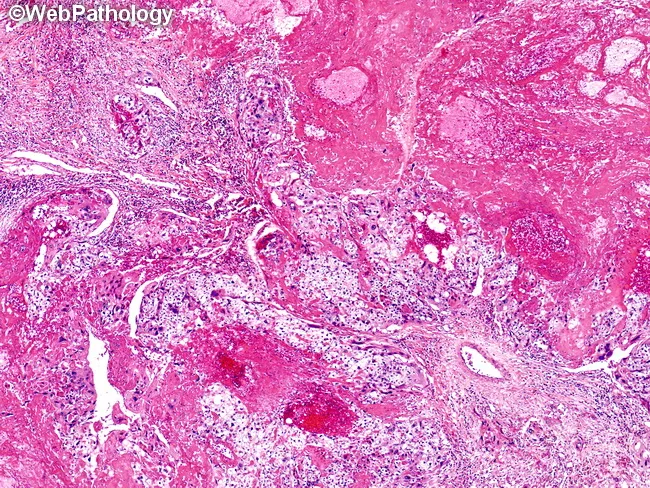
Histopathology image, prepared as a hematoxylin and eosin (H&E) stained tissue section, depicting choriocarcinoma within endometrial/uterine tissue. The lesion displays islands of viable malignant trophoblastic cells embedded in extensive hemorrhage and coagulative necrosis. Trophoblastic proliferation is biphasic, with cytotrophoblasts forming polygonal nests and clusters and syncytiotrophoblasts showing multinucleated, eosinophilic cytoplasm adjacent to fragile hemorrhagic stroma. Nuclear atypia and brisk mitotic activity are evident in viable foci. The surrounding matrix is fibromyxoid with necrotic debris and residual vascular channels, reflecting aggressive, vascular tumor growth. The lack of well-formed villi distinguishes choriocarcinoma from other gestational trophoblastic lesions. Areas of extensive hemorrhage contribute to the gross appearance of reddish, necrotic tissue in vivo. In immunohistochemistry, tumor cells typically express human chorionic gonadotropin (hCG) and cytokeratins, supporting trophoblastic lineage; p63 or inhibin may be variably positive, depending on subtype. Clinically, this histology corresponds to a highly vascular, metastasis-prone neoplasm that requires prompt evaluation of serum hCG levels and imaging for metastatic disease. The diagnostic significance lies in distinguishing choriocarcinoma from non-neoplastic gestational tissue and other germ cell tumors, guiding management including chemotherapy and monitoring for beta-hCG decline. This image is illustrative for educational reference, highlighting histologic hallmarks and diagnostic pitfalls in trophoblastic neoplasia today.
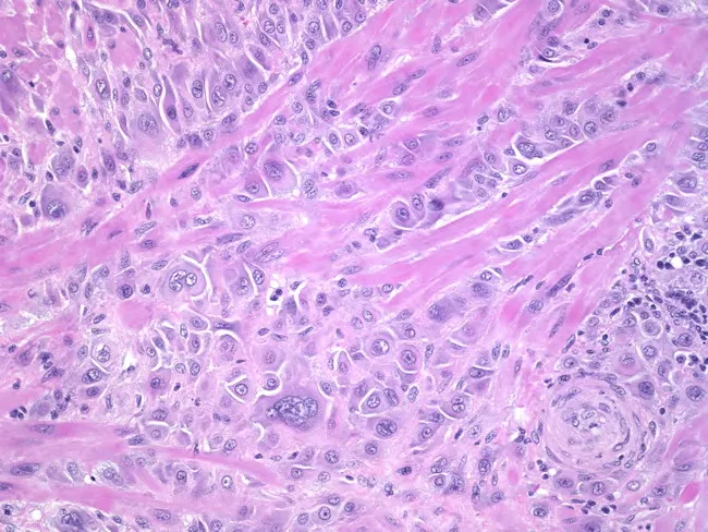
Light microscopy of hematoxylin and eosin–stained uterine tissue demonstrates invasive trophoblastic proliferation. The primary neoplastic element comprises intermediate trophoblastic cells with pleomorphic, enlarged nuclei and conspicuous nucleoli, dispersed in coherent sheets and singly scattered within the deep myometrium at the placental bed. Multinucleated giant cells are present, along with occasional clusters of cells showing subtle cytoplasmic eosinophilia. The tumor demonstrates deep myometrial invasion with irregular nests extending well away from the endometrial–myometrial junction, with minimal accompanying stromal desmoplasia. Cellular morphology includes moderate cytoplasm, nuclear variability, and occasional mitotic figures, though mitotic activity is not prominent. The architectural pattern lacks diffuse cytotrophoblastic and abundant syncytial proliferation seen in choriocarcinoma, and there is relative preservation of placental barrier features in otherwise infiltrative tissue. These findings are most compatible with an invasive trophoblastic neoplasm arising from placental site trophoblastic tumor (PSTT) or epithelioid trophoblastic tumor (ETT) spectrum, rather than classic choriocarcinoma. Clinically, such histology correlates with gestational trophoblastic disease and necessitates correlation with serum hCG, pregnancy history, and imaging. Differential diagnoses include PSTT, ETT, choriocarcinoma, and invasive mole; the prognosis and treatment vary, with PSTT/ETT often requiring surgical management and close follow-up with surveillance.
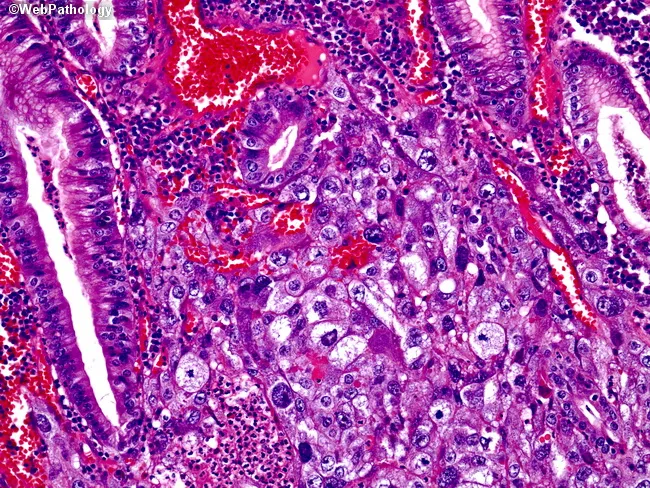
This histopathology slide shows a gastric carcinoma with choriocarcinomatous differentiation. Hematoxylin and eosin stained tissue from the stomach demonstrates a biphasic neoplasm: conventional adenocarcinoma glands forming malignant epithelial elements intermingled with a focal trophoblastic component composed of cytotrophoblasts (polygonal cells with distinct borders and vesicular nuclei) and intermediate trophoblasts arranged in sheets and nests. The trophoblastic component invades the gastric mucosa and submucosa; there is frequent hemorrhage and areas of necrosis; tumor cells show marked pleomorphism and mitotic activity. The histology is identical to gonadal choriocarcinoma, lending support to the diagnosis of primary gastric choriocarcinoma arising in association with adenocarcinoma; serum beta-hCG levels are typically elevated in patients with trophoblastic differentiation. Immunohistochemistry would reveal positivity for HCG in the trophoblastic cells, with cytokeratin expression in the adenocarcinoma component. Clinically, this entity portends an extremely poor prognosis and aggressive behavior, necessitating prompt systemic therapy and careful tumor staging to exclude metastatic germ cell disease. This image is relevant for pathology education, differential diagnosis in gastric tumors, and studies of trophoblastic differentiation in non-gestational contexts. Potential use cases include diagnostic pathology review, teaching slide collections, and research into gastric choriocarcinoma biology, trophoblastic markers, and therapeutic targeting.
Approach to Diagnosis of Gestational Trophoblastic Disease (GTD) / Tumors
Definition and Classification
| Category | Entity |
|---|---|
| Benign / Premalignant | Complete Hydatidiform Mole, Partial Hydatidiform Mole |
| Locally Invasive (GTN) | Invasive Mole |
| Malignant (GTN) | Gestational Choriocarcinoma |
| Rare Malignant | Placental Site Trophoblastic Tumor (PSTT), Epithelioid Trophoblastic Tumor (ETT) |
Clinical Presentation - When to Suspect GTD
- Hyperemesis gravidarum disproportionate to gestational age
- Uterus large for dates (classic sign)
- Persistent irregular vaginal bleeding after pregnancy (normal, ectopic, or molar)
- Markedly elevated serum hCG for gestational age
- Early-onset preeclampsia (before 20 weeks)
- Bilateral theca lutein ovarian cysts (caused by hCG stimulation)
- Hyperthyroidism (hCG has mild TSH-like activity)
- Respiratory failure or neurological symptoms - suggests metastatic GTN
Diagnostic Approach
1. Clinical History
- Prior molar pregnancy, abortion, ectopic pregnancy, or live birth
- Time elapsed since last pregnancy (important for risk stratification)
- Age (risk highest at extremes of reproductive life: teens and >40 years)
- Symptoms as above
2. Serum hCG (The Cornerstone Marker)
- Quantitative serum beta-hCG must be measured in ALL cases
- In complete mole: hCG markedly elevated above levels expected for gestational age
- In partial mole: hCG less elevated, but above normal
- Choriocarcinoma: typically very high (may exceed mole levels), though necrotic tumors may have low levels
- PSTT/ETT: only mildly elevated hCG (key distinguishing feature - these tumors produce human placental lactogen/HPL more than hCG)
- Persistent elevation after evacuation = persistent GTN, requires further management
- A urine pregnancy test for hCG is a useful first step in any suspected case
3. Pelvic Ultrasonography
- First-line imaging tool
- Complete mole: classic "snowstorm" appearance - uterine cavity filled with multiple echogenic foci without a gestational sac or fetus
- Partial mole: ratio of transverse to AP dimension of gestational sac >1.5, Swiss-cheese appearance of placenta, possible fetal parts
- Bilateral theca lutein cysts (>6 cm) indicate very high hCG - associated with high-risk disease
- Invasive mole: myometrial invasion visible; Doppler shows increased vascularity
- Used to detect disease extent and uterine size
4. Gross Pathology
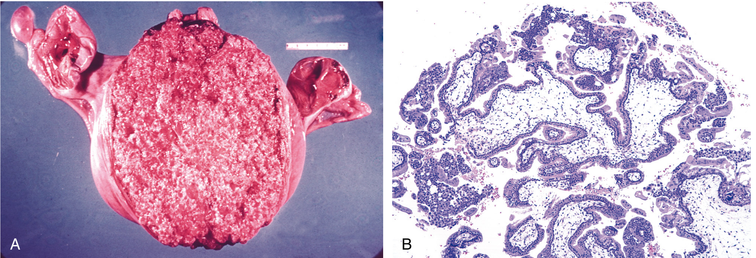
- Uterus markedly enlarged, filled with thin-walled, translucent, cystic, grape-like vesicles
- Delicate friable mass of hydropic (edematous) villi
- No fetal parts
- Only some villi are enlarged; fetal parts may be present
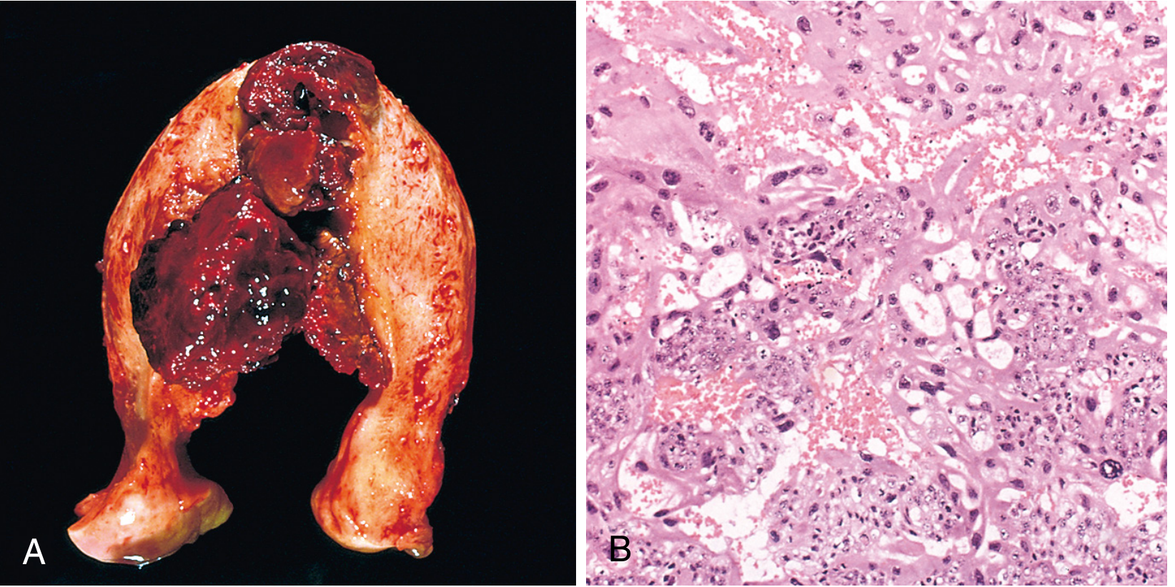
- Soft, fleshy, yellow-white bulky mass
- Large areas of pale necrosis and extensive hemorrhage
- Invades myometrium, penetrates blood vessels
5. Histopathology (Definitive Diagnosis)
A. Complete Hydatidiform Mole
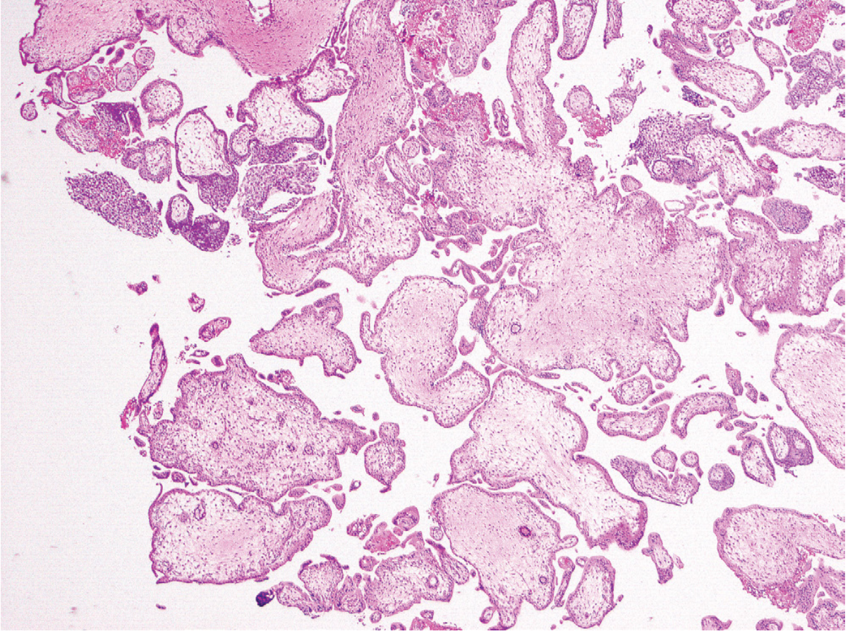
- All chorionic villi enlarged, smooth, oval
- Central cistern (central cavitation/cisternae) formation
- Marked villous edema (hydropic change)
- Diffuse, circumferential trophoblastic hyperplasia - both cytotrophoblasts AND syncytiotrophoblasts
- No fetal vasculature in the villi (avascular villi)
- Nuclear atypia in trophoblasts
B. Partial Hydatidiform Mole
- Only some villi enlarged - two population of villi (large + small)
- Villous outlines irregular/scalloped (unlike smooth round complete mole)
- Focal, slight trophoblastic hyperplasia - predominantly syncytiotrophoblasts only
- Prominent trophoblastic inclusions within villi (characteristic)
- Fetal vasculature and fetal RBCs may be present
- Central cisterns less pronounced
| Feature | Complete Mole | Partial Mole |
|---|---|---|
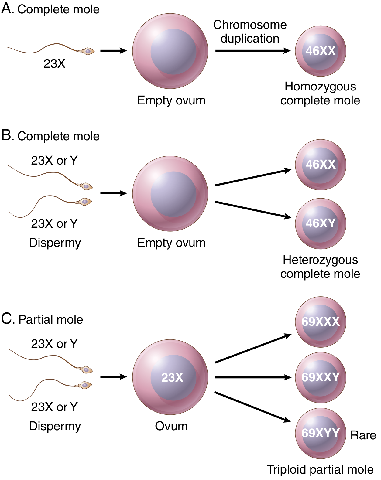
| Karyotype | Diploid (46,XX or 46,XY) | Triploid (69,XXY most common) |
| Villous edema | All villi | Some villi |
| Trophoblast proliferation | Diffuse; circumferential | Focal; slight |
| Serum hCG | Markedly elevated | Less elevated |
| Tissue hCG | ++++ | + |
| Fetal/embryonic tissue | Absent | May be present |
| Risk of choriocarcinoma | 2.5% | Rare |
C. Invasive Mole
- Hydropic chorionic villi with proliferating cytotrophoblast and syncytiotrophoblast
- Invades myometrium - the villi penetrate the myometrial wall (key diagnostic feature)
- May perforate the uterus
- Emboli to distant sites (lungs ~5%) but the villi remain intact
- Not true carcinoma - villi are present (distinguishes from choriocarcinoma)
D. Gestational Choriocarcinoma
- No chorionic villi (absolute hallmark - absence of villi distinguishes from invasive mole)
- Biphasic proliferation: malignant cytotrophoblasts (polygonal, mononuclear, pale cytoplasm) + syncytiotrophoblasts (large, multinucleate, dark, eosinophilic)
- Abundant mitoses, often abnormal
- Extensive hemorrhage and necrosis throughout the tumor
- Deep myometrial invasion and vascular penetration
- IHC: hCG strongly positive in syncytiotrophoblasts; cytokeratin positive
E. Placental Site Trophoblastic Tumor (PSTT)
- Composed of polygonal mononuclear or binucleated extravillous (intermediate) trophoblasts
- Abundant eosinophilic cytoplasm
- Invades myometrium by splaying apart smooth muscle fibers without frank destruction or hemorrhage (unique feature)
- Shows tropism for maternal blood vessels
- hCG only mildly elevated; HPL (human placental lactogen) strongly positive - IHC key diagnostic marker
- No villi; no biphasic pattern
- 10-15% mortality from disseminated disease
F. Epithelioid Trophoblastic Tumor (ETT)
- Sheets of mononuclear trophoblast cells with abundant clear cytoplasm
- Associated with characteristic pink hyaline material (eosinophilic matrix) - pathognomonic feature
- Most commonly intrauterine or cervical; extrauterine cases occur
- Lacks overt invasive features of PSTT
- Capable of metastasis to lungs and bone
- IHC: p63 positive (unlike PSTT); cyclin E positive
6. Immunohistochemistry (IHC) Panel
| Marker | Complete Mole | Choriocarcinoma | PSTT | ETT |
|---|---|---|---|---|
| hCG | ++++ | ++ (syncytia) | + | + |
| HPL | - | - | ++++ | + |
| p63 | - | - | - | ++++ |
| Inhibin | + | + | + | - |
| Cytokeratin | + | + | + | + |
| Ki-67 | Variable | High | Low-moderate | Moderate |
7. Cytogenetics / Molecular Studies
- Flow cytometry or FISH: ploidy analysis - confirms diploid (complete) vs. triploid (partial)
- Short tandem repeat (STR) analysis / DNA fingerprinting: distinguishes gestational from non-gestational choriocarcinoma (gestational tumors have paternal DNA); also proves origin from a molar pregnancy
- p57KIP2 immunostaining: extremely useful differentiator
- p57 is a paternally imprinted, maternally expressed gene
- Complete mole: p57 negative (absent maternal genome - no expression in cytotrophoblasts/stromal cells)
- Partial mole and hydropic abortion: p57 positive
- This is the most practical tool to distinguish complete mole from partial mole and non-molar hydropic abortion on formalin-fixed tissue
8. Staging and Risk Stratification (for Malignant GTN)
- Chest X-ray / CT chest (lungs most common metastatic site - 50%)
- CT abdomen and pelvis
- MRI brain (if neurological symptoms or high-risk score)
- Bone scan if clinically indicated
Summary Diagnostic Algorithm
Suspect GTD (vaginal bleeding + elevated hCG + clinical features)
|
Serum beta-hCG (quantitative)
Pelvic ultrasound
|
Uterine evacuation (suction curettage)
|
Histopathology of all products of conception
|
Molar pregnancy confirmed?
|
YES → Monitor hCG weekly post-evacuation
→ Persistent/rising hCG = GTN
→ FIGO staging + WHO risk scoring
→ CT chest/abdomen/pelvis ± MRI brain
Key Diagnostic Points for Exam
- hCG is the single most important marker - monitored serially post-evacuation
- Histology remains the gold standard for typing - all evacuation material must be examined
- Absence of villi = choriocarcinoma (villi present = mole or invasive mole)
- p57KIP2 negativity = complete mole (paternally imprinted gene, no maternal genome)
- PSTT/ETT produce HPL > hCG - important because hCG alone underestimates disease
- Gestational vs. non-gestational choriocarcinoma: distinguished by paternal DNA (STR analysis) - prognostic significance since gestational type is highly chemosensitive
- Persistent hCG after evacuation defines GTN and mandates systemic chemotherapy workup
- Near 100% cure rate with appropriate chemotherapy (methotrexate for low-risk; EMA-CO for high-risk)