
trigeminal neuralgia
"trigeminal neuralgia"[MeSH Terms] AND treatment
trigeminal neuralgia anatomy trigeminal nerve branches
trigeminal nerve distribution face V1 V2 V3
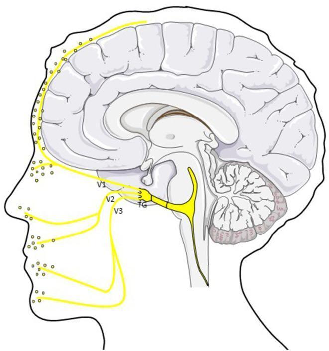
An anatomical diagram in sagittal profile illustrating the trigemino-vascular system and its role in migraine pathophysiology. The central focus is the trigeminal ganglion (TG), from which three primary divisions of the trigeminal nerve (CN V) emerge: the ophthalmic (V1), maxillary (V2), and mandibular (V3) branches. These peripheral afferents are shown innervating the scalp, forehead, mid-face, and jaw regions, with yellow dots indicating the release of Calcitonin Gene-Related Peptide (CGRP) at nerve terminals. Intracranially, the diagram highlights the ophthalmic division's (V1) innervation of the dura mater and cerebral vasculature. Centrally, a primary nerve tract is depicted traveling from the trigeminal ganglion toward the brainstem and trigeminocervical complex (TCC), representing the pathway for nociceptive signal transmission. The illustration combines a stylized mid-sagittal section of the human brain—including the cerebrum, cerebellum, and brainstem—with a facial outline to demonstrate the neuroanatomical connection between intracranial meningeal structures and peripheral facial sensory distribution.
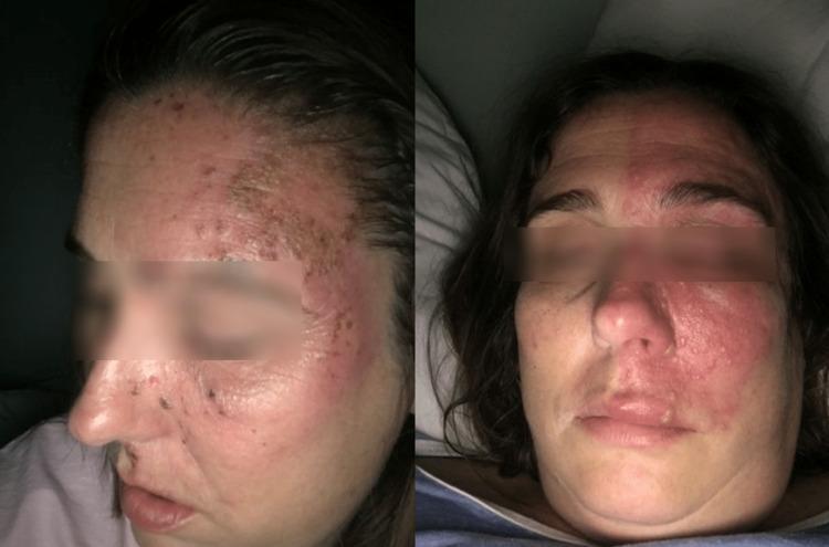
Clinical photograph of a patient's face demonstrating skin manifestations of herpes zoster (shingles) with a classic dermatomal distribution. The image consists of two views: a lateral profile of the left side and an anterior-posterior view. On the left side of the face, there is a cluster of erythematous, vesicular, and hemorrhagic crusted lesions. These lesions follow the sensory distribution of the ophthalmic (V1), maxillary (V2), and mandibular (V3) branches of the trigeminal nerve. A sharp midline demarcation is visible, where the intense erythema and papular lesions end abruptly at the facial midline, characteristic of varicella-zoster virus reactivation. The affected area involves the left forehead, eyelid, nose, cheek, and upper lip. The presence of lesions on the tip and side of the nose (Hutchinson's sign) indicates involvement of the nasociliary branch of the ophthalmic nerve, raising clinical concern for ocular complications. This image serves as an educational example of trigeminal zoster and associated post-herpetic trigeminal neuralgia manifestations.
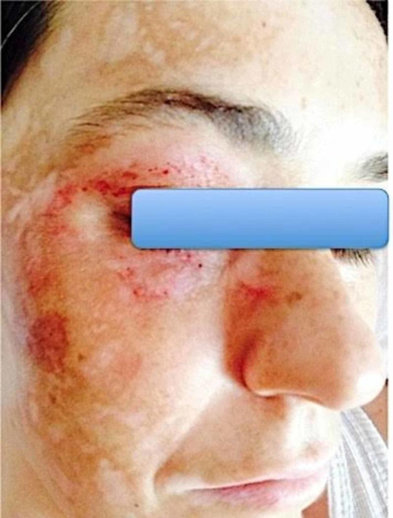
This clinical photograph shows a close-up frontal view of a patient's face, primarily focusing on the right side. The skin exhibits complex dermatological changes following stereotactic radiosurgery for trigeminal neuralgia. The lesions are localized in a distribution corresponding to the branches of the right trigeminal nerve (V1, V2, and V3). Morphological features include multiple erythematous, petechial-like punctate lesions and maculopapular eruptions around the orbital region and right cheek. Additionally, there are significant areas of dyschromia, characterized by large, irregular patches of hypopigmentation (leukoderma) interspersed with hyperpigmented macules, giving the skin a mottled appearance. The skin texture appears uneven with areas of thinning and possible scarring. This image demonstrates the dermatological manifestations of Trigeminal Trophic Syndrome (TTS), a rare complication where self-inflicted trauma or dysregulated sensory feedback leads to chronic skin lesions in the anesthetic distribution of the nerve. It serves as an educational example of the clinical relationship between neuropathic pain disorders and secondary cutaneous pathology.
Trigeminal Neuralgia
Definition
Classification (ICHD-3)
| Type | Description |
|---|---|
| Classical TN | MRI or surgical evidence of neurovascular compression with nerve root atrophy or displacement |
| Secondary TN | Caused by an underlying disease (multiple sclerosis, space-occupying lesion); may have continuous background pain in addition to paroxysms |
| Idiopathic TN | No causative lesion identified and no clear morphological nerve change on imaging |
Epidemiology
- Incidence: ~4 per 100,000 population
- Onset after age 40 in 90% of patients; peak incidence 50-70 years
- Slightly more common in women (ratio ~1.5:1)
- In younger patients, always consider multiple sclerosis as a secondary cause
- Rare familial cases exist, suggesting a genetic contribution in some families
-
- Bradley and Daroff's Neurology in Clinical Practice; Goldman-Cecil Medicine
Pathogenesis
- Superior cerebellar artery (most common offending vessel)
- Anterior/posterior inferior cerebellar arteries
- Superior petrosal vein
- Ectopic neuronal discharges
- Ephaptic transmission (cross-talk between neighboring axons)
- Paroxysmal bursts of pain from trivial stimuli
Clinical Features
Characteristic Pain
- Quality: Electric shock-like, lancinating, shooting - often described as the worst pain imaginable
- Duration: Seconds (rarely up to 2 minutes); attacks may cluster and blur together
- Distribution: Most commonly V2 (maxillary) and V3 (mandibular) divisions; V2+V3 combination is most frequent. V1 (ophthalmic) alone is extremely rare.
- Laterality: Unilateral (bilateral is very rare)
- Timing: Mostly diurnal; attacks during sleep are uncommon but do occur
Trigger Factors
- Light touch to the trigger zone (often near the nasolabial fold, remote from pain)
- Chewing, talking, brushing teeth
- Cool air or breeze on the face
- A refractory period typically follows each volley
What is Absent (Classical TN)
- No sensory loss on examination
- No motor weakness of masticatory muscles
- The presence of sensory deficit or weakness should prompt consideration of secondary TN / trigeminal neuropathy from a mass or lesion at the gasserian ganglion, main sensory root, or REZ
Trigeminal Nerve Anatomy
Diagnosis
- Paroxysmal attacks of pain lasting 1 second to 2 minutes
- Affecting one or more trigeminal divisions
- Intensely sharp, stabbing, or triggered by a stimulus
- Stereotypical attacks
- No other neurological deficits (in classical TN)
Investigations
- MRI brain (mandatory): To exclude secondary causes - MS plaques, pontine lacunar infarct, meningioma, schwannoma, skull base malignancy, vascular loops
- High-resolution MRI/MRA: May identify neurovascular compression
- EMG/blink reflex studies: Normal in idiopathic TN
Differential Diagnosis
- Trigeminal autonomic cephalalgias (have autonomic features - lacrimation, conjunctival injection, nasal congestion)
- Atypical facial pain / persistent idiopathic facial pain
- Idiopathic stabbing headache
- Tolosa-Hunt syndrome
- Dental pain, TMJ disorder
- Post-herpetic neuralgia
Treatment
Medical (First-line)
| Drug | Dose | Notes |
|---|---|---|
| Carbamazepine | 600-1200 mg/day in divided doses | First-line; sodium channel blocker; initiate at 50-100 mg and titrate slowly |
| Oxcarbazepine | 300-1800 mg/day | Better tolerated than carbamazepine; watch for hyponatremia |
- Monitor CBC, LFTs, serum sodium at baseline, during first months, and annually (risk of agranulocytosis with carbamazepine)
- Once pain is controlled, try periodic tapering to assess for spontaneous remission
Medical (Second-line)
- Gabapentin (900-1800 mg/day) - favorable side-effect profile, often tried early
- Pregabalin
- Baclofen (50-60 mg/day)
- Lamotrigine (100-400 mg)
- Phenytoin (200-300 mg)
- Valproate, clonazepam, topiramate also used
- Botulinum toxin injections - evidence of benefit
Acute Severe Attack
- IV fosphenytoin 15-20 mg phenytoin equivalents/kg
- Anesthesia of the ipsilateral conjunctival sac with proparacaine (ophthalmic local anesthetic) can provide relief for hours to days
Surgical / Procedural Options (for medically refractory cases)
| Procedure | Mechanism | Best For | Key Points |
|---|---|---|---|
| Microvascular Decompression (MVD) | Craniotomy; offending vessel separated from nerve + Teflon pad placed | Young, fit patients | 70% pain-free at 10 years (Jannetta series); preserves sensation; mortality ~1%; risk to CN IV, VII, VIII |
| Percutaneous Radiofrequency Thermocoagulation | Heat ablation of trigeminal ganglion | Elderly, medically unfit | Effective; produces sensory loss (corneal numbness risk) |
| Percutaneous Balloon Compression | Mechanical compression of gasserian ganglion | Elderly/comorbid | Relatively simple; produces sensory loss |
| Glycerol Rhizotomy | Chemical destruction of trigeminal nerve | Elderly/comorbid | Produces sensory loss |
| Gamma Knife Radiosurgery (stereotactic) | Focused radiation to REZ | Poor surgical candidates | Non-invasive; delayed onset of pain relief (weeks-months) |
| Peripheral nerve block (alcohol) | Peripheral branch block of V1/V2/V3 | Temporary relief, elderly | Relief in high proportion; relapse in 6-18 months; repeatable 1-2x |
- Young, healthy patient + refractory to medications → MVD (only procedure addressing the underlying mechanism)
- Elderly or significant comorbidities → Percutaneous procedures at the gasserian ganglion (radiofrequency thermocoagulation or balloon compression)
Course and Prognosis
- Spontaneous remissions can occur, especially early in the disease
- Frequency of attacks tends to increase over time
- Medical therapy controls symptoms in the majority initially, but long-term control may diminish
- Surgical outcomes are best with MVD (longest-lasting pain relief)
- Secondary TN (MS-related) may be harder to control and often requires procedural management; recent systematic reviews confirm radiosurgery and minimally invasive rhizotomy are the most used approaches in MS patients (PMID 40220576; PMID 38648730)
Recent Evidence (2024-2026)
- A 2025 systematic review/meta-analysis (PMID 39780062) found TENS (transcutaneous electrical nerve stimulation) may improve TN symptoms - an emerging non-invasive adjunct
- Two 2024-2025 systematic reviews confirm that in MS-associated TN, minimally invasive procedures (radiosurgery, radiofrequency rhizotomy) are effective but recurrence rates are higher than in classical TN