
A 26-year-old male. A 26-year-old male. A 26-year-old male. A 26-year-old male. A 26-year-old male. Admitted with a history of... Admitted with a history of... Admitted with a history of... Admitted with a history of... Admitted with a history of... Fever for one month. Fever for one month. Fever for one month. Fever for one month. Fever for one month. Vet-ııı, ilacı. Vet-ııı, ilacı. Vet, eee, ilacı. Vet-ııı, iyi. Difficulty in breathing. Difficulty in breathing. With, अ, इ-- Difficulty in breathing. With history of difficulty in breathing. Difficulty in breathing. With history of difficulty in breathing. Difficulty in breathing. With history of difficulty in breathing. With history of difficulty in breathing. With history of difficulty in breathing. On admission, his vitals was BP 110/80, sa- uh, pulse rate 120, and saturation is 80% in room air and improved with nasal oxygen. He was started on injection doxycycline, injection hydrocortisone 50 milligram IV TDS. On admission, his vitals was BP 110/80, sa- uh, pulse rate 120, and saturation is 80% in room air and improved with nasal oxygen. He was started on injection doxycycline, injection hydrocortisone 50 milligram IV TDS, And others have nasal oxygen and other support measures. On admission, his vitals was BP 110/80, sa- uh, pulse rate 120, and saturation is 80% in room air and improved with nasal oxygen. He was started on injection doxycycline, injection hydrocortisone 50 milligram IV TDS, And others have nasal oxygen and other support measures. On admission, his vitals was BP 110/80, sa- uh, pulse rate 120, and saturation is 80% in room air and improved with nasal oxygen. He was started on injection doxycycline, injection hydrocortisone 50 milligram IV TDS, Later, after two days, his, uh... Another sup- nasal oxygen and other support measures. Another sup- nasal oxygen and other support measures. Later, after two days, his, uh... Later, after two days, his, uh... Later, after two days, his, uh... On admission, his vitals was BP 110/80, sa- uh, pulse rate 120, and saturation is 80% in room air and improved with nasal oxygen. He was started on injection doxycycline, injection hydrocortisone 50 milligram IV TDS, Fever settled down. His CT... Fever settled down. His CT... Fever settled down. His CT... Fever settled down. His CT... Another sup- nasal oxygen and other support measures. Later, after two days, his, uh... Fever settled down. His CT chest showed bilateral multifocal patchy consolidation,, he was discharged with oral prednisolone for 5 days. Again he came for review with history of fever for past 2 days,, how to workup in this case
bilateral consolidation fever young adult workup differential diagnosis
"scrub typhus" AND "pneumonia" AND "doxycycline"
fever consolidation immunocompromised recurrent differential diagnosis workup
fever unknown origin young adult HIV pulmonary consolidation workup
Clinical Workup: 26-Year-Old Male, Recurrent Fever After Discharge
Clinical Summary (What We Know)
| Parameter | Finding |
|---|---|
| Age/sex | 26-year-old male |
| Duration of initial illness | 1 month fever |
| Vitals on admission | BP 110/80, HR 120, SpO2 80% on room air |
| Initial CT chest | Bilateral multifocal patchy consolidation |
| Treatment given | Inj. doxycycline + hydrocortisone 50 mg IV TDS + nasal O2 |
| Response | Fever settled in 2 days; discharged on oral prednisolone 5 days |
| Current presentation | Recurrent fever for 2 days after completing prednisolone |
Step 1 - Think About the Top Differentials
A. Rickettsial Disease (Most Likely, Inadequately Treated)
- Subacute fever for weeks, tachycardia, hypoxia
- Bilateral consolidation / interstitial pneumonia (develops in severe cases due to vascular endothelial injury - Harrison's 22E, Section 192)
- Responds to doxycycline
- BUT: treatment duration matters - requires 7-15 days oral doxycycline; if discharged early on a short course, relapse is expected
- In severe scrub typhus, combination doxycycline + azithromycin is superior to monotherapy (Harrison's 22E); some strains (especially in South/Southeast Asia) are doxycycline-resistant and respond only to azithromycin + rifampin
B. HIV / Immunodeficiency with Opportunistic Infection
- 26-year-old male: HIV must be excluded first
- PJP (Pneumocystis jirovecii Pneumonia): bilateral diffuse infiltrates, severe hypoxia disproportionate to clinical appearance, responds to steroids acutely, relapses without adequate TMP-SMX treatment
- CMV pneumonitis, cryptococcal disease, atypical mycobacteria (MAC) - all possible in undiagnosed HIV
- The use of steroids without HIV testing/PJP treatment is potentially dangerous
C. Pulmonary Tuberculosis ± Co-infection
- 1-month fever, young male, bilateral consolidation in a TB-endemic region
- Sputum smear/GeneXpert may be negative early
- Steroid-induced immune suppression can unmask TB
D. Fungal Infection
- Pulmonary histoplasmosis, aspergillosis, or mucormycosis (especially if immunocompromised or diabetic)
- Can mimic bacterial pneumonia, respond partially to antibiotics, then relapse
E. Autoimmune / Cryptogenic
- Cryptogenic organizing pneumonia (COP): fever, patchy consolidation, good steroid response but classic relapse on steroid taper
- Hypersensitivity pneumonitis
- Eosinophilic pneumonia
Step 2 - Workup on Re-presentation
Tier 1 - MUST DO Immediately
| Test | Rationale |
|---|---|
| HIV ELISA (or 4th gen Ag/Ab combo) | Exclude AIDS; changes entire management |
| Sputum for AFB smear x 3 + GeneXpert MTB/RIF | Exclude TB; steroids without anti-TB = catastrophe |
| Blood cultures x 2 (aerobic/anaerobic) | Bacteremia, typhoid |
| Full blood count with differential | Leukopenia + thrombocytopenia = rickettsial; eosinophilia = fungi/parasites/eosinophilic lung disease |
| LFT, renal function, electrolytes | Hyponatremia + elevated transaminases = rickettsial hallmark |
| Repeat CXR or CT chest | Compare with baseline; worsening vs. resolving consolidation |
| LDH (lactate dehydrogenase) | Elevated in PJP (often markedly), lymphoma |
| Weil-Felix test (OXK) + IgM/IgG scrub typhus serology (IFA or ELISA) | Confirm/refute rickettsial etiology - note: single acute titre may be negative; convalescent titre at 2-3 weeks needed |
Tier 2 - High Yield if HIV Positive or Immunocompromised
| Test | Rationale |
|---|---|
| CD4 count + HIV viral load | Stage HIV disease |
| Induced sputum or BAL for Pneumocystis jirovecii (PCR or GMS stain) | Diagnose PJP |
| Beta-D-glucan (1,3-β-d-glucan) | Elevated in PJP and fungal infections |
| Serum cryptococcal antigen (CrAg) | Rapid, cheap, highly sensitive for cryptococcal disease |
| CMV PCR (blood) | CMV pneumonitis in HIV |
| BAL for fungal culture + AFB | Broad screening if HIV+ |
Tier 3 - If Rickettsial Relapse Suspected
| Test | Rationale |
|---|---|
| PCR for Orientia tsutsugamushi (blood/eschar biopsy) | Most sensitive in acute phase; look for eschar again at repeat exam |
| Paired serology (IFA) | 4-fold rise in titres between acute and convalescent samples = confirmatory |
| Azithromycin sensitivity/resistance pattern | If from Thailand or Southeast Asia where doxy-resistance is documented (Harrison's 22E) |
Tier 4 - If Autoimmune / COP Suspected
| Test | Rationale |
|---|---|
| ANA, ANCA, anti-CCP, RF | Connective tissue disease with ILD |
| BAL differential (CD4:CD8 ratio) | Hypersensitivity pneumonitis (high CD4:CD8), sarcoidosis |
| Transbronchial biopsy or VATS lung biopsy | If BAL non-diagnostic and autoimmune process likely |
Step 3 - Examine the Patient Again (Do Not Skip This)
- Eschar (painless black scab, often missed - axilla, groin, behind ear, hairline) - pathognomonic of scrub typhus
- Lymphadenopathy - scrub typhus, HIV, lymphoma
- Hepatosplenomegaly - rickettsial disease, HIV, lymphoma, TB
- Rash - maculopapular (rickettsial, measles); oral thrush (HIV/immunocompromise)
- Weight loss history - TB, HIV, malignancy
- Risk factors for HIV - sexual history, IV drug use, transfusion history
Step 4 - Immediate Management While Awaiting Results
- Do NOT restart steroids blindly - if PJP or TB is possible, steroids alone will worsen the patient
- Continue/resume doxycycline (100 mg PO BD) if rickettsial relapse is suspected, but for a full 14-day total course with azithromycin combination if severe
- If HIV+ with CD4 <200 and PJP suspected: Start TMP-SMX (15-20 mg/kg/day in 3-4 divided doses) + adjunctive prednisolone only if PaO2 <70 mmHg
- Empirical TB treatment only if AFB positive or strong clinical suspicion confirmed by GeneXpert
- Repeat SpO2 and blood gas to assess current respiratory status
Diagnostic Algorithm Summary
Re-presented fever
       │
       ├── HIV test (MANDATORY FIRST)
       │        │
       │        ├── HIV+ → CD4, viral load, PJP workup, CrAg, CMV
       │        │
       │        └── HIV– → Continue below
       │
       ├── CBC: Leukopenia + thrombocytopenia? → Rickettsia/Scrub typhus serology
       │
       ├── Look for eschar → Scrub typhus PCR + serology
       │
       ├── Sputum AFB + GeneXpert → TB excluded?
       │
       ├── LDH markedly elevated → PJP even if HIV–? (consider BAL)
       │
       └── Steroid taper relapse + no infection found → COP → Lung biopsy
Key Teaching Points in This Case
-
A 5-day prednisolone course after rickettsial disease may suppress fever temporarily but does not treat the underlying infection - relapse is predictable if doxycycline was not continued for a full course (7-15 days total)
-
Severe scrub typhus with pneumonitis benefits from combination doxycycline + azithromycin, not monotherapy, per a 2023 NEJM trial cited in Harrison's 22E
-
Bilateral patchy consolidation + severe hypoxia + young male = always exclude HIV/PJP before attributing to any other cause
-
Cryptogenic organizing pneumonia is a classic steroid-taper-relapse diagnosis - but only made after infections are excluded
-
Do not discharge a febrile patient with hypoxia on short steroid course without a confirmed diagnosis - this creates exactly the scenario seen here
- Harrison's Principles of Internal Medicine 22E (2025), Chapter 192 "Rickettsial Diseases" - scrub typhus clinical features, diagnosis, and combination therapy recommendation
- Goldman-Cecil Medicine, Chapter on Pneumocystis jirovecii pneumonia
- Scrub typhus combination antibiotic therapy review (Chung et al., Infect Chemother, 2023 - PMID 37407239)
- Scrub typhus presenting as lobar pneumonia (Vihari et al., Natl Med J India, 2024 - PMID 39448532)
45 year old non diabetic non hypertensive postmenopausal female came with complaints of generalised swelling of body, dryness of skin, constipation, slowness of speech and walk, loss of appetite, hair loss for past 4 years her symptoms worsened for past 1 week.. previously evaluated in various hospitals as per oral records everything is normal like RFT, thyroid values all reports were normal before 3 months, how to evaluate this case,, what may be the cause
hypothyroidism normal TSH subclinical diagnosis postmenopausal
central hypothyroidism pituitary diagnosis TSH normal low
Clinical Evaluation: 45-Year-Old Postmenopausal Female with Generalised Swelling, Dry Skin, Constipation, Slow Speech/Gait, Hair Loss
The Core Diagnostic Problem
- Was it TSH only (the most common "thyroid test" done at peripheral hospitals)?
- Was free T4 measured?
- Was free T3 measured?
- What assay was used, and what reference range was applied?
Step 1 - Recognise the Syndrome
| Her Symptom | Myxoedema Explanation |
|---|---|
| Generalised body swelling | Mucopolysaccharide accumulation in subcutaneous tissue (non-pitting myxoedema), ± serous effusions |
| Dry skin | Reduced sweating, reduced sebaceous secretion |
| Constipation | Reduced GI motility from hypothyroid state |
| Slowness of speech | Fluid accumulation in vocal cords and tongue - hoarse, slow voice |
| Slowness of walk | Myopathy, cerebellar ataxia, delayed tendon reflex relaxation |
| Loss of appetite | Hypometabolism |
| Hair loss | Diffuse alopecia; classically outer third of eyebrow shed (madarosis) |
| Postmenopausal female, 45 yrs | Hashimoto thyroiditis peaks in 5th decade, female:male ratio 10-14:1 |
Step 2 - Why "Normal Thyroid Tests" Should NOT Reassure You
A. Central (Secondary/Tertiary) Hypothyroidism
- Caused by pituitary or hypothalamic failure
- TSH is low, normal, or only mildly elevated - NOT high
- Free T4 is low
- This is the most dangerous pitfall - a "normal TSH" completely misses it
- Cummings Otolaryngology table: "Pituitary hypothyroidism: Free T4 = Low; TSH = Normal, low, or slightly high"
- Harrison's 22E: "Secondary or central hypothyroidism is usually diagnosed in the context of other anterior pituitary hormone deficiencies; TSH levels may be low, normal..."
B. Subclinical (Early) Hypothyroidism - Progressed to Overt Now
- 3 months ago: TSH was borderline high but within lab "normal range" (lab normals vary widely)
- Today: disease has progressed, TSH is now clearly elevated
- The disease progressed - she has had symptoms for 4 years; 3 months ago may have been early stage
C. Euthyroid Sick Syndrome / Non-Thyroidal Illness
- Seen in patients with chronic systemic illness (nephrotic syndrome, heart failure, malignancy)
- T3 is low, T4 and TSH may be "normal" - mimics hypothyroidism clinically
- These patients have an underlying primary illness causing both the sick euthyroid picture and the oedema/symptoms
Step 3 - Structured Workup (Tier by Tier)
Tier 1 - Thyroid Panel (Repeat, Complete)
| Test | What to Look For |
|---|---|
| Serum TSH (sensitive assay) | High = primary hypothyroidism; Normal/low = central hypothyroidism |
| Free T4 (fT4) | Low in overt hypothyroidism of any cause |
| Free T3 (fT3) | Low in severe/prolonged cases |
| Anti-TPO antibodies (TPOAb) | Positive in >95% of Hashimoto's thyroiditis |
| Anti-thyroglobulin antibodies (TgAb) | Additional autoimmune marker |
| Thyroid ultrasound | Hashimoto's: heterogeneous, hypoechoic gland; goitre; nodules |
Tier 2 - Confirm/Exclude Central Hypothyroidism (If TSH is Normal/Low with Low fT4)
| Test | Rationale |
|---|---|
| MRI pituitary with contrast | Pituitary adenoma, Sheehan syndrome (if history of PPH), empty sella, craniopharyngioma |
| IGF-1, prolactin, morning cortisol, LH, FSH | Full anterior pituitary axis evaluation |
| TRH stimulation test | Traditionally used to distinguish pituitary vs hypothalamic cause (less used now) |
Tier 3 - Exclude Non-Thyroid Causes of the Same Clinical Picture
| Condition | Tests |
|---|---|
| Nephrotic syndrome | Urine albumin:creatinine ratio, 24hr urine protein, serum albumin, renal biopsy |
| Congestive cardiac failure | Echocardiogram, BNP/pro-BNP, chest X-ray |
| Liver disease / cirrhosis | LFT, serum albumin, prothrombin time, ultrasonogram abdomen |
| Hypoalbuminaemia (any cause) | Serum albumin, total protein |
| Anaemia | CBC, peripheral smear, serum B12, folate, iron studies |
| Lymphoedema | Clinical exam for regional vs generalised; Doppler |
Tier 4 - Associated Autoimmune Workup (If Hashimoto's Confirmed)
- Pernicious anaemia (anti-intrinsic factor antibody, serum B12)
- Addison's disease - Schmidt's syndrome (morning cortisol, ACTH stimulation test)
- Type 1 diabetes (fasting glucose, HbA1c)
- Vitiligo (clinical exam)
- Coeliac disease (anti-tissue transglutaminase IgA, total IgA)
- Rheumatoid arthritis, SLE (ANA, anti-dsDNA, RF, anti-CCP)
Diagnostic Algorithm to Follow (from Harrison's 22E)
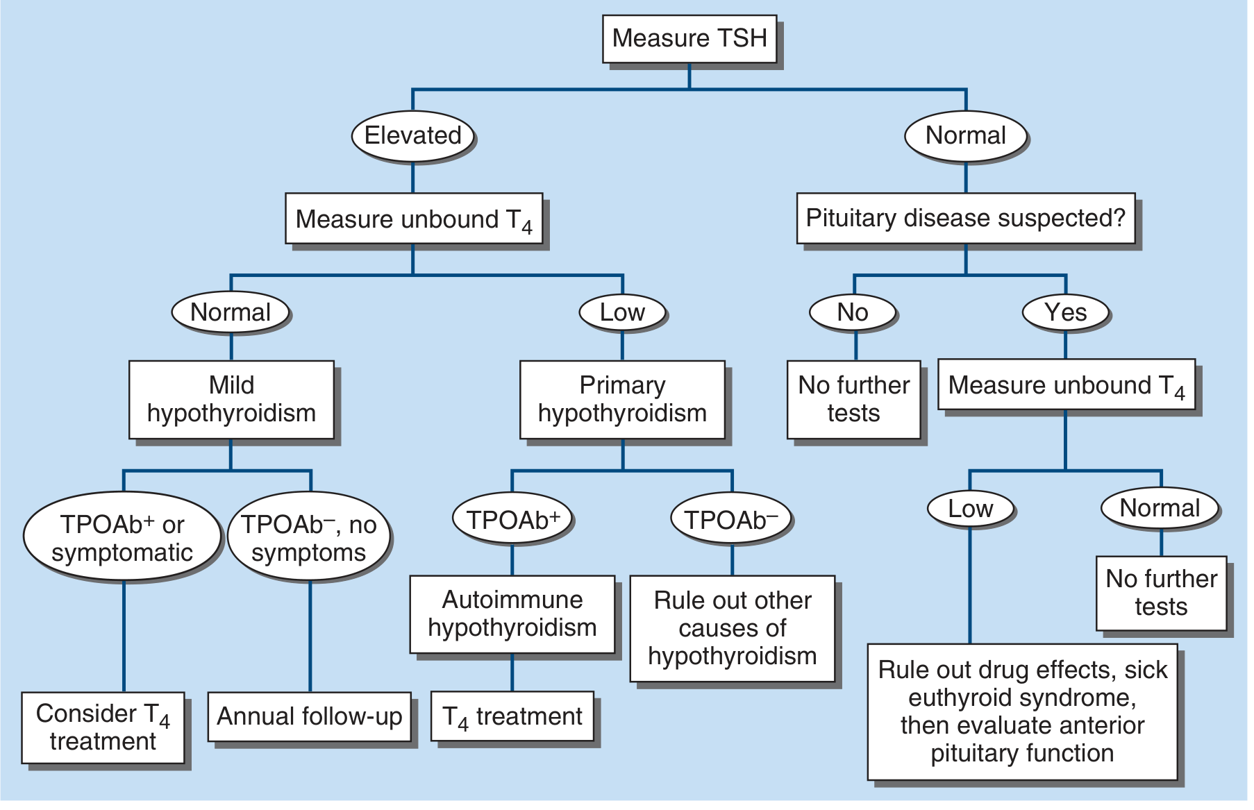
Step 4 - Clinical Examination Points Not to Miss
- Periorbital puffiness - characteristic myxoedematous facies
- Puffy hands and feet (non-pitting oedema - press firmly; true myxoedema does not pit)
- Madarosis - loss of outer third of eyebrows (highly specific for hypothyroidism)
- Thyroid size - goitre suggests Hashimoto's; impalpable gland = atrophic thyroiditis or central
- Delayed relaxation of Achilles tendon reflex - pathognomonic of hypothyroidism
- Bradycardia - resting HR <60 in a symptomatic patient
- Hoarse, slow, low-pitched voice - myxoedema voice
- Dry, rough, cool skin with yellowish tint (carotenaemia due to reduced conversion of beta-carotene)
- Diffuse hair thinning - check scalp, axillae, pubic area (for panhypopituitarism)
- Signs of pericardial effusion - muffled heart sounds, raised JVP
- Visual fields - bitemporal hemianopia if pituitary macroadenoma
- Signs of Addison's - hyperpigmentation, postural hypotension (autoimmune polyendocrinopathy)
Step 5 - Why She Worsened in the Last 1 Week
- Infection (any intercurrent illness)
- Cold exposure
- Unrelated medication (sedatives, diuretics)
- Non-compliance with any pre-existing thyroid therapy she may not have disclosed
- Alert: If she develops altered sensorium, hypothermia, bradycardia, hypoventilation, hyponatraemia - this is myxoedema coma, a medical emergency requiring ICU admission, IV T3 or T4, IV hydrocortisone, passive rewarming, and treatment of the precipitant.
Most Probable Diagnosis
- Only TSH was checked and it was subclinically elevated (borderline), or
- The test was done at a different lab with a wider "normal" range, or
- The disease was subclinical then and has progressed to overt now
Summary of Immediate Tests to Order
- TSH + free T4 + free T3 (repeat, complete panel - not TSH alone)
- Anti-TPO + anti-Tg antibodies
- Thyroid ultrasound
- Serum albumin + urine protein (to exclude nephrotic/hypoalbuminaemic oedema)
- CBC, LFT, RFT, lipids (hypothyroidism raises LDL and triglycerides; CPK often elevated)
- ECG (bradycardia, low-voltage complexes if pericardial effusion)
- Echocardiogram (pericardial effusion occurs in ~30% of hypothyroid patients)
- Morning cortisol (if pituitary disease considered or if any suggestion of Addison's)