I now have all the image URLs I need from the textbooks. Let me compile the complete illustrated study notes.Here are your complete illustrated study notes for the Anatomy course (Accident & Emergency Care Technology):
📚 ANATOMY — Complete Illustrated Study Notes
Accident & Emergency Care Technology
UNIT 1: INTRODUCTION TO ANATOMICAL TERMS & ORGANIZATION OF THE HUMAN BODY
1.1 Anatomical Terminology
Anatomical Position: Body erect, face forward, arms at sides with palms facing forward. All descriptions refer to this standard position.
Planes of Section:
- Sagittal plane - divides body into left and right
- Coronal (frontal) plane - divides body into anterior and posterior
- Transverse (horizontal) plane - divides body into superior and inferior
Terms of Direction:
| Term | Meaning |
|---|
| Anterior / Ventral | Toward the front |
| Posterior / Dorsal | Toward the back |
| Superior / Cranial | Toward the head |
| Inferior / Caudal | Toward the feet |
| Medial | Toward the midline |
| Lateral | Away from the midline |
| Proximal | Closer to the origin |
| Distal | Further from the origin |
| Superficial | Closer to the surface |
| Deep | Further from the surface |
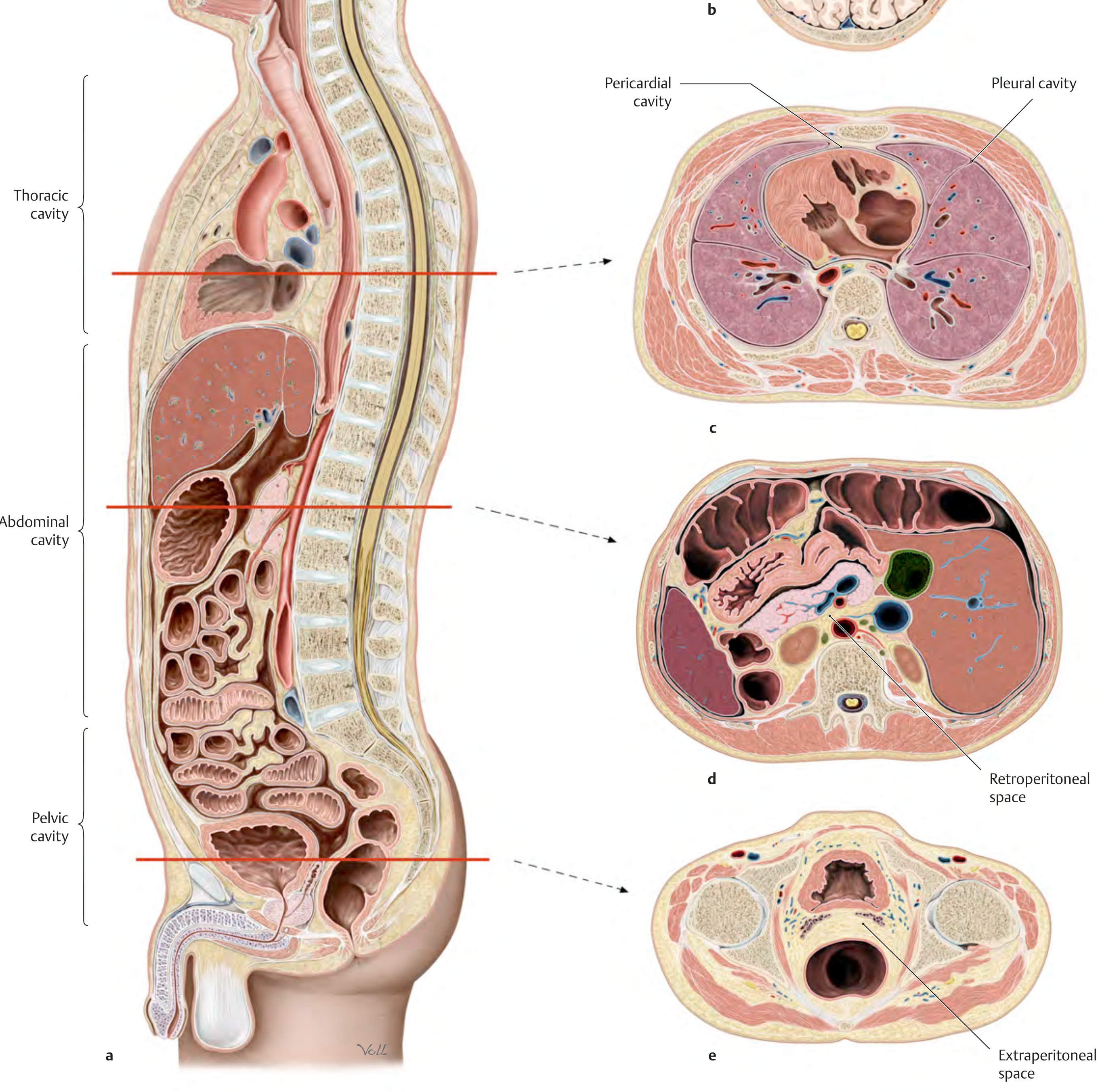
1.2 Body Cavities & Serous Membranes
The body contains enclosed serous cavities lined by membranes:
Thoracic cavity:
- Pleural cavity (right and left) - surrounds each lung
- Pericardial cavity - surrounds the heart
Abdominopelvic cavity:
- Peritoneal cavity (abdomen + pelvis)
Each serous cavity has two layers: a visceral layer (directly invests the organ) and a parietal layer (lines the cavity wall). A small amount of fluid lies between them to reduce friction.
Body cavities: pericardial, pleural, abdominal, retroperitoneal spaces - General Anatomy & Musculoskeletal System (THIEME Atlas)
1.3 The Human Cell
The cell is the basic structural and functional unit of life.
Key organelles and their functions:
| Organelle | Function |
|---|
| Nucleus | Contains DNA; controls cell activity |
| Cell membrane | Selectively permeable barrier; controls entry/exit |
| Mitochondria | Energy production (ATP) - "powerhouse of the cell" |
| Rough ER | Protein synthesis (studded with ribosomes) |
| Smooth ER | Lipid synthesis, detoxification |
| Golgi apparatus | Packaging and secretion of proteins |
| Lysosomes | Digestion of waste / foreign material |
| Ribosomes | Site of protein synthesis |
| Cytoskeleton (neurofibrils) | Structural support and transport |
The cell's internal milieu: Cells are bathed by extracellular fluid. The bloodstream links the interstitium to organs (lungs, kidneys, digestive tract) that absorb nutrients and excrete metabolic products, maintaining homeostasis.
"The cells of a multicellular organism are bathed by extracellular fluid... the internal milieu would change very quickly if the intercellular space (interstitium) were not linked via the bloodstream to organs such as the lungs, kidneys, and digestive tract." - THIEME Atlas of General Anatomy
1.4 Tissues
Definition: A tissue is a group of similar cells that perform a specific function.
Four primary tissue types:
| Type | Characteristics | Location | Function |
|---|
| Epithelial | Closely packed cells, avascular, rests on basement membrane | Skin, lining of organs, glands | Protection, absorption, secretion, sensation |
| Connective | Widely spaced cells in matrix; has blood supply | Bone, cartilage, blood, fat, fascia | Support, binding, transport, protection |
| Muscle | Contractile cells with actin and myosin | Skeletal: voluntary muscles; Cardiac: heart wall; Smooth: hollow organs | Movement, circulation, peristalsis |
| Nervous | Neurons + neuroglia | Brain, spinal cord, nerves | Communication, control, integration |
Epithelial sub-classification:
- Simple (one layer): squamous, cuboidal, columnar
- Stratified (multiple layers): squamous (skin), columnar, transitional (bladder)
- Pseudostratified: appears layered but is single-layer (e.g., respiratory tract)
1.5 Membranes
Types:
- Serous membranes - line closed body cavities (pleura, pericardium, peritoneum); secrete watery fluid
- Mucous membranes (mucosa) - line passages opening to exterior (digestive, respiratory, urogenital tracts); secrete mucus
- Synovial membranes - line joint cavities; secrete synovial fluid for lubrication
- Cutaneous membrane (skin) - covers body surface; protection
1.6 Glands
Glands are secretory structures derived from epithelium.
| Type | Secretion route | Examples |
|---|
| Exocrine | Via ducts to surface | Sweat, salivary, pancreatic (digestive enzymes), sebaceous |
| Endocrine | Directly into bloodstream (ductless) | Thyroid, pituitary, adrenal, pancreatic islets |
UNIT 2: BONES & JOINTS
2.1 Overview of Bones
Functions of bone:
- Support and structural framework
- Protection of vital organs (skull, thoracic cage)
- Movement (levers for muscles)
- Haemopoiesis (blood cell formation in red bone marrow)
- Mineral storage (calcium and phosphorus)
Classification of bones:
- Long bones - humerus, femur, tibia (tubular shape)
- Short bones - carpals, tarsals (cube-shaped)
- Flat bones - skull, sternum, scapula (broad, protective)
- Irregular bones - vertebrae, hip bone
- Sesamoid bones - patella (embedded in tendons)
2.2 Upper Limb Bones
Clavicle (Collar Bone)
- S-shaped long bone; only bony link between upper limb and thoracic skeleton
- Medial end: articulates with manubrium of sternum (sternoclavicular joint)
- Lateral end: articulates with acromion of scapula (acromioclavicular joint)
- Prominent features: conoid tubercle (lateral surface), subclavian groove (inferior surface)
- Applied: Most commonly fractured bone in the body; fracture usually at the junction of middle and lateral thirds
Scapula (Shoulder Blade)
- Flat, triangular bone on the posterior thoracic wall (ribs 2-7)
- Key features:
- Acromion - forms the "shoulder point"; articulates with clavicle
- Coracoid process - projects anteriorly; attachment for muscles and ligaments
- Glenoid cavity - pear-shaped socket for articulation with head of humerus
- Spine of scapula - prominent ridge on posterior surface
- Supraspinous and infraspinous fossae - above and below the spine
- Subscapular fossa - concave anterior surface
Humerus
- Long bone of the arm (between shoulder and elbow)
- Upper end:
- Head - articulates with glenoid cavity (ball-and-socket joint)
- Greater tubercle (lateral) - attachment of rotator cuff muscles (supraspinatus, infraspinatus, teres minor)
- Lesser tubercle (anterior) - attachment of subscapularis
- Anatomical neck - groove between head and tubercles
- Surgical neck - common fracture site, axillary nerve at risk
- Shaft: Deltoid tuberosity (lateral), radial groove (spiral groove for radial nerve)
- Lower end:
- Capitulum - articulates with head of radius
- Trochlea - articulates with trochlear notch of ulna
- Medial epicondyle (ulnar nerve passes posterior to it)
- Lateral epicondyle
- Olecranon fossa (posterior), Coronoid fossa (anterior)
Radius
- Lateral forearm bone (thumb side)
- Upper end: Head (circular - articulates with capitulum of humerus and radial notch of ulna), neck, radial tuberosity (insertion of biceps brachii)
- Lower end: Broader; articulates with scaphoid and lunate (wrist joint); styloid process (lateral)
- Applied: Colles' fracture - most common fracture of distal radius after a fall on outstretched hand
Ulna
- Medial forearm bone (little finger side)
- Upper end:
- Olecranon - forms the point of the elbow; insertion of triceps brachii
- Trochlear (semilunar) notch - large concavity articulating with trochlea of humerus
- Coronoid process - projects anteriorly from trochlear notch
- Radial notch - articulates with head of radius
- Lower end: Head (articulates with ulnar notch of radius), styloid process (medial)
2.3 Lower Limb Bones
Hip Bone (Os Coxae)
- Large, irregular bone formed by fusion of three bones at the acetabulum:
- Ilium (superior - broad, fan-like)
- Ischium (posteroinferior)
- Pubis (anteroinferior)
- Key features:
- Iliac crest - superior border of ilium; runs from anterior superior iliac spine (ASIS) to posterior superior iliac spine (PSIS)
- Acetabulum - deep cup-shaped socket for head of femur (hip joint)
- Obturator foramen - large opening between ischium and pubis
- Ischial tuberosity - "sitting bone"; bears weight when seated
- Greater and lesser sciatic notches
- Two hip bones + sacrum + coccyx = pelvis
Femur (Thigh Bone)
- Longest, strongest bone in the body
- Upper end:
- Head - articulates with acetabulum (ball-and-socket)
- Neck - angled ~125° to shaft; common fracture site in elderly (neck of femur fracture)
- Greater trochanter (lateral) - attachment of gluteal muscles
- Lesser trochanter (medial inferior) - insertion of iliopsoas
- Intertrochanteric crest (posterior), intertrochanteric line (anterior)
- Shaft: Linea aspera (posterior ridge; muscle attachment)
- Lower end: Medial and lateral condyles (articulate with tibia); medial and lateral epicondyles; patellar surface (articulates with patella); intercondylar notch
Sacrum
- Triangular bone formed by fusion of 5 sacral vertebrae
- Articulates superiorly with L5 (lumbosacral joint) and laterally with hip bones (sacroiliac joints)
- Anterior (pelvic) surface: concave, smooth, 4 pairs of anterior sacral foramina
- Posterior surface: convex, rough, 4 pairs of posterior sacral foramina; median sacral crest
- Sacral hiatus (inferior opening of sacral canal) - site for caudal anaesthesia
- Sacral promontory - superior anterior margin; obstetric landmark
Tibia (Shin Bone)
- Medial, weight-bearing bone of the leg
- Upper end: Medial and lateral condyles; tibial plateau; tibial tuberosity (insertion of patellar tendon - quadriceps mechanism)
- Shaft: Sharp anterior border (subcutaneous - no overlying muscle); medial surface subcutaneous
- Lower end: Medial malleolus; inferior articular surface for talus (ankle joint)
- Applied: Tibia is most commonly fractured long bone
Fibula
- Slender lateral bone of the leg; non-weight-bearing
- Upper end: Head (articulates with lateral condyle of tibia)
- Lower end: Lateral malleolus (forms lateral boundary of ankle mortice)
- Applied: Lateral malleolus fracture is most common ankle fracture
Vertebral Column
Structure: 33 vertebrae total:
- 7 Cervical (C1-C7)
- 12 Thoracic (T1-T12)
- 5 Lumbar (L1-L5)
- 5 Sacral (fused = sacrum)
- 4 Coccygeal (fused = coccyx)
Curvatures:
- Cervical and lumbar: Lordosis (concave posteriorly) - secondary curves
- Thoracic and sacral: Kyphosis (convex posteriorly) - primary curves
Typical vertebra features:
- Body (anterior, weight-bearing)
- Vertebral arch (posterior) = pedicles + laminae
- Vertebral foramen (forms vertebral canal for spinal cord)
- Spinous process (posterior)
- Transverse processes (lateral, 2)
- Superior and inferior articular processes (facet joints)
Intervertebral discs: fibrocartilaginous; nucleus pulposus (gel core) + annulus fibrosus (fibrous ring). Prolapse ("slipped disc") can compress nerve roots.
UNIT 3: THORAX
3.1 Bony Thoracic Cage
Components: Sternum (anterior), 12 pairs of ribs, 12 thoracic vertebrae (posterior)
Functions: Protects heart and lungs; facilitates breathing via changes in volume
Sternum
Three parts:
- Manubrium (superior): articulates with clavicles and 1st/2nd ribs; jugular (suprasternal) notch
- Body (middle): articulates with ribs 2-7
- Xiphoid process (inferior): cartilaginous in young, ossifies with age
- Sternal angle (angle of Louis): Junction of manubrium and body; T4/T5 level; marks attachment of 2nd rib - clinical landmark
- Applied: Sternal bone marrow aspiration is done through the flat subcutaneous body of the sternum
Ribs
12 pairs: True ribs (1-7), False ribs (8-10), Floating ribs (11-12)
- True ribs: Connect directly to sternum via costal cartilages
- False ribs: Costal cartilages join to rib above, not sternum directly
- Floating ribs: No anterior cartilage attachment
- Each typical rib has: head (2 articular facets), neck, tubercle, shaft with subcostal groove (lodges neurovascular bundle - vein, artery, nerve - VAN from above)
- Applied (rib fractures): Single fractures are painful but manageable. Multiple fractures at two+ sites per rib = flail chest - paradoxical movement of the flail segment impairs ventilation and may require assisted ventilation.
Thoracic Vertebrae
- Body + costal facets for rib articulation
- Long spinous processes pointing inferiorly
- Articulate with ribs at demifacets on vertebral bodies (head of rib) and facets on transverse processes (tubercle of rib)
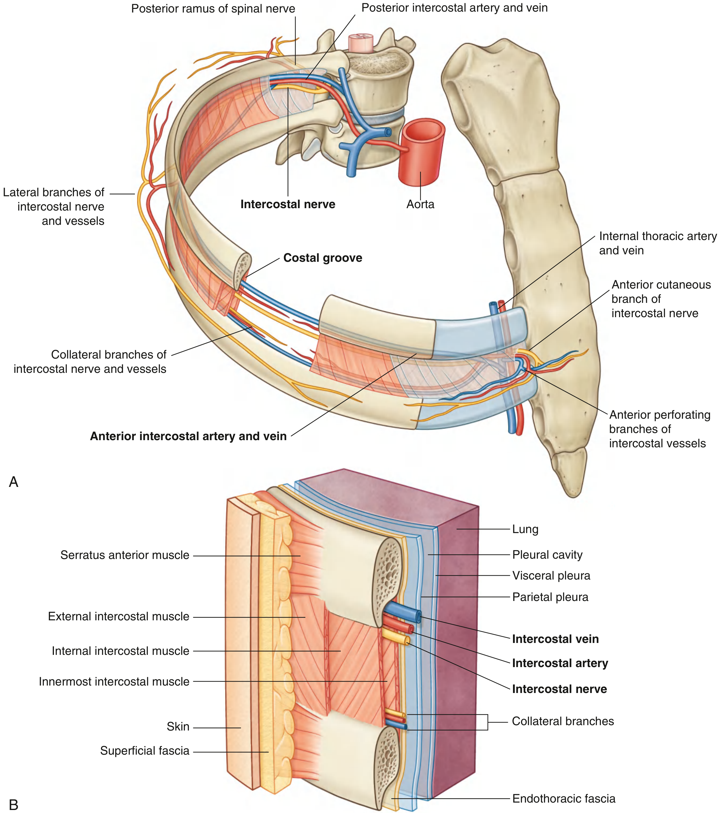
3.2 Intercostal Space
Each intercostal space contains (from superficial to deep):
- External intercostal muscle (fibres run downward and forward)
- Internal intercostal muscle (fibres run downward and backward)
- Innermost intercostal muscle
Between internal and innermost muscles runs the intercostal neurovascular bundle: intercostal vein, artery, and nerve (VAN - from superior to inferior in the subcostal groove).
Clinical note: When performing thoracentesis (pleural tap) or placing a chest drain, insert the needle/drain just above the superior border of the rib below to avoid the neurovascular bundle.
Fig. 3.27 Intercostal Space showing posterior ramus of spinal nerve, arteries, nerves, and muscles - Gray's Anatomy for Students
3.3 Pleura
The pleura is a serous membrane investing each lung:
- Visceral pleura: Tightly covers the lung surface
- Parietal pleura: Lines the thoracic wall, diaphragm, and mediastinum
- Pleural cavity: Potential space between the two layers; contains a small amount of fluid (lubrication)
- Costodiaphragmatic recess: The lowest point of the pleural cavity; the site where fluid accumulates (pleural effusion)
Parts of parietal pleura: Costal, diaphragmatic, mediastinal, cervical (cupola - extends above clavicle into neck)
Applied:
- Pneumothorax: Air in pleural cavity → lung collapse
- Haemothorax: Blood in pleural cavity (trauma)
- Pleural effusion: Fluid in pleural cavity
3.4 Diaphragm
- Dome-shaped musculotendinous sheet separating thorax from abdomen
- Main respiratory muscle: Contraction flattens the domes, increasing thoracic volume (inspiration)
- Innervation: Phrenic nerve (C3, C4, C5) - "C3, 4, 5 keeps the diaphragm alive"
- Three main openings:
- Aortic hiatus (T12) - aorta + thoracic duct
- Oesophageal hiatus (T10) - oesophagus + vagus nerves
- Caval opening (T8) - inferior vena cava
3.5 Muscles of the Thorax
| Muscle | Origin | Insertion | Action |
|---|
| External intercostals | Lower border of rib above | Upper border of rib below | Elevate ribs (inspiration) |
| Internal intercostals | Lower border of rib above | Upper border of rib below | Depress ribs (expiration) |
| Innermost intercostals | Inner surface of rib | Inner surface of rib below | Assist internal intercostals |
| Diaphragm | Xiphoid, lower 6 costal cartilages, lumbar vertebrae | Central tendon | Main muscle of inspiration |
| Serratus anterior | Outer surfaces ribs 1-8 | Medial border of scapula | Protracts/rotates scapula |
| Pectoralis major | Clavicle, sternum, upper 6 costal cartilages | Greater tubercle of humerus (lateral lip of bicipital groove) | Flexion, adduction, medial rotation of arm |
| Pectoralis minor | Ribs 3-5 | Coracoid process of scapula | Depresses scapula; elevates ribs |
UNIT 4: HEART
4.1 Surface Anatomy of the Heart
The heart lies in the middle mediastinum, enclosed in the pericardium. It is the size of a closed fist.
- Apex: Left 5th intercostal space, midclavicular line (8-9 cm from midsternal line) - site of apex beat
- Base: Posterior surface, opposite vertebrae T5-T8, formed by atria
- Right border: Right atrium
- Left border: Mainly left ventricle (some left atrium)
- Superior border: Aorta, pulmonary trunk emerging
- Inferior border: Right ventricle
4.2 Pericardium
The pericardium is a fibroserous sac enclosing the heart.
- Fibrous pericardium (outer): tough, inextensible; fused with central tendon of diaphragm
- Serous pericardium (inner): two layers
- Parietal layer (lines fibrous pericardium)
- Visceral layer / epicardium (adheres to heart muscle)
- Pericardial cavity: Small space between parietal and visceral layers; contains ~20-50 mL of serous fluid
- Applied: Cardiac tamponade - fluid accumulation in pericardial cavity compresses the heart, reducing cardiac output. The rigid fibrous pericardium prevents outward expansion. Emergency pericardiocentesis is required.
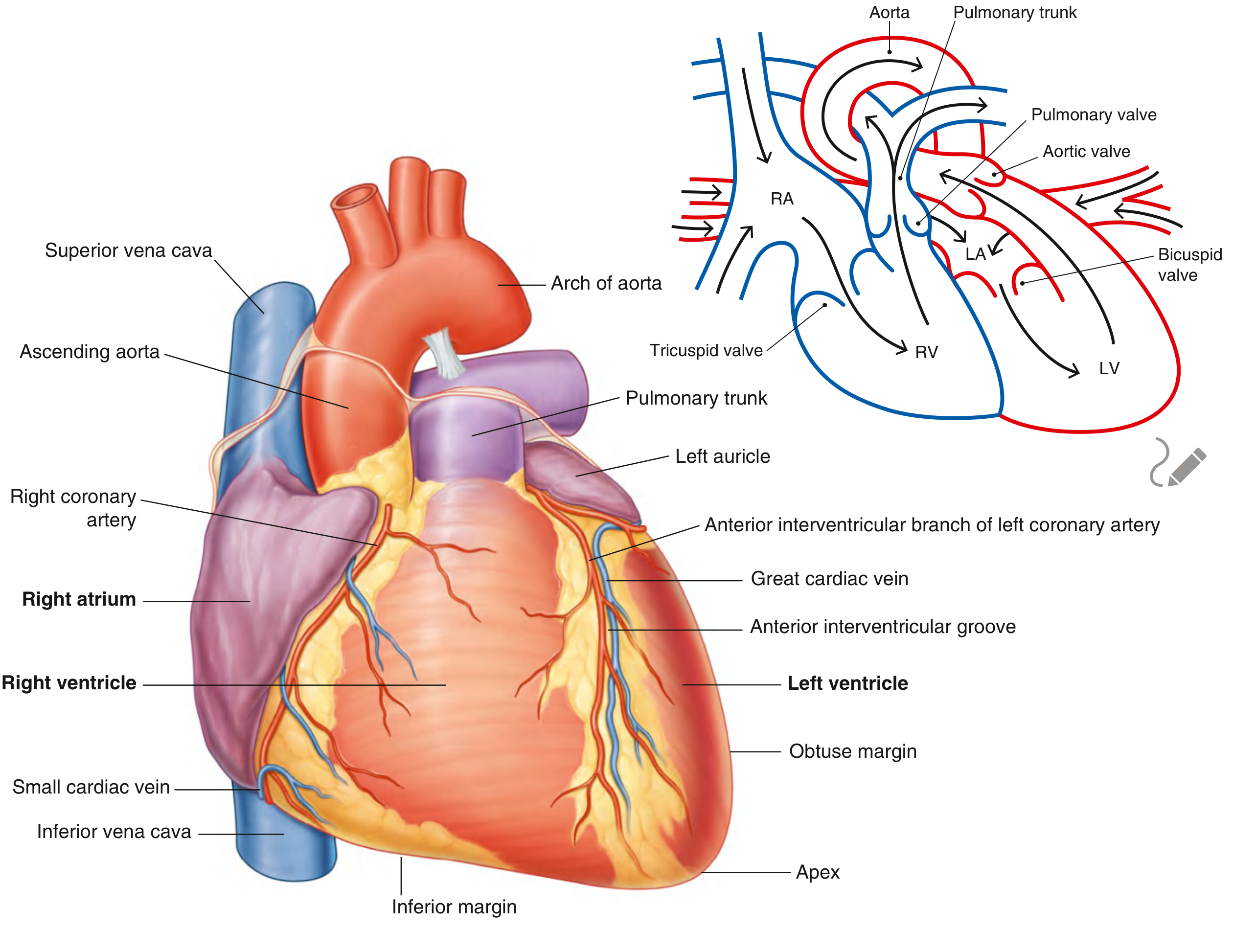
4.3 Chambers of the Heart
Fig. 3.67 Anterior Surface of the Heart - LA (left atrium), LV (left ventricle), RA (right atrium), RV (right ventricle) - Gray's Anatomy for Students
The heart has 4 chambers:
Right Atrium (RA):
- Receives deoxygenated blood from the body via:
- Superior vena cava (SVC) - from upper body
- Inferior vena cava (IVC) - from lower body
- Coronary sinus - from heart muscle itself
- Internal features: crista terminalis, pectinate muscles, fossa ovalis (remnant of foramen ovale)
- Opens into right ventricle via the tricuspid (right atrioventricular) valve
Right Ventricle (RV):
- Pumps blood to lungs via pulmonary trunk (pulmonary circulation)
- Crescent-shaped in cross-section
- Trabeculae carneae on interior wall; moderator band (contains right bundle branch of conduction system)
- Separated from left ventricle by the interventricular septum
- Tricuspid valve (3 cusps): anterior, posterior, septal
- Pulmonary valve (3 semilunar cusps) guards exit into pulmonary trunk
Left Atrium (LA):
- Receives oxygenated blood from the 4 pulmonary veins (2 right, 2 left)
- Smooth-walled (except left auricle which has pectinate muscles)
- Opens into left ventricle via the mitral (bicuspid/left atrioventricular) valve
Left Ventricle (LV):
- Pumps oxygenated blood to entire body via aorta (systemic circulation)
- Thick-walled (3x thicker than RV - generates higher pressure)
- Circular in cross-section; heavily trabeculated
- Mitral valve (2 cusps): anterior and posterior
- Aortic valve (3 semilunar cusps) guards exit into aorta
4.4 Valves of the Heart
| Valve | Location | Type | Function |
|---|
| Tricuspid (right AV) | Between RA and RV | 3 cusps + chordae tendineae + papillary muscles | Prevents backflow into RA during ventricular contraction |
| Pulmonary | Base of pulmonary trunk | 3 semilunar cusps (no chordae) | Prevents backflow into RV after ejection |
| Mitral/Bicuspid (left AV) | Between LA and LV | 2 cusps + chordae tendineae + papillary muscles | Prevents backflow into LA during ventricular contraction |
| Aortic | Base of aorta | 3 semilunar cusps (no chordae) | Prevents backflow into LV after ejection |
Heart sounds: S1 ("lub") = AV valves closing; S2 ("dub") = semilunar valves closing. Murmurs indicate valve dysfunction.
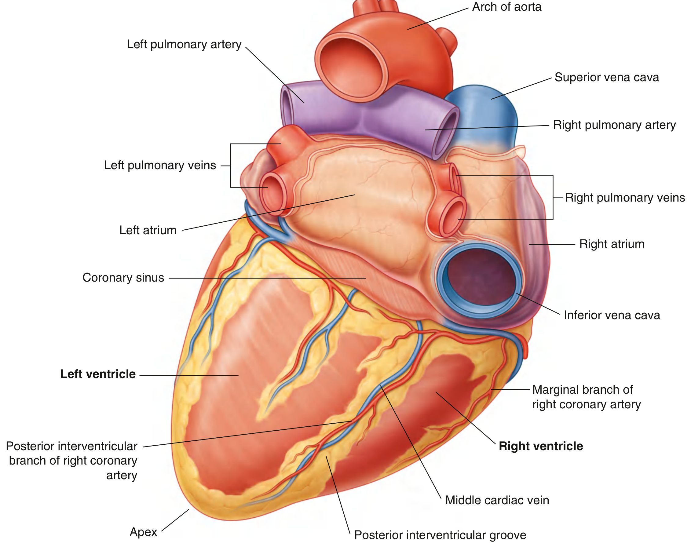
4.5 Major Blood Vessels of the Heart
- Aorta - carries oxygenated blood from LV to systemic circulation
- Pulmonary trunk - carries deoxygenated blood from RV; divides into right and left pulmonary arteries
- Superior vena cava (SVC) - returns blood from head, neck, upper limbs
- Inferior vena cava (IVC) - returns blood from lower body
- Pulmonary veins (4) - carry oxygenated blood from lungs to LA
Posterior/inferior surfaces of the heart with labeled arteries, veins, and chambers - Gray's Anatomy for Students
4.6 Coronary Arteries
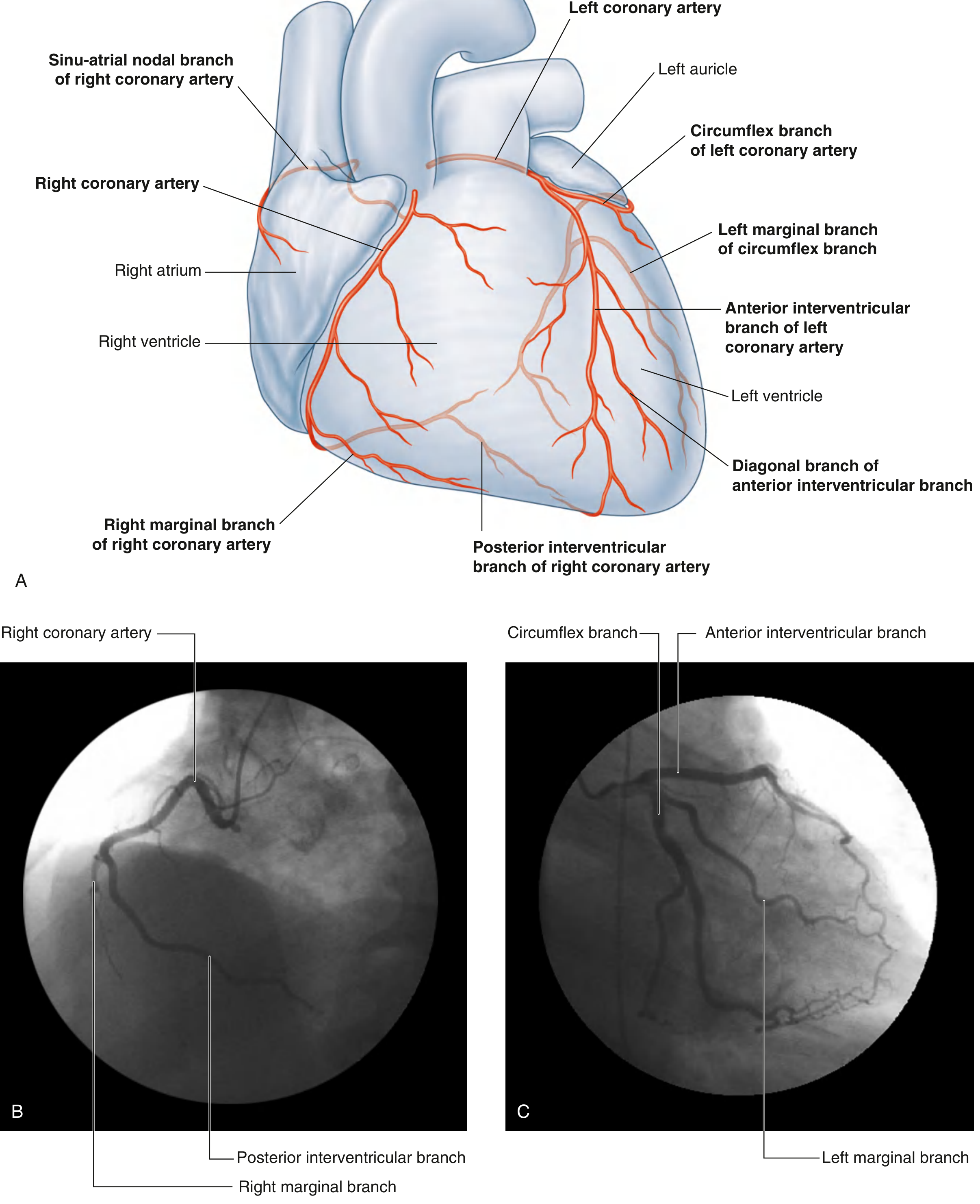
The myocardium is supplied by the right and left coronary arteries, which arise from the aortic sinuses just above the aortic valve.
Left Coronary Artery (LCA):
- Short trunk (left main stem)
- Divides into:
- Anterior interventricular artery (LAD - Left Anterior Descending) - supplies anterior LV, anterior septum, apex
- Circumflex artery - winds round in the coronary groove to supply lateral and posterior LV
Right Coronary Artery (RCA):
- Runs in the right atrioventricular groove
- Main branches:
- Marginal artery - supplies right border of heart
- Posterior interventricular artery (PDA - Posterior Descending Artery) - supplies posterior septum and inferior LV (in right-dominant hearts)
- SA nodal artery - supplies the sinoatrial node (in ~60%)
- AV nodal artery - supplies the atrioventricular node
Coronary arteries and heart chambers with labeled branches - Gray's Anatomy for Students
Applied: Coronary artery disease (CAD) - atherosclerotic blockage leads to myocardial ischaemia (angina) or infarction (MI / heart attack). LAD occlusion is the most dangerous ("widow-maker").
4.7 Conduction System of the Heart
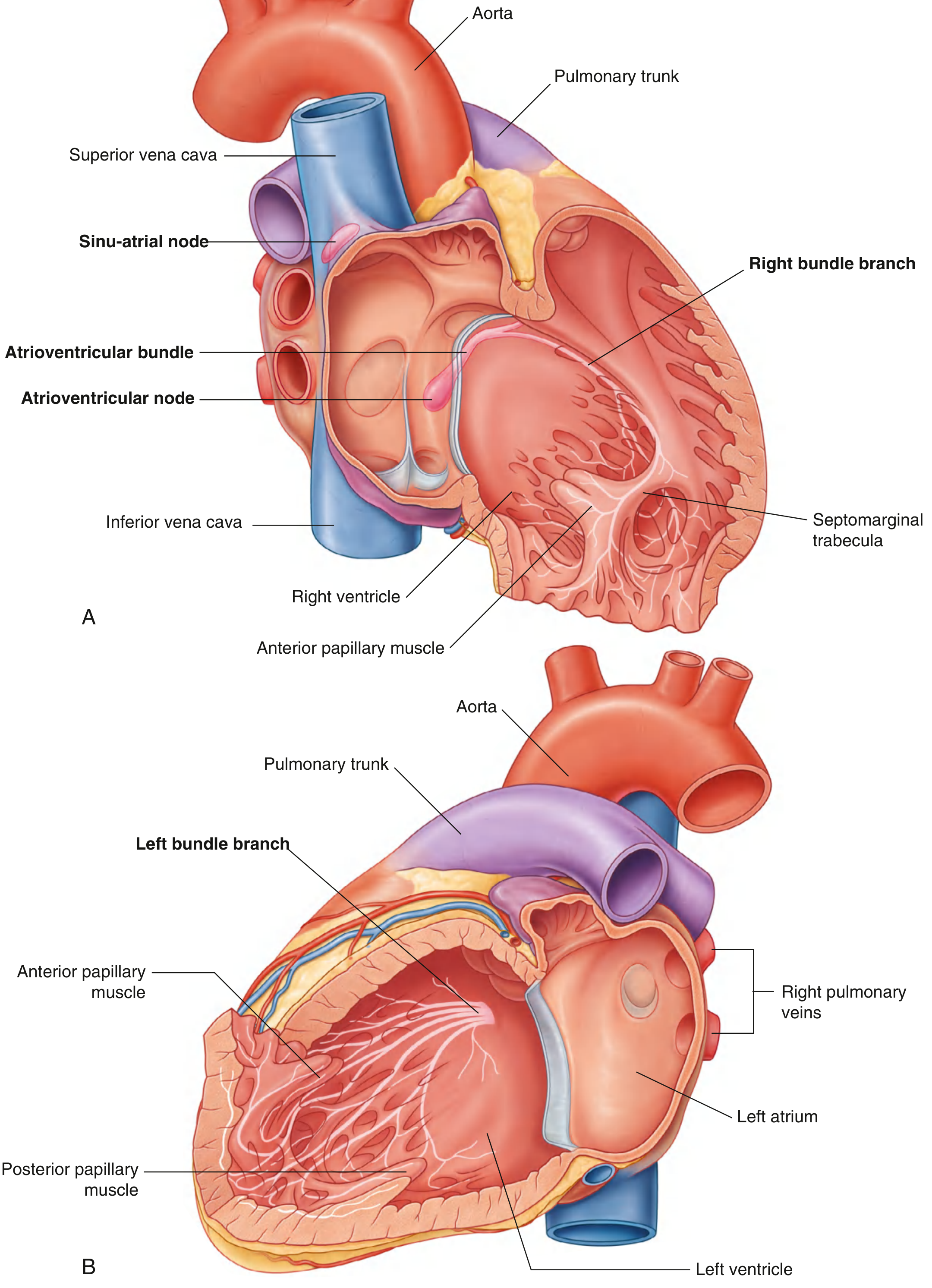
Fig. 3.86 Conduction System of the Heart showing SA node, AV node, Bundle of His, and bundle branches - Gray's Anatomy for Students
Pathway of electrical impulse:
- SA node (sinoatrial node) - right atrium near SVC; "natural pacemaker" (60-100 bpm)
- AV node (atrioventricular node) - between atria and ventricles; delays impulse
- Bundle of His - passes through AV junction into interventricular septum
- Right and Left bundle branches - travel down each side of septum
- Purkinje fibres - spread impulse through ventricular myocardium
UNIT 5: MUSCLES OF THE THORAX AND UPPER LIMB
5.1 Muscles of the Upper Limb (Arm)
Muscles of the Arm
Flexors (Anterior compartment):
| Muscle | Origin | Insertion | Action | Nerve |
|---|
| Biceps brachii | Long head: supraglenoid tubercle; Short head: coracoid process | Radial tuberosity + bicipital aponeurosis | Flexion of elbow; supination of forearm | Musculocutaneous (C5, C6) |
| Brachialis | Anterior surface of distal humerus | Coronoid process + ulnar tuberosity | Flexion of elbow (pure flexor) | Musculocutaneous (C5, C6) |
| Coracobrachialis | Coracoid process | Middle of medial humerus | Flexion + adduction of arm | Musculocutaneous (C6, C7) |
Extensors (Posterior compartment):
| Muscle | Origin | Insertion | Action | Nerve |
|---|
| Triceps brachii | Long head: infraglenoid tubercle; Lateral head: above radial groove; Medial head: below radial groove | Olecranon of ulna | Extension of elbow | Radial nerve (C7, C8) |
| Anconeus | Lateral epicondyle of humerus | Lateral surface of olecranon | Assists elbow extension | Radial nerve |
5.2 Muscles of the Forearm
Flexor Group (Anterior Compartment):
Superficial layer (all originate from medial epicondyle of humerus - "common flexor origin"):
- Pronator teres - pronation of forearm
- Flexor carpi radialis - flexion and radial deviation of wrist
- Palmaris longus - flexion of wrist (often absent)
- Flexor carpi ulnaris - flexion and ulnar deviation of wrist
- Flexor digitorum superficialis - flexion of PIP joints of fingers 2-5
Deep layer:
- Flexor digitorum profundus - flexion of DIP joints of fingers 2-5
- Flexor pollicis longus - flexion of thumb IP joint
- Pronator quadratus - pronation
Nerve supply: Median nerve (most muscles); Ulnar nerve (flexor carpi ulnaris + medial half of FDP)
Extensor Group (Posterior Compartment):
All originate from lateral epicondyle of humerus - "common extensor origin":
- Extensor carpi radialis longus and brevis - extension + radial deviation of wrist
- Extensor digitorum - extension of fingers 2-5
- Extensor carpi ulnaris - extension + ulnar deviation of wrist
- Extensor pollicis longus and brevis - extension of thumb
- Abductor pollicis longus - abduction of thumb
- Supinator - supination of forearm
Nerve supply: Radial nerve and its branches
UNIT 6: EXCRETORY SYSTEM
6.1 Kidneys
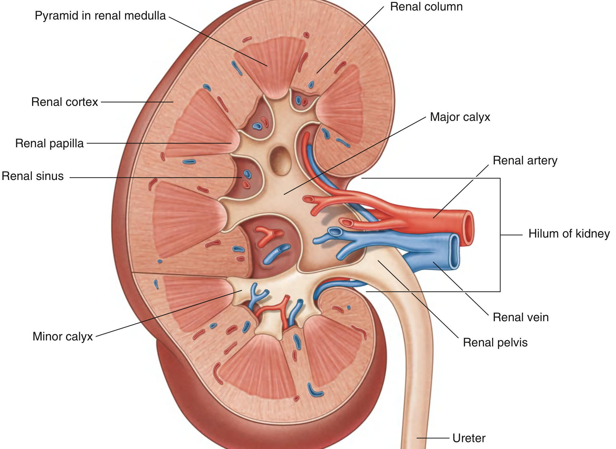
Fig. 4.156 Internal Structure of the Kidney showing cortex, medulla, renal pyramids, calyces, pelvis, artery, vein, and ureter - Gray's Anatomy for Students
Location: Retroperitoneal, on posterior abdominal wall
- Right kidney: opposite T12-L3; lies slightly lower (due to liver above it)
- Left kidney: opposite T11-L2
Dimensions: ~10-12 cm long, 5-6 cm wide, 3-4 cm thick
Structure:
- Enclosed in fibrous renal capsule
- Hilum (medial margin): entry/exit for renal vessels, ureter, lymphatics, nerves
- Renal cortex (outer): continuous band; contains glomeruli and convoluted tubules
- Renal medulla (inner): renal pyramids (cone-shaped, striped); separated by renal columns (cortex extensions)
- Renal papilla: apex of each pyramid; opens into minor calyx
- Minor calyces → major calyces (2-3) → renal pelvis → ureter
Blood supply:
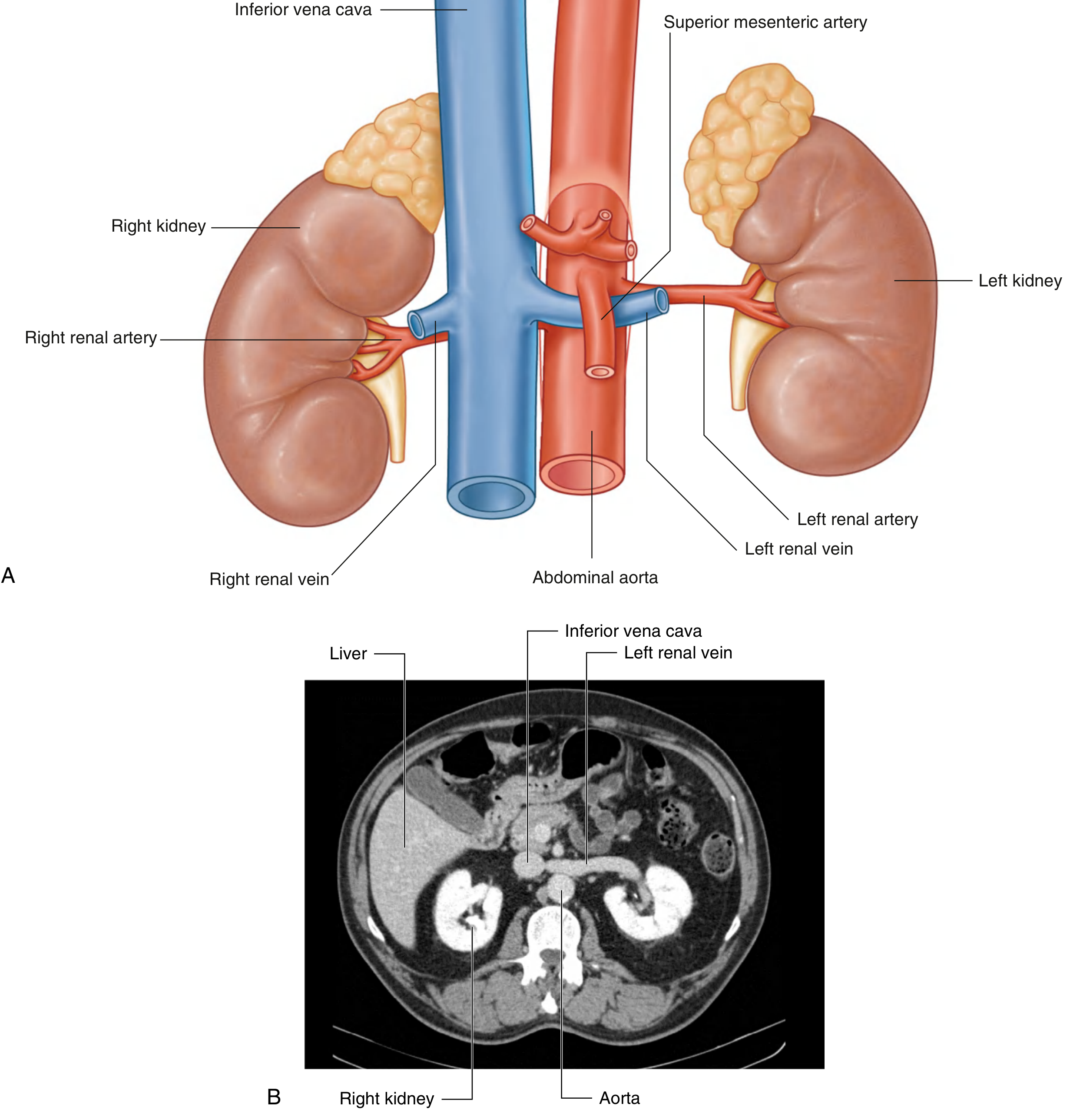
- Renal artery (direct branch of abdominal aorta, L1-L2)
- Right renal artery longer; passes posterior to IVC
- Left renal vein longer; crosses midline anterior to aorta
Functions: Filtration, reabsorption, secretion → urine production; regulation of blood pressure (renin), erythropoiesis (EPO), calcium/phosphate metabolism (vitamin D activation)
Renal vasculature showing kidneys, aorta, veins, and ureters - Gray's Anatomy for Students
6.2 Ureters
- Muscular tubes, ~25-30 cm long, ~5 mm diameter
- Carry urine from renal pelvis to urinary bladder
- Course: descends retroperitoneally on psoas muscle → crosses pelvic brim → enters bladder
- Three sites of physiological narrowing (where stones commonly lodge):
- Pelviureteric junction (PUJ)
- Crossing the pelvic brim (over bifurcation of common iliac artery)
- Vesicoureteric junction (VUJ) - where ureter enters bladder
Nerve supply: Ureteric pain is referred to the groin/testicle (T10-L1 dermatomes)
6.3 Urinary Bladder
- Hollow muscular organ (detrusor muscle = smooth muscle)
- Location: Pelvis, behind the pubic symphysis (empty); rises into abdomen when full
- Capacity: ~300-500 mL normally; can hold more
- Trigone: Triangular smooth area between the two ureteric orifices and internal urethral orifice; always smooth (no rugae)
- Neck of bladder: Where bladder opens into urethra; surrounded by internal urethral sphincter (smooth muscle, involuntary)
Applied: Bladder is palpable/percussible above the pubic symphysis when full. Urinary retention (inability to void) is a common A&E presentation, often requiring urethral catheterization.
UNIT 7: NERVOUS SYSTEM
7.1 Overview
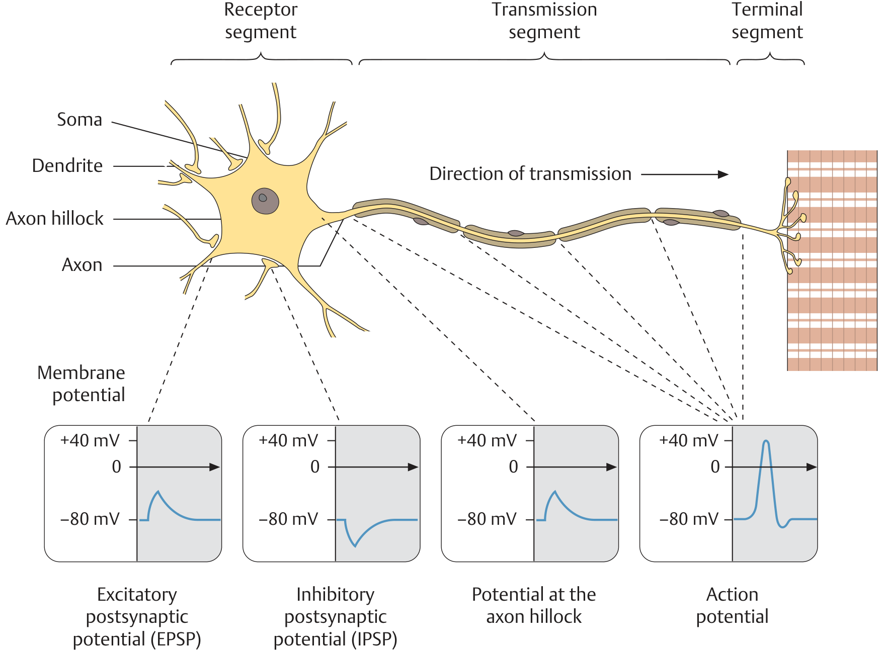
Neuron structure, labeled segments, and transmission direction - General Anatomy & Musculoskeletal System (THIEME Atlas)
The nervous system is divided into three functionally related subdivisions:
| Subdivision | Components | Special Features |
|---|
| Central (CNS) | Brain + spinal cord | Oligodendrocytes provide myelin; axons CANNOT regenerate |
| Peripheral (PNS) | Peripheral ganglia, sensory receptors, cranial and spinal nerves | Schwann cells provide myelin; axons CAN regenerate |
| Autonomic (ANS) | Portions of CNS + PNS; regulates visceral function | Sympathetic + parasympathetic divisions |
"All elements of the nervous system work closely together in a way that has no clear boundaries." - Medical Physiology (Boron & Boulpaep)
7.2 Central Nervous System (CNS)
Components: Brain + spinal cord
Meninges (3 protective membranes covering CNS):
- Dura mater (outer): tough, fibrous
- Arachnoid mater (middle): delicate, web-like
- Pia mater (inner): delicate, adherent to brain/cord surface
- CSF (cerebrospinal fluid) circulates in subarachnoid space (between arachnoid and pia)
Brain regions:
- Cerebrum: Largest part; higher functions (thought, memory, speech, voluntary movement); divided into lobes (frontal, parietal, temporal, occipital)
- Brainstem: Midbrain + Pons + Medulla oblongata; vital centres (cardiovascular, respiratory, consciousness)
- Cerebellum: Coordination, balance, fine motor control
Spinal cord:
- Extends from foramen magnum to L1/L2 (conus medullaris)
- 31 pairs of spinal nerves (8 cervical, 12 thoracic, 5 lumbar, 5 sacral, 1 coccygeal)
- Gray matter (H-shaped, centre): neuron cell bodies
- White matter (surrounding): myelinated axons (ascending sensory + descending motor tracts)
7.3 Peripheral Nervous System (PNS)
- All nervous tissue outside the CNS dura mater
- Includes: sensory receptors, peripheral portions of spinal and cranial nerves, peripheral ganglia
Nerve fiber types:
- Afferent (sensory) fibres: Carry signals FROM periphery TO CNS
- Efferent (motor) fibres: Carry signals FROM CNS TO effectors (muscles/glands)
Spinal nerves:
- 31 pairs; each formed by dorsal root (sensory) + ventral root (motor) combining at the intervertebral foramen
- Divide into: dorsal ramus (back muscles/skin), ventral ramus (limbs + anterior trunk)
Cranial nerves: 12 pairs (I-XII); emerge directly from brain
7.4 Autonomic Nervous System (ANS)
The ANS controls visceral (involuntary) functions - heart rate, blood pressure, digestion, temperature.
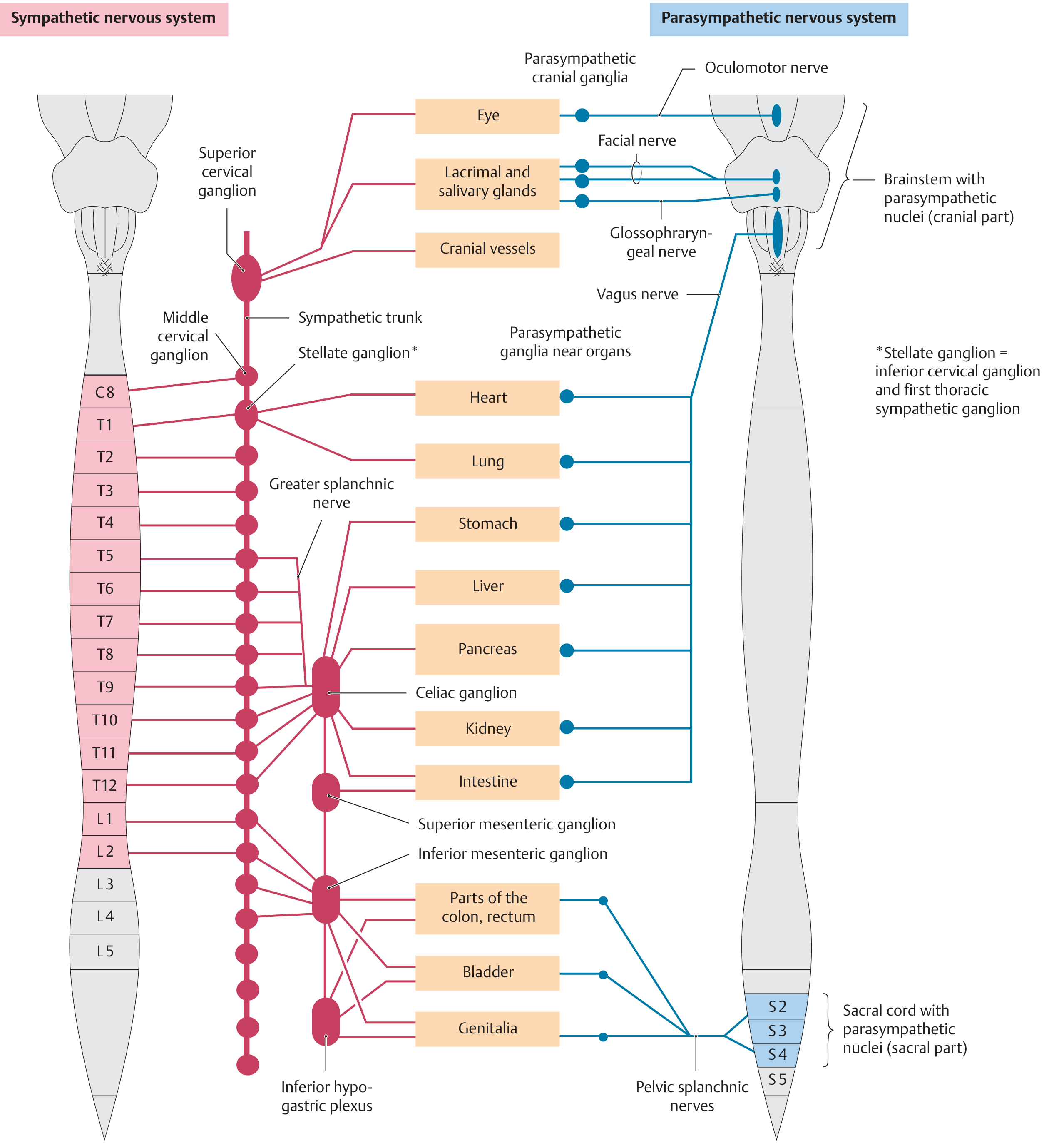
Structure of the autonomic nervous system showing sympathetic (red) and parasympathetic (blue) connections to organs - General Anatomy & Musculoskeletal System (THIEME Atlas)
Two-neuron pathway (both divisions): Preganglionic (CNS) neuron → ganglion → postganglionic neuron → target organ
| Feature | Sympathetic | Parasympathetic |
|---|
| Function | "Fight or flight" | "Rest and digest" |
| Origin | Lateral horns T1-L2 (thoracolumbar) | Brainstem + S2-S4 (craniosacral) |
| Ganglia location | Near spinal cord (paravertebral chains) | Near/within target organs |
| Preganglionic fibre length | Short | Long |
| Postganglionic fibre length | Long | Short |
| Neurotransmitter at target | Norepinephrine (noradrenaline) | Acetylcholine |
Effects on organs:
| Organ | Sympathetic | Parasympathetic |
|---|
| Heart rate | Increases | Decreases |
| Bronchi | Dilates | Constricts |
| Pupils | Dilates (mydriasis) | Constricts (miosis) |
| Salivary glands | Scant, viscous secretion | Copious, watery secretion |
| GI motility | Decreases | Increases |
| Blood vessels | Vasoconstriction | (minimal effect) |
| Urinary bladder | Relaxes detrusor; contracts sphincter (retention) | Contracts detrusor; relaxes sphincter (voiding) |
| Sweat glands | Increases sweating | - |
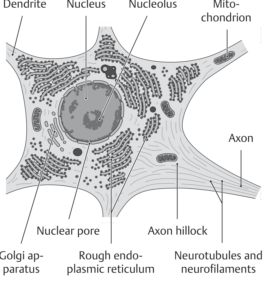
Neuron structure (relevant to nervous system histology):
Electron microscopy of the neuron showing nucleus, axon, Golgi apparatus, rough ER (Nissl substance), neurofibrils - General Anatomy & Musculoskeletal System (THIEME Atlas)
Types of neurons:
- Multipolar (most common): multiple dendrites + 1 axon; motor neurons, interneurons
- Bipolar: 1 dendrite + 1 axon; retina, cochlea, olfactory epithelium
- Pseudounipolar: 1 process divides into central + peripheral; sensory (dorsal root ganglia)
QUICK-REFERENCE APPLIED ANATOMY (A&E Focus)
| Condition | Anatomy Basis |
|---|
| Fractured clavicle | Junction of middle and outer thirds; brachial plexus/subclavian vessels at risk |
| Surgical neck of humerus fracture | Axillary nerve injury → deltoid paralysis, loss of sensation lateral arm |
| Midshaft humerus fracture | Radial nerve injury → wrist drop |
| Medial epicondyle fracture | Ulnar nerve injury → "claw hand" |
| Colles' fracture | Distal radius; median nerve at risk |
| Flail chest | Multiple rib fractures; paradoxical movement; requires ventilatory support |
| Tension pneumothorax | Air in pleural cavity under pressure → shifts mediastinum; needle decompression 2nd ICS midclavicular line |
| Chest drain insertion | 4th/5th ICS, anterior axillary line; always above superior rib border |
| Neck of femur fracture | Elderly females; avascular necrosis risk; limb shortened and externally rotated |
| Cardiac tamponade | Pericardial fluid → Beck's triad (hypotension, JVD, muffled heart sounds); pericardiocentesis |
| Myocardial infarction | LAD occlusion most common; STEMI vs NSTEMI |
| Urinary retention | Bladder distension above pubis; catheterization |
| Ureteric colic | Loin-to-groin pain; haematuria; stones at 3 sites of narrowing |
Sources: Gray's Anatomy for Students (Elsevier), General Anatomy and Musculoskeletal System - THIEME Atlas, Medical Physiology (Boron & Boulpaep). All diagrams from the medical library.