
Thigh anatomy in doppler
thigh femoral artery vein anatomy Doppler ultrasound
deep vein thrombosis femoral vein duplex ultrasound thigh
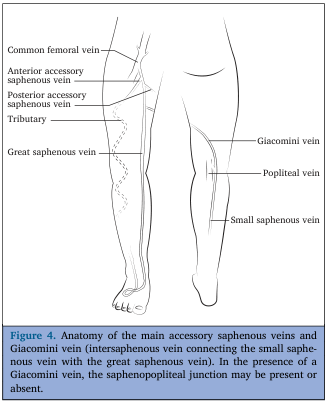
lower limb venous anatomy diagram femoral vein saphenous vein
femoral artery duplex ultrasound waveform superficial femoral artery
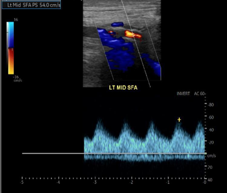
This diagnostic image is a duplex ultrasound of the left mid-superficial femoral artery (SFA), combining B-mode imaging, color Doppler, and spectral Doppler waveform analysis. The top panel shows a color Doppler box over the longitudinal view of the vessel, displaying turbulent flow with a mixture of red and blue hues, indicating variations in velocity and direction. The lower panel displays a spectral Doppler waveform indicating monophasic flow, characterized by a dampened systolic peak and a total lack of the normal early diastolic reversal and late diastolic forward flow seen in healthy peripheral arteries. The vertical axis measures velocity in cm/s, with the peak systolic velocity (PSV) measured at approximately 54.0 cm/s as indicated in the top-left annotation. Such monophasic, low-velocity waveforms are characteristic findings in peripheral artery disease (PAD), specifically suggesting hemodynamically significant proximal stenosis or occlusion. The clinical context relates to vascular assessment in a patient with lower extremity ulcers and suspected limb ischemia.
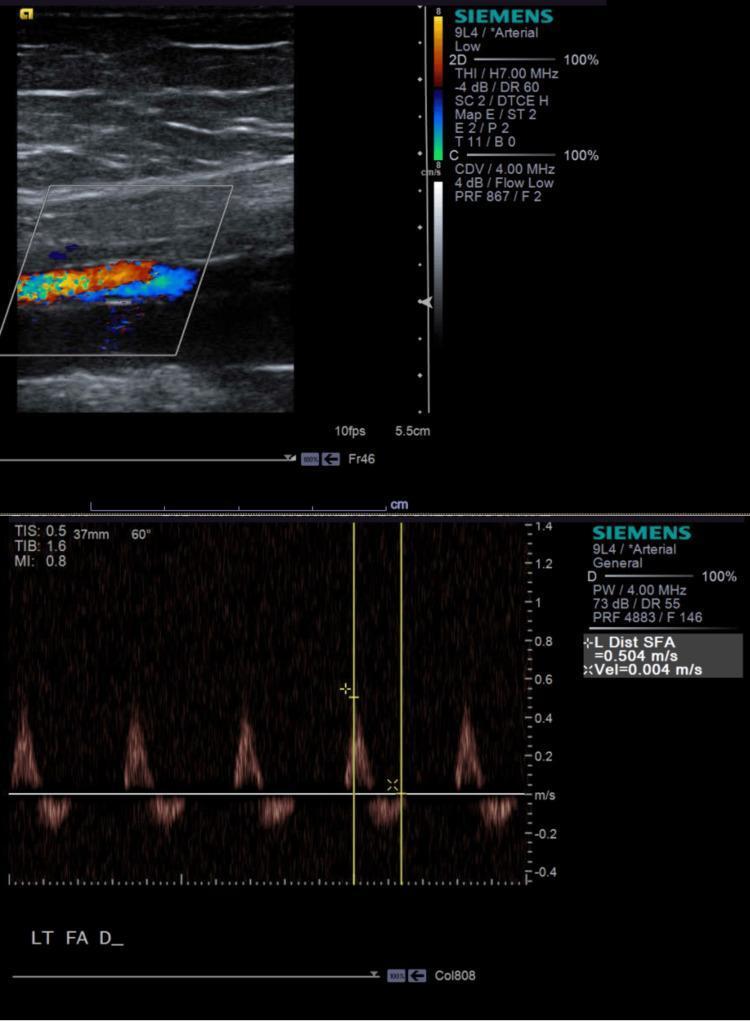
This composite image displays a lower extremity arterial duplex ultrasound of the left superficial femoral artery (SFA). The top panel features a color flow Doppler image of the distal SFA, showing a heterogeneous mosaic color pattern (mixture of red, blue, and yellow) within the lumen. This color aliasing and turbulence are indicative of localized high-velocity flow, consistent with arterial stenosis. The bottom panel presents a spectral Doppler waveform analysis of the same segment. The waveform is triphasic, characterized by a sharp systolic peak, a brief period of early diastolic flow reversal, and a small late diastolic forward component. Quantitative measurements indicate a peak systolic velocity (PSV) of 0.504 m/s. The ultrasound is used to evaluate peripheral arterial disease (PAD) and assess the degree of luminal narrowing in the lower extremity vasculature. Key educational points include the recognition of turbulent flow patterns on color Doppler and the interpretation of spectral waveforms in the context of arterial resistance.
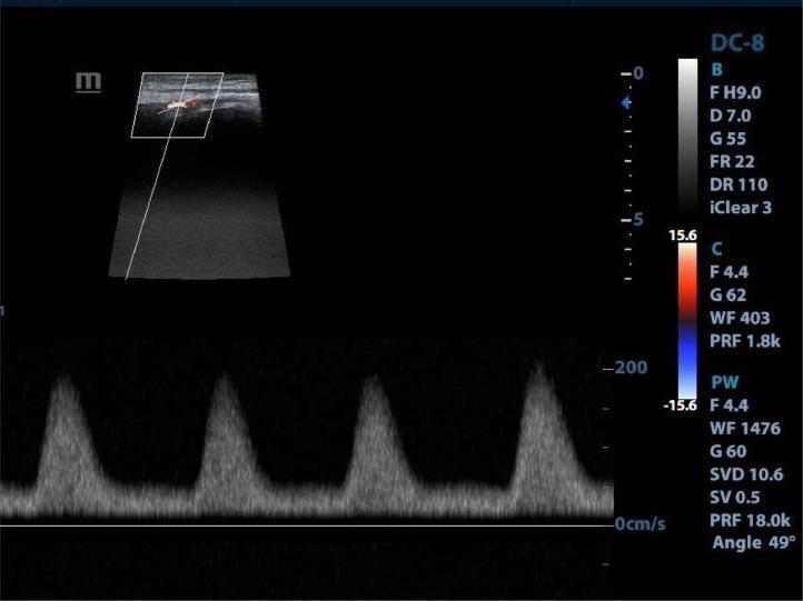
This diagnostic image is a duplex ultrasound of the left lower limb, specifically targeting the superficial femoral artery (SFA). The top portion shows a B-mode ultrasound with a color Doppler overlay, displaying a mixture of red and blue signals within the vessel lumen, suggesting turbulent or complex flow patterns. The lower portion of the image contains a pulse-wave (PW) spectral Doppler display showing a monophasic waveform pattern. This waveform is characterized by a loss of the normal triphasic arterial signal, showing instead a slow upstroke and a single, broad peak followed by a gradual decay without a diastolic reversal component. Four distinct cardiac cycles are visible. Such monophasic waveforms in the peripheral arteries are clinically significant as they often indicate significant proximal arterial stenosis or occlusion, reflecting a dampened 'tardus parvus' flow distal to a high-grade obstruction. The image includes technical parameters such as a pulse repetition frequency (PRF) of 18.0k and an insonation angle of 49 degrees.
Thigh Vascular Anatomy on Doppler/Duplex Ultrasound
Arterial anatomy
- Common femoral artery (CFA): The external iliac artery becomes the CFA as it passes beneath the inguinal ligament, at a point midway between the anterior superior iliac spine and the pubic symphysis.
- Deep femoral (profunda femoris) artery: Branches off the CFA roughly 3-4 cm distal to the inguinal ligament. It runs posterolateral to the superficial femoral artery, supplies the hamstrings, and terminates in the distal thigh with branches to the adductor magnus.
- Superficial femoral artery (SFA): Continues along the anteromedial thigh, passes through the adductor canal, and exits via the adductor hiatus to become the popliteal artery at the junction of the middle and lower thirds of the thigh.
- The femoral head/neck receive supply mainly from the medial and lateral circumflex femoral arteries (branches of the femoral artery), forming an extracapsular ring, plus a minor contribution from the obturator artery via the ligamentum teres.
Venous anatomy
- Common femoral vein (CFV): At the inguinal ligament it lies posterior/medial to the CFA, then shifts lateral to it more distally.
- Femoral vein (previously called the "superficial femoral vein" - this term is now discouraged because it wrongly implies a superficial location and has caused mistaken DVT triage; it is a deep vein): Runs alongside the SFA.
- Deep femoral (profunda femoris) vein: Runs parallel to the deep femoral artery and joins the femoral vein just distal to the inguinal ligament.
- Great saphenous vein (GSV): A superficial vein arising from the dorsal foot, ascending medially up the leg and thigh within its own fascial sheath (saphenous sheath), terminating at the saphenofemoral junction, where it drains into the CFV after receiving the superficial circumflex iliac, superficial epigastric, and external pudendal veins.
- Standard "deep venous nomenclature" for the thigh: common femoral vein -> femoral vein -> profunda femoris (deep femoral) vein, medial/lateral circumflex femoral veins, deep femoral communicating veins.
Doppler/duplex scanning technique (DVT protocol)
- Patient supine, leg externally rotated, knee slightly flexed.
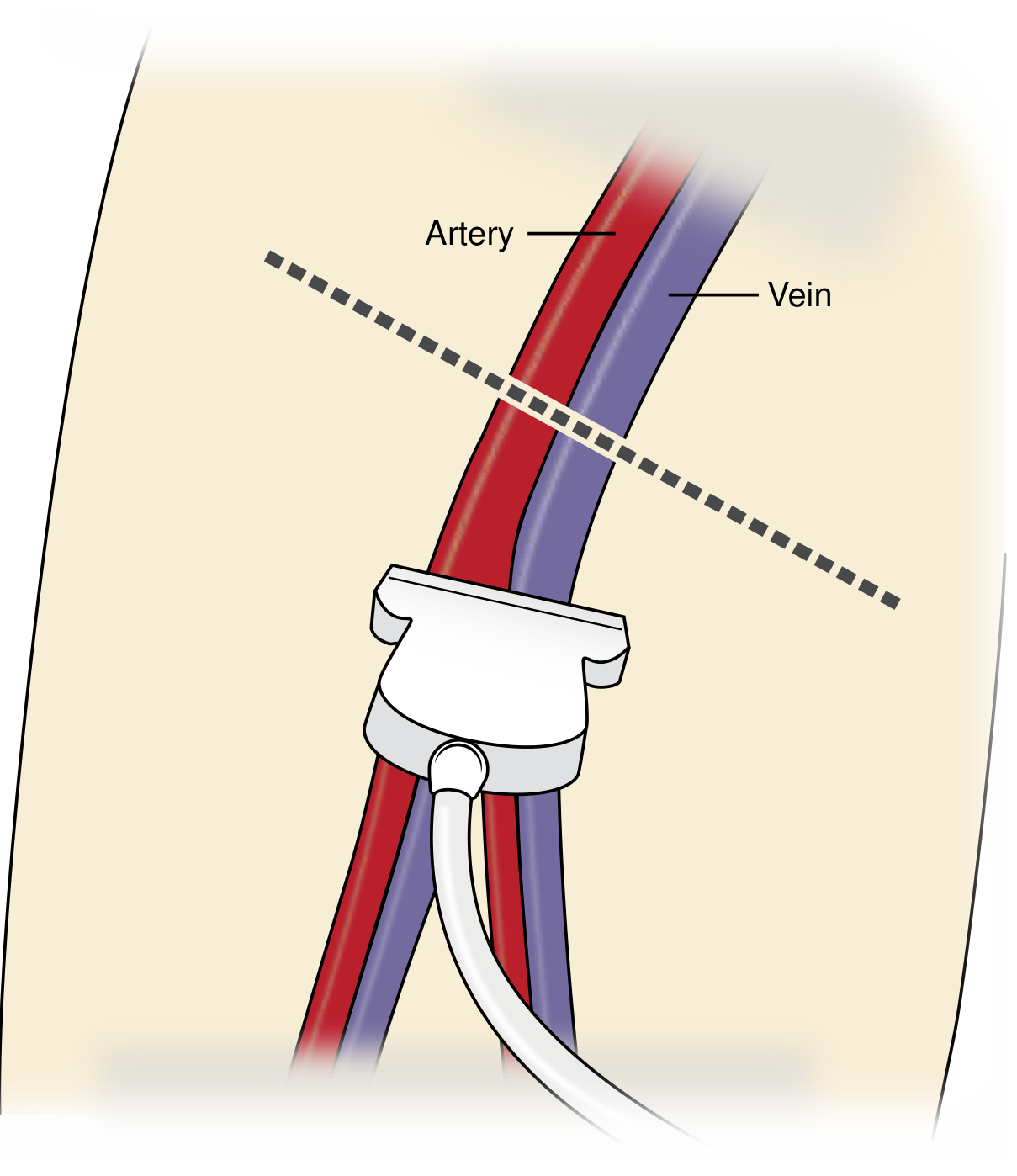
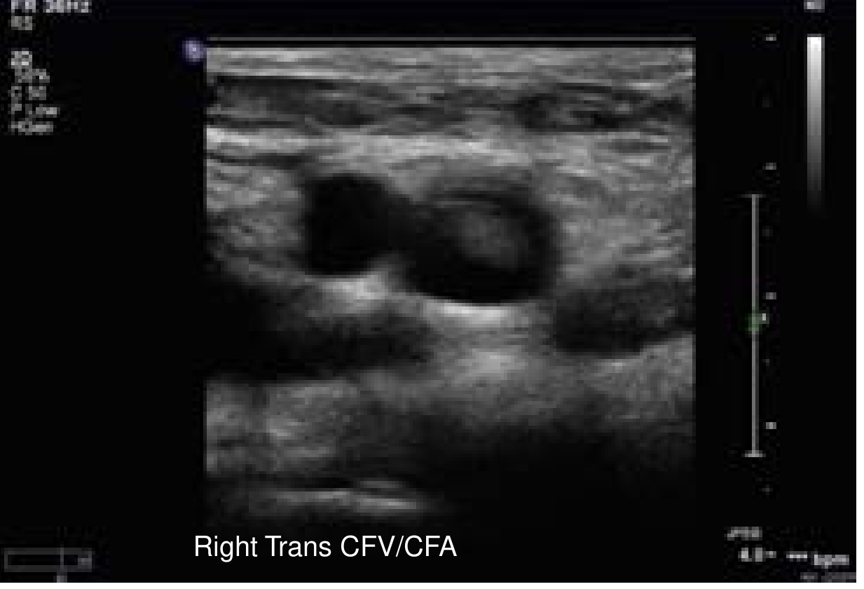
- High-frequency linear transducer (7-12 MHz) placed transversely just below the inguinal ligament to identify the CFV.
- Compression is applied every 1-2 cm while sliding distally through the femoral triangle to visualize the deep femoral and femoral veins, then followed into the popliteal fossa; imaging is repeated in the sagittal plane.
- A normal vein fully compresses with gentle probe pressure. Findings diagnostic of DVT: non-compressibility, hyperechoic intraluminal material (clot), venous distension, and absent spectral/color Doppler signal (acute); partial compressibility with echogenic thrombus and collaterals with some flow (chronic).
Key clinical points
- A complete lower-extremity venous duplex exam evaluates the CFV, femoral vein, deep femoral vein, popliteal vein, posterior tibial veins, GSV, and small saphenous vein.
- Compression ultrasonography (with or without color/spectral Doppler) is the standard non-invasive test for DVT because of its accuracy for popliteal and more proximal veins; sensitivity is lower for isolated calf-vein thrombus, so a repeat scan in 5-7 days is advised if only proximal veins were scanned initially and the exam is negative but suspicion remains.
- Arterial duplex of the thigh is used mainly to grade stenosis/occlusion in peripheral artery disease, using peak systolic velocity ratios and waveform morphology (triphasic -> biphasic -> monophasic as disease severity increases).
How to identyfy
How to Identify the Thigh Vessels on Doppler/Duplex Ultrasound
1. General principles: artery vs. vein
| Feature | Artery | Vein |
|---|---|---|
| Compressibility | Does not collapse with gentle probe pressure | Fully collapses/flattens with light compression |
| Pulsatility | Visible pulsation in B-mode, especially with probe held still | No pulsation (may show slow respiratory variation) |
| Wall/shape | Thicker wall, round, less distensible | Thinner wall, more oval, distensible |
| Doppler signal | Pulsatile, high-velocity, triphasic/multiphasic spectral waveform | Continuous, low-velocity, phasic with respiration; augments with distal limb compression |
2. Step-by-step scanning protocol for the thigh
- Position: patient supine, leg externally rotated, knee slightly flexed to open the femoral triangle.
- Transducer: high-frequency linear probe (7-12 MHz for compression/venous exam; lower frequency 3-5 MHz curvilinear may be needed for deep/large thighs on arterial exam).
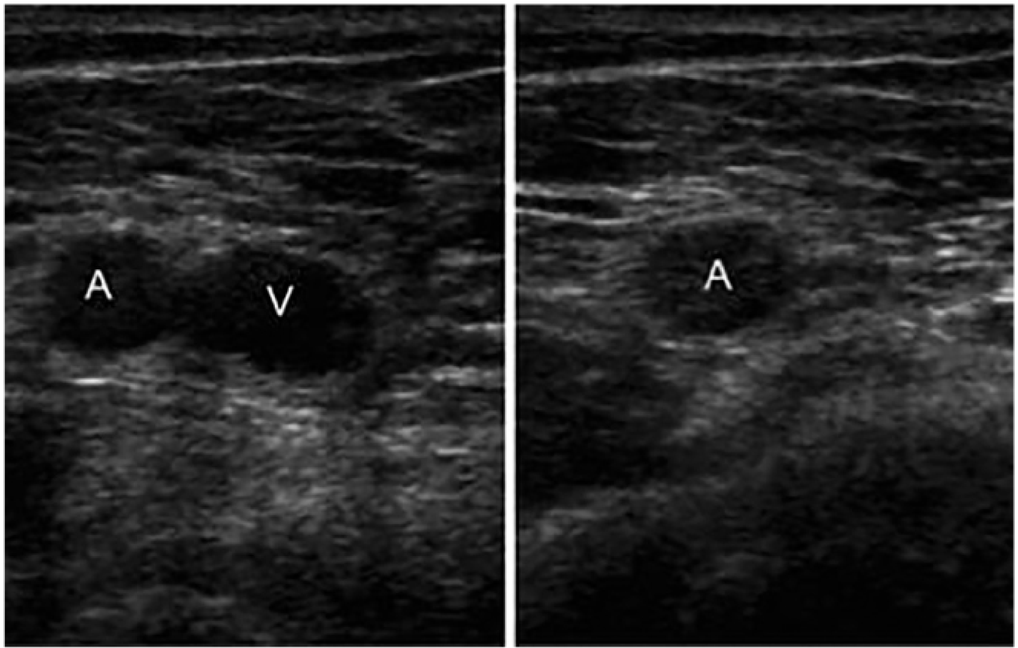
- Start point: place the probe transversely just below the inguinal ligament. The common femoral vein (CFV) lies medial to the common femoral artery (CFA) at this level - this medial position is your anatomic landmark.
- Compress every 1-2 cm as you slide the probe distally. Confirm the vein by watching it collapse fully; the artery remains round and pulsatile.
- Continue distally until the CFA/CFV bifurcate: the artery splits into the superficial femoral artery (SFA) (continues anteromedially) and the deep femoral (profunda femoris) artery (courses posterolaterally); the vein splits similarly into the femoral vein and deep femoral vein.
- Identify the saphenofemoral junction: the great saphenous vein joins the CFV from the superficial/medial side, just distal to the inguinal ligament - a classic superficial landmark ("Mickey Mouse" sign in transverse view, with the CFA and CFV as the head and the GSV as one ear).
- Follow the femoral vessels down the medial thigh through the adductor canal; loss of the SFA signal here as it dives through the adductor hiatus marks its transition to the popliteal artery behind the knee.
- Repeat everything in the sagittal (longitudinal) plane to confirm continuity and rule out a focal, non-compressible segment (clot) that might be missed transversely.
3. Color and spectral Doppler cues
- Color Doppler immediately separates the two vessels by flow direction/color assignment and confirms patency without needing to compress - useful when anatomy is distorted (e.g., large hematoma, aneurysm, prior surgery, or a pseudoaneurysm producing "pseudo-pulsation" that can mimic the artery).
- Spectral Doppler waveform:
- Normal thigh artery (CFA/SFA) = triphasic: sharp systolic upstroke, brief early diastolic flow reversal, small late diastolic forward flow.
- Diseased/stenotic segment = monophasic, dampened, slow-rising ("tardus parvus") waveform - a sign of proximal stenosis or occlusion.
- Normal vein = low-velocity, respirophasic flow that augments (increases) with manual compression of the calf, confirming forward flow and patency.
4. Practical pitfalls
- In hypovolemic or critically ill patients, veins can appear rounder and less compressible, and arteries less pulsatile - always confirm with color/spectral Doppler, not B-mode alone.
- A pseudoaneurysm at a prior puncture site can show pseudo-pulsation on B-mode; transient compression and spectral analysis (to-and-fro "yin-yang" flow) differentiate it from the true artery.
- Anatomic variants (duplicated femoral vein, high bifurcation of the profunda) are common - always trace vessels continuously rather than relying on a single cross-section.