
Copd pink buffer
- Pink = maintains near-normal oxygenation by hyperventilating (not cyanotic)
- Puffer = pursed-lip breathing, barrel chest, accessory muscle use
- Thin/cachectic, tachypneic, minimal sputum
- Contrasts with the "blue bloater" (chronic bronchitis type)
pink puffer emphysema COPD barrel chest
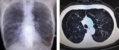
This dual-panel image displays diagnostic thoracic imaging from a patient with Chronic Obstructive Pulmonary Disease (COPD). The left panel is a posterior-anterior (PA) chest X-ray showing classic features of severe emphysema, including hyperinflated lung fields, sparse bronchovascular markings, and a widened chest diameter consistent with a barrel chest deformity. Diaphragmatic flattening and a narrowed mediastinal silhouette are also visible. The right panel is an axial CT scan of the chest (lung window) at the level of the upper lobes, revealing extensive bullous emphysema. Multiple large, thin-walled bullae are visible, particularly in the subpleural regions, along with architectural distortion of the lung parenchyma. These findings demonstrate advanced obstructive lung disease and are clinically significant for explaining symptoms of chronic shortness of breath and impaired gas exchange in elderly patients with a significant smoking history.
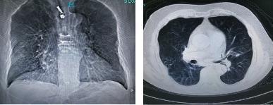
This diagnostic image pair displays thoracic radiology findings in an 80-year-old male with Chronic Obstructive Pulmonary Disease (COPD). The left panel shows a frontal chest radiograph (X-ray) exhibiting hallmark signs of hyperinflation, including a widened anteroposterior diameter and flattened diaphragmatic domes. Both lung fields demonstrate sparse bronchovascular markings and increased lucency, indicative of diffuse emphysema. The right panel shows an axial CT scan of the thorax, further characterizing the pulmonary pathology. It reveals significant emphysematous destruction of the lung parenchyma with the presence of multiple bullae and areas of decreased attenuation. Additionally, subtle interstitial lesions are visible. The imaging illustrates the classic presentation of severe COPD, characterized by permanent enlargement of airspaces distal to the terminal bronchioles and associated structural changes to the chest wall often described as a barrel chest.
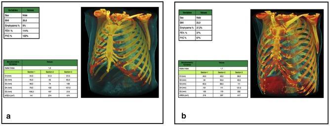
This diagnostic comparison uses 3D CT reformations and morphometric data to demonstrate rib cage remodeling in Chronic Obstructive Pulmonary Disease (COPD). Image A shows a normal rib cage of a 58-year-old male smoker with 0% emphysema, characterized by an elongated, conical shape with a relatively narrow upper aperture and gradual lower expansion. Image B illustrates a 61-year-old male with severe emphysema (37.2%) exhibiting a classic 'barrel chest' configuration. Key morphologic changes in the COPD model include a significant increase in upper rib cage dimensions (larger transverse and anteroposterior diameters) and a relative decrease in lower rib cage area compared to the control. The ribs in the emphysematous chest show a more horizontal orientation and increased curvature, reflecting hyperinflation and mechanical displacement of the thoracic wall. Accompanied tables provide quantitative variables, including Haller index, sectional diameters (D1-D5), and cross-sectional areas, correlating visual remodeling with objective functional decline (decreased FEV1/FVC ratios).
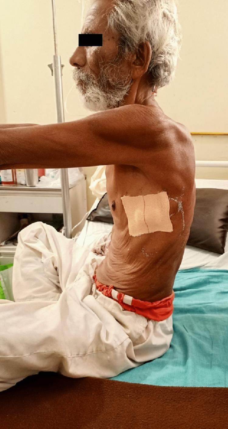
This clinical photograph provides a lateral view of an elderly male patient in a seated position, demonstrating a classic barrel-shaped chest deformity. This morphological change is characterized by a significant increase in the anterior-posterior (AP) diameter of the thoracic cage, commonly associated with chronic obstructive pulmonary disease (COPD) or severe emphysema. Visible physical findings include significant wasting of the pectoral and shoulder musculature (cachexia) and thin, wrinkled skin over the thorax. A nasal cannula is visible, indicating the need for supplemental oxygen therapy. A square medical dressing is applied to the lateral chest wall, likely at a site following the removal of an intercostal drainage (ICD) tube. The patient exhibits a 'tripod' posture or forward-leaning tendency, which is a common compensatory mechanism used to engage accessory muscles of respiration. A bedside IV pole is visible in the background, suggesting an inpatient clinical setting. This image serves as a teaching tool for physical examination findings in chronic respiratory failure and obstructive lung disease.
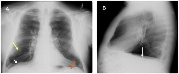
This diagnostic image displays a two-panel chest roentgenogram (X-ray) from a 65-year-old male, highlighting radiographic features of severe pulmonary emphysema and chronic obstructive pulmonary disease (COPD). Panel A presents a postero-anterior (PA) view, while Panel B provides a lateral view. Key findings include significant hyperinflation of the lung fields with marked flattening of the hemidiaphragms (indicated by white arrows), which loss their normal convex shape. In Panel A, a yellow arrow identifies areas of increased translucency and attenuation of vascular markings consistent with emphysematous parenchymal changes. A red arrow highlights the decreased zone of apposition, representing the diminished area where the diaphragm sits parallel to the inner rib cage. Panel B further illustrates the flattened diaphragmatic contour and an increased retrosternal clear space, reflecting an increased anterior-posterior thoracic diameter (barrel chest). These visual markers are clinically significant as they correlate with the mechanical basis of Hoover's sign, where the horizontal orientation of the diaphragm causes inward rib cage retraction during inspiration.
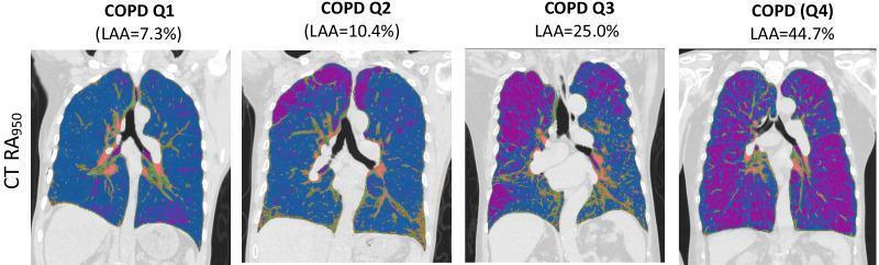
This diagnostic image series presents four coronal chest CT scans illustrating emphysema quantification in patients with varying severity of Chronic Obstructive Pulmonary Disease (COPD). The quantification is based on the RA950 metric (relative area of CT density histogram voxels < -950 HU), which identifies Low Attenuation Areas (LAA) characteristic of emphysematous tissue. A color-coded overlay is used to differentiate tissue types: healthy lung parenchyma is represented in blue, while emphysematous regions are highlighted in purple. The four panels (labeled Q1 through Q4) demonstrate a progressive increase in LAA percentage, ranging from 7.3% in early-stage disease to 44.7% in advanced emphysema. Visually, the purple areas transition from sparse focal distributions in Q1 to widespread, confluent regions in Q4, often appearing more prominent in the upper lung zones. Central mediastinal and hilar structures are visible, with some orange/red markers indicating vascular or hilar regions. This comparison chart serves as an educational tool for understanding radiological progression and automated volumetric quantification of lung destruction in COPD patients.
COPD: The "Pink Puffer" (Emphysema Type)
What Is a Pink Puffer?
- Pink = maintains near-normal arterial oxygenation by markedly increasing respiratory drive and minute ventilation - stays pink (not cyanotic)
- Puffer = works extremely hard to breathe, with visible pursed-lip breathing, accessory muscle use, and tachypnea
"In others, parenchymal destruction predominates producing the emphysema phenotype (the 'pink puffer')." - Frameworks for Internal Medicine
Pathophysiology
- Loss of elastic recoil - airways collapse on exhalation (dynamic airway collapse), causing air trapping
- Loss of alveolar surface area - reduced gas exchange capacity
- Hyperinflation - air trapping causes lung overinflation, driving up the residual volume and total lung capacity
- Increased work of breathing - to compensate, the respiratory center drives hyperventilation to maintain PaO2
Clinical Features
| Feature | Pink Puffer (Emphysema) | Blue Bloater (Chronic Bronchitis) |
|---|---|---|
| Body habitus | Thin, cachectic | Overweight, edematous |
| Cyanosis | Absent (pink) | Present (blue) |
| Breathing pattern | Pursed-lip, hyperventilation | Normal/shallow |
| Sputum | Minimal | Copious |
| PaO2 | Near-normal (high respiratory drive) | Low (hypoxemic) |
| PaCO2 | Low-normal (blows off CO2) | Elevated (CO2 retainer) |
| Barrel chest | Prominent | Less prominent |
| Cor pulmonale | Late/less common | Early/more common |
| Emphysema type | Panlobular | Centrilobular |
Physical Examination Findings
- Inspection: pursed-lip breathing, barrel chest (increased AP diameter), use of accessory muscles, cachexia/muscle wasting, forward-leaning "tripod" posture
- Percussion: hyperresonant due to hyperinflation; reduced diaphragmatic excursion
- Auscultation: decreased breath sounds globally; prolonged expiration (>6 seconds on forced exhalation); expiratory wheeze may be present; heart sounds muffled by hyperinflated lungs
- No clubbing - its presence should prompt investigation for lung cancer
Pulmonary Function Tests (PFTs)
| Parameter | Finding in Emphysema |
|---|---|
| FEV1 | Decreased |
| FVC | Decreased (less than FEV1 reduction) |
| FEV1/FVC ratio | < 0.70 (post-bronchodilator) = obstructive |
| Total Lung Capacity (TLC) | Increased (due to air trapping) |
| Residual Volume (RV) | Markedly increased |
| DLCO | Decreased (lost alveolar surface area) |
Chest Radiology
- Hyperinflated lung fields with increased radiolucency
- Flattened hemidiaphragms (most reliable sign)
- Increased AP diameter (barrel chest)
- Widened retrosternal clear space (lateral view)
- Reduced vascular markings (arterial deficiency pattern)
- Bullae formation on CT
Mechanism of "Pursed-Lip" Breathing
Key Mnemonic: Pink Puffer vs. Blue Bloater
- Pink Puffer = Parenchymal destruction = Panlobular emphysema = Puffs to stay pink
- Blue Bloater = Bronchitis = Big/obese + Big sputum = Blue from hypoxia
Note: In clinical practice, most COPD patients have a mixed phenotype of both emphysema and chronic bronchitis. The classic pink puffer / blue bloater dichotomy is a didactic simplification used to understand the two extremes.