
1. Otitis Media Must revise ● Acute otitis media ● CSOM ● Safe vs unsafe CSOM ● ● Complications of otitis media Mastoiditis Frequently repeated ● Cholesteatoma ● Atticoantral disease ● Tubotympanic disease
cholesteatoma tympanic membrane pars flaccida retraction pocket
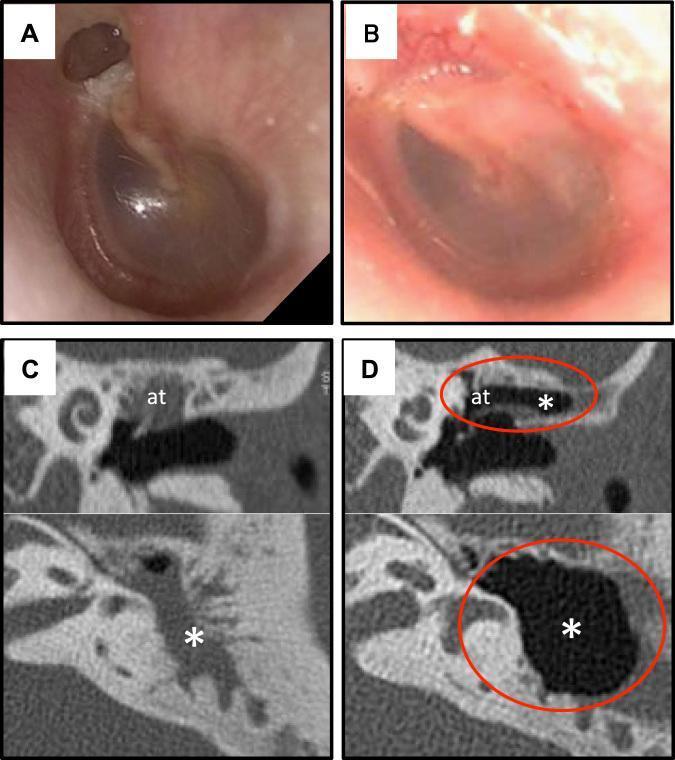
Educational comparison of preoperative and 12-month postoperative findings for pars flaccida cholesteatoma treated with cell sheet transplantation. (A) Otoscopic clinical photograph showing preoperative retraction of the attic and visible cholesteatoma. (B) Postoperative clinical photograph demonstrating a healthy tympanic membrane with no recurrence or retraction. (C) Preoperative computed tomography (CT) in coronal (top) and axial (bottom) views showing opacified attic (at) and mastoid cavity (*) with absent pneumatization due to disease. (D) 12-month postoperative CT scans highlighting successful restoration of air-filled spaces (black) in the attic and mastoid regions (circled in red), indicating effective mucosal regeneration and cavity ventilation. This composite image serves as a clinical case study in otolaryngology for regenerative surgical techniques in middle ear disease.
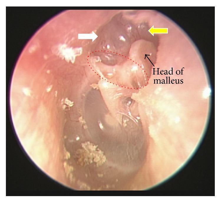
This endoscopic clinical photograph displays the left tympanic membrane, illustrating signs of chronic middle ear disease. The image focuses on the superior attic region (pars flaccida). A white arrow identifies an attic retraction pocket, where the tympanic membrane is invaginated medially. A red dotted circle highlights an atelectatic Prussak's space, indicating a localized collapse of the membrane. Adjacent to this, a yellow arrow points to an eroded scutum, showing loss of the bony lateral attic wall. The head of the malleus is visible and labeled due to the loss of overlying structural integrity. These findings are characteristic precursors or manifestations of acquired cholesteatoma, reflecting chronic negative middle ear pressure and inflammatory bone resorption. The image serves as a clinical example for otolaryngology education regarding the diagnosis of middle ear pathology and the assessment of structural damage to the ossicular chain and surrounding bony landmarks.
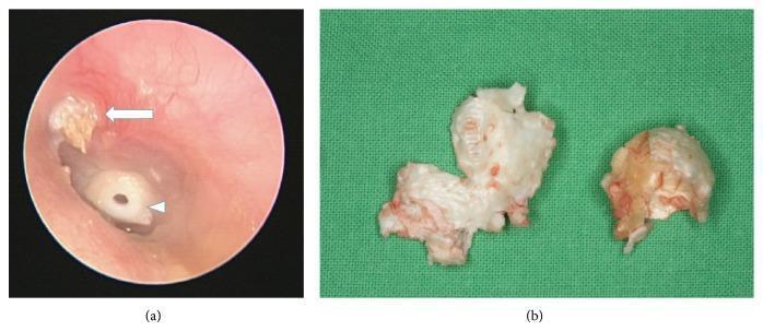
This composite image illustrates the clinical presentation and gross pathology of an acquired cholesteatoma. Image (a) is an otoscopic view of a left ear showing a keratinous debris accumulation in the attic region (white arrow), characteristic of a cholesteatoma originating from a retraction pocket in the pars flaccida. A tympanostomy tube or grommet (white arrowhead) is visible in the tympanic membrane, indicating a history of middle ear ventilation issues. Image (b) shows the macroscopic appearance of the dissected cholesteatoma tissue. The specimen exhibits a characteristic pearly-white, greasy-looking thin wall with a friable, pultaceous internal substance. This visual provides an educational comparison between the in situ endoscopic appearance of middle ear pathology and the gross morphological features of the excised squamous epithelial mass. It is relevant for otolaryngology training regarding chronic otitis media and surgical planning for mastoidectomy.
Otitis Media - Complete Revision Notes
1. ACUTE OTITIS MEDIA (AOM)
Definition & Pathogenesis
- Viral URTI causes nasopharyngeal inflammation
- Impaired mucociliary clearance of the Eustachian tube (ET)
- ET dysfunction → negative middle ear pressure → effusion → bacterial superinfection
- Viruses involved: Rhinovirus, RSV (most common), adenovirus, coronavirus, PIV
Causative Organisms (Bacteria)
| Organism | Notes |
|---|---|
| Streptococcus pneumoniae | Most common; most severe; more mastoidectomies |
| Haemophilus influenzae | Second most common; non-typeable strains |
| Moraxella catarrhalis | High spontaneous resolution rate |
| S. pyogenes (Group A Strep) | Less otalgia |
| Pseudomonas aeruginosa | Especially in children with ventilation tubes; less aggressive course |
Clinical Features
- Otalgia (ear pain) - often severe, throbbing
- Fever
- Hearing loss (conductive)
- Bulging, erythematous, opaque tympanic membrane (TM) - classic finding
- Infants: irritability, ear pulling, poor feeding
- Spontaneous perforation → otorrhea → pain relief
Stages of AOM
- Congestion - hyperemia of TM, no exudate yet
- Exudation - outpouring of exudate, TM bulges; severe pain
- Suppuration - pus forms, TM perforates → relief of pain and fever
- Resolution - TM heals, effusion resolves
Diagnosis
- Clinical: otoscopy showing bulging, opaque, hyperemic TM with reduced or absent light reflex
- Pneumatic otoscopy: reduced or absent TM mobility
- Tympanometry: type B (flat) curve
Management
- Watchful waiting (48-72 hrs) in mild cases (age >2 yrs, unilateral, no severe symptoms)
- Amoxicillin is first-line antibiotic (80-90 mg/kg/day in divided doses)
- Amoxicillin-clavulanate if: failure after 48-72 hrs, recurrent AOM, prior antibiotics
- Analgesia: paracetamol/ibuprofen
- Myringotomy if: severe bulging, extreme pain, toxic child, or needed for culture
Recurrent AOM (rAOM)
- ≥3 episodes in 6 months or ≥4 in 12 months
- Management: Grommet/ventilation tube insertion
2. CHRONIC SUPPURATIVE OTITIS MEDIA (CSOM)
Definition
Pathogenesis - Smyth's Theory
- Stable "healed scar" - tympanosclerosis
- Permanent perforation of pars tensa
- Atelectatic TM (drapes over medial wall)
- Retraction pocket → cholesteatoma (if self-cleansing fails)
Microbiology of CSOM
- Pseudomonas aeruginosa (most common)
- Staphylococcus aureus
- Proteus mirabilis
- Klebsiella, E. coli, anaerobes
3. SAFE vs UNSAFE CSOM
| Feature | TUBOTYMPANIC (Safe/Benign) | ATTICOANTRAL (Unsafe/Dangerous) |
|---|---|---|
| Also called | Mucosal disease / Benign CSOM | Squamous disease / Dangerous CSOM |
| Perforation site | Central (pars tensa); margins covered by stratified squamous epithelium | Attic (pars flaccida) OR posterior-superior quadrant of pars tensa |
| True perforation? | Yes - permanent perforation | Retraction pocket (not a true perforation) - like the cut finger of a surgical glove |
| Tissue affected | Mucosa only | Mucosa + bone (mucoperiosteum AND underlying bone) |
| Bone involvement | Absent | Regular osteitic erosion of scutum, posterior-superior bony canal wall, ossicles |
| Cholesteatoma | Absent | Present (defines "unsafe") |
| Exudate | Mucopurulent (polymorphs, lymphocytes, plasma cells) | Purulent, creamy |
| Cholesterol granuloma | Frequently associated | Unusual |
| Tympanosclerosis | May occur (chalky white plaques on TM) | Unusual |
| Mucosal changes | Cuboidal/high columnar ciliated; goblet cell proliferation; polyp may form | Cholesteatoma sac advances, inducing osteitis + granulation |
| Complications | Unusual (can occur if neglected) | Common - intratemporal and intracranial |
| Discharge | Profuse, odourless (mucoid) or mucopurulent | Scanty, foul-smelling (keratin breakdown) |
| Hearing loss | Conductive (TM perforation) | Conductive ± sensorineural (ossicular erosion/labyrinthine fistula) |
| Surgery | Myringoplasty/tympanoplasty (elective) | Mastoidectomy (urgent/mandatory) |
Key mnemonic: "Safe = Central perforation, no cholesteatoma; Unsafe = Attic/posterior-superior perforation + cholesteatoma"
4. CHOLESTEATOMA
Definition
Types
| Type | Details |
|---|---|
| Congenital | Occurs anywhere in temporal bone; behind intact TM; white pearly mass anterior to malleus; rare (~2%) |
| Acquired (~98%) | Complication of chronic otitis media |
Acquired Cholesteatoma - Two Subtypes
- Arises from invagination of the pars flaccida (retraction pocket)
- Keratin accumulates in the lateral epitympanic (Prussak's) space
- Causes: scutum erosion, medial displacement of the ossicular chain, ossicular erosion
- Enlarges lateral to the head of malleus and body of incus
- Arises from a retraction pocket or perforation of the posterior-superior quadrant
- Located medial to the ossicular chain
- Causes early erosion of the long process of incus (LPI)
- More erosion of inner ear structures
- No scutum erosion (unlike pars flaccida type)
- Tends toward labyrinthine complications
Imaging
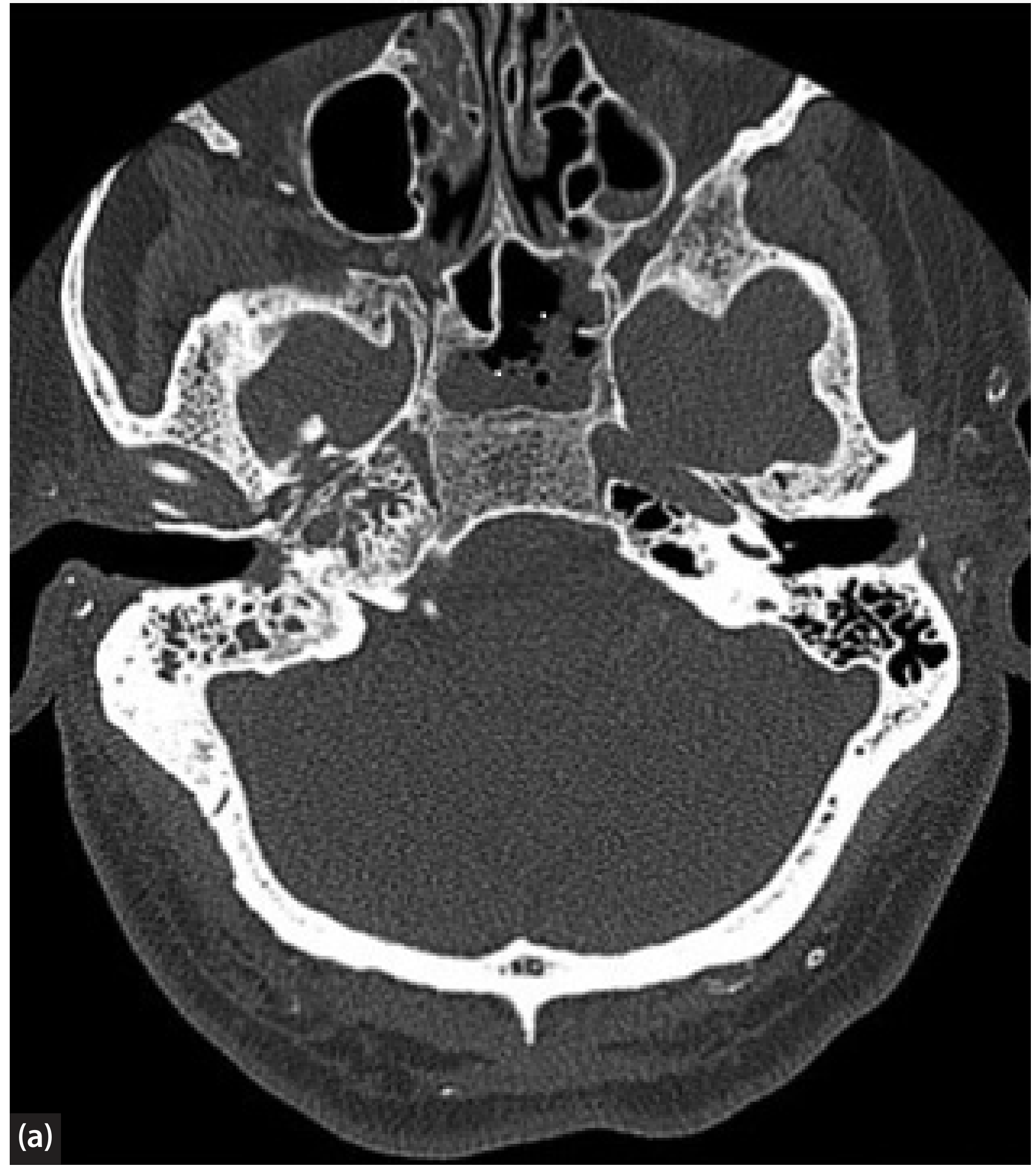
- CT scan: First choice - shows middle ear opacification, bone erosions of scutum and ossicles; high NPV when normal; cannot differentiate cholesteatoma from other soft tissue masses (granulation, cholesterol granuloma)
- MRI (DWI - Diffusion Weighted Imaging): Distinguishes cholesteatoma (restricted diffusion = tightly packed keratin, poor water mobility) from inflammatory disease (good water mobility); replaces routine second-look surgery in many centres
CT - Images
Clinical Images
Treatment
- Mastoidectomy is mandatory
- Canal wall up (CWU) mastoidectomy vs Canal wall down (CWD) mastoidectomy
- Aim: eradication of disease + reconstruction
5. MASTOIDITIS
Four Stages/Classes
| Stage | Description | Features |
|---|---|---|
| 1. Mastoid effusion | Infection extends into mastoid cavity during AOM; radiological finding only | No clinical signs of mastoiditis; NOT a complication |
| 2. Acute mastoiditis with periostitis | Spread via emissary veins to mastoid periosteum | Postauricular crease fullness, pinna pushed forward, mild swelling/erythema/tenderness; no abscess |
| 3. Acute mastoid osteitis (Coalescent mastoiditis) | Bone destruction of mastoid air cells | Subperiosteal abscess may develop postauricularly; destruction of bony septa |
| 4. Masked (Subacute) Mastoiditis | Incompletely treated AOM after 10-14 days | Subtle signs; persistent otalgia and fever; can still progress to serious complications |
Subperiosteal Abscess Variants
- Postauricular abscess: Most common location - pushes pinna forward and downward
- Zygomatic abscess: Above and in front of pinna
- Bezold abscess: Perforation of medial mastoid cortex → tracks down sternomastoid into posterior triangle
- Retropharyngeal/Parapharyngeal abscess: Via peritubal cells
Bacteriology
- Streptococcus pneumoniae (most severe; more mastoidectomies)
- Pseudomonas aeruginosa (especially post-ventilation tube; less aggressive)
- Streptococcus pyogenes (less otalgia)
- Staphylococcus aureus, H. influenzae, Moraxella catarrhalis
Investigations
- FBC, CRP, blood cultures
- CT mastoid: recommended when intracranial complications suspected, not improving on antibiotics, or pre-operatively; shows osteitis, abscesses, intracranial complications
- MRI: better for specific intracranial pathology
Management
- IV antibiotics: 3rd-generation cephalosporin or aminopenicillin + β-lactamase inhibitor
- If Pseudomonas suspected: ciprofloxacin, piperacillin or fosfomycin
- Myringotomy ± ventilation tube: for drainage and culture
- Cortical mastoidectomy indicated for:
- Suspected intracranial complication
- Subperiosteal abscess
- No response to conservative treatment within 24-72 hours
- Unwell/deteriorating child
Important: Intracranial complications from acute mastoiditis develop in 6-17% of cases - early surgery is preferred over delayed intervention.
6. COMPLICATIONS OF OTITIS MEDIA
Classification (Cummings)
- Acute mastoiditis
- Coalescent mastoiditis
- Chronic mastoiditis
- Masked mastoiditis
- Postauricular abscess
- Bezold abscess
- Temporal abscess
- Petrous apicitis (Gradenigo's syndrome: triad of otorrhea + ipsilateral VI nerve palsy + retro-orbital pain)
- Labyrinthine fistula (most common intratemporal complication in cholesteatoma - lateral semicircular canal most affected)
- Facial nerve paralysis
- Acute suppurative labyrinthitis
- Encephalocele and CSF leakage
- Meningitis (most common overall intracranial complication)
- Brain abscess - temporal lobe (from middle ear) or cerebellar (from mastoid)
- Subdural empyema
- Epidural abscess
- Lateral (sigmoid) sinus thrombosis - presents with "picket fence" fever, Tobey-Ayer test positive
- Otitic hydrocephalus (raised ICP without ventricular dilatation, usually with lateral sinus thrombosis)
Key Complication Notes
- Lateral semicircular canal most susceptible (most prominent in aditus, in path of enlarging cholesteatoma)
- 7% of COM cases
- Symptoms: vertigo (especially with Valsalva), hearing loss, facial paresis
- Positive fistula test (nystagmus on external pressure)
- Nystagmus beats toward the diseased ear (if fistula on non-ampullated arc)
- Diagnosis confirmed by thin-section bone-window CT
- Triad: 1) Otorrhea, 2) Pain behind eye (retro-orbital/V nerve), 3) Ipsilateral VI nerve (abducens) palsy → diplopia
- In AOM: usually incomplete; TM appears red and bulging; pathology = erosion of bony Fallopian canal or congenital dehiscence; treatment = myringotomy + antibiotics ± cortical mastoidectomy; full recovery usual
- In CSOM: very uncommon; more common in developing world; associated with cholesteatoma erosion
Quick-Reference Summary Table: CSOM
| Tubotympanic (Safe) | Atticoantral (Unsafe) | |
|---|---|---|
| Perforation | Central pars tensa | Attic / posterosuperior pars tensa |
| Type | True perforation | Retraction pocket |
| Cholesteatoma | No | Yes |
| Bone erosion | No | Yes |
| Discharge | Profuse, mucoid, odourless | Scanty, foul-smelling |
| Hearing loss | CHL | CHL + SNHL |
| Complications | Rare | Frequent |
| Surgery | Myringoplasty (elective) | Mastoidectomy (urgent) |