Markings on a Bone
Reading File
Finding Sources
Finding Sources
Finding Sources
Finding Sources
Finding Sources
Finding Sources
Finding Sources
Finding Sources
Reading File
Finding Sources
Finding Sources
Finding Sources
Finding Sources
Finding Sources
Finding Sources
Finding Sources
Reading File
Reading File
Reading File
Finding Sources
Finding Sources
Loading Image
Here is a thorough overview of bone markings - the named features found on the surface of bones.
Markings on a Bone
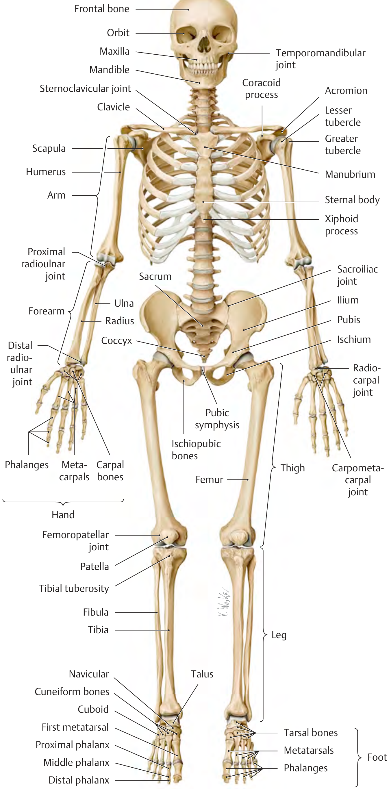
Bone markings fall into three broad functional categories: projections (elevated areas), depressions (sunken areas), and openings (passages for nerves and vessels). Each has a specific anatomical name depending on its shape, size, and function.
1. Projections (Prominences)
These are raised areas that project above the surface. They serve as attachment sites for muscles, tendons, and ligaments, or as articular surfaces.
Processes (Large Named Projections)
| Term | Description | Example |
|---|---|---|
| Process | Any bony prominence or projection | Coracoid process of scapula, transverse process of vertebra |
| Spine (Spinous process) | Sharp, slender projection | Spine of scapula, spinous process of vertebrae |
| Ramus | Arm-like extension that forms an angle with the main body of bone | Ramus of mandible, inferior ramus of pubis |
Knob-like and Rounded Projections
| Term | Description | Example |
|---|---|---|
| Condyle | Large, rounded articular knuckle at the end of a bone | Medial/lateral condyle of femur, occipital condyles |
| Epicondyle | Projection above (epi-) a condyle; non-articular; for tendon/ligament attachment | Medial/lateral epicondyle of humerus |
| Head | Rounded, ball-like articular end, often connected to the shaft by a neck | Head of femur, head of humerus, head of radius |
| Neck | Constricted region connecting head to shaft | Anatomical neck of humerus, neck of femur |
Bumps and Ridges for Muscle/Tendon Attachment
| Term | Description | Example |
|---|---|---|
| Tubercle | Small, rounded projection | Greater and lesser tubercle of humerus, adductor tubercle of femur |
| Tuberosity | Large, rough, rounded projection | Tibial tuberosity (attachment of patellar tendon), ischial tuberosity, deltoid tuberosity of humerus |
| Trochanter | Very large, blunt, irregular projection (unique to femur) | Greater trochanter, lesser trochanter |
| Malleolus | Rounded, hammer-shaped projection | Medial malleolus (tibia), lateral malleolus (fibula) |
| Crest | Prominent ridge or border of bone | Iliac crest, sacral crest, intertrochanteric crest |
| Line (Linea) | Low, narrow ridge - less prominent than a crest | Linea aspera of femur, soleal line of tibia |
| Ridge | Narrow elevated border | Interosseous ridge of radius/ulna |
| Trochanterion / Trochlea | Pulley-shaped articular surface | Trochlea of humerus (for articulation with ulna), trochlea of talus |
| Facet | Small, flat articular surface | Costal facets on vertebrae, facets of carpals |
Blade-like and Pointed Structures
| Term | Description | Example |
|---|---|---|
| Acromion | Flat, lateral extension | Acromion of scapula |
| Styloid process | Long, pointed, spike-like projection | Styloid process of radius, ulna, temporal bone |
| Mastoid process | Large, rounded, nipple-like process | Mastoid process of temporal bone |
| Olecranon | Large, blunt proximal projection of ulna | Tip of elbow |
2. Depressions (Concavities)
These are hollowed or sunken areas, often receiving the projections of adjacent bones or housing tendons and nerves.
| Term | Description | Example |
|---|---|---|
| Fossa | Shallow, bowl-shaped depression or pit | Infraspinous fossa of scapula, olecranon fossa and coronoid fossa of humerus, iliac fossa |
| Sulcus (Groove) | Furrow or channel on the bone surface | Bicipital groove (intertubercular sulcus) of humerus, sulcus for radial nerve |
| Notch (Incisure) | A deep indentation or cleft at the edge of a bone | Greater/lesser sciatic notch, trochlear notch of ulna, jugular notch |
| Facet | Small, flat, shallow articular depression | Articular facets of vertebrae |
| Pit (Fovea) | Small, pit-like depression | Fovea capitis of the femoral head (attachment of ligament of head) |
| Sinus | Air-filled cavity within a bone, lined by mucosa | Frontal sinus, maxillary sinus, ethmoid sinus |
| Meatus | Canal or tunnel running through a bone | External acoustic meatus, internal acoustic meatus |
| Fissure | Narrow, slit-like opening | Superior orbital fissure, inferior orbital fissure |
3. Openings (Foramina and Passageways)
These allow nerves, blood vessels, and other structures to pass through or into bones.
| Term | Description | Example |
|---|---|---|
| Foramen (pl. foramina) | Rounded hole or aperture through a bone | Foramen magnum, obturator foramen, mental foramen, nutrient foramen |
| Canal | Tunnel running the length of bone (or through it) | Haversian canal (within osteons), condylar canal, carotid canal |
| Fissure | Narrow, elongated cleft or slit | Superior/inferior orbital fissure |
| Meatus | Tube-like passage | External acoustic meatus |
Quick Reference Summary Table
| Category | Terms |
|---|---|
| Large projections | Process, spine, ramus, trochanter, malleolus |
| Articular projections | Head, condyle, epicondyle, facet, trochlea |
| Rough attachment sites | Tuberosity, tubercle, crest, line, ridge |
| Depressions | Fossa, sulcus/groove, notch, fovea, pit |
| Air spaces | Sinus, cell |
| Openings/passageways | Foramen, meatus, canal, fissure |
Why Bone Markings Matter
- Muscle/tendon attachment: Tuberosities, tubercles, crests, and lines are roughened to increase the surface area gripping tendons and ligaments (e.g., the tibial tuberosity anchors the patellar tendon - attachment of the quadriceps femoris).
- Articulation: Condyles, heads, facets, and trochleae form the smooth surfaces of joints, covered by hyaline cartilage in life.
- Neurovascular passageways: Foramina and canals guide nerves and blood vessels to their targets without injury from movement (e.g., the nutrient foramen admits the nutrient artery that supplies compact bone).
- Palpable landmarks: Many markings (iliac crest, medial epicondyle, mastoid process, tibial tuberosity, acromion) are used clinically to locate underlying structures, plan injections, and assess injuries.
- Forensic/anthropological identification: Patterns of markings reflect musculature, activity level, sex, and age in skeletal remains.
Sources: General Anatomy and Musculoskeletal System, THIEME Atlas of Anatomy; Gray's Anatomy for Students
Bones and cartilages: Skeleton, Bones, function of bones, bone tissue and it's types, classification of bones, markings on a bone, gross anatomy of an adult typical long bone, ossification, diaphysis and epiphysis, Bone growth and Remodeling, blood supply of long bones, Bone Injury and repair
Reading File
Finding Sources
Finding Sources
Finding Sources
Finding Sources
Finding Sources
Reading File
Reading File
Reading File
Reading File
Reading File
Reading File
Reading File
Reading File
Finding Sources
Reading File
Reading File
Reading File
Reading File
Reading File
Finding Sources
Reading File
Reading File
Reading File
Loading Image
Loading Image
Loading Image
Loading Image
Loading Image
Reading File
Reading File
Your credits are exhausted. Your daily credits reset tomorrow, or you can purchase a top-up for more.
This is a shared conversation. Sign in to Orris to start your own chat.