
Fragile x syndrome
I'll look up Fragile X syndrome from the medical library for you.
Searching sources...
Reading File
Now let me search for Fragile X syndrome in the library and PubMed in parallel.
Finding Sources
Searching PubMed
"fragile X syndrome"[MeSH Terms]
Excellent results. Now let me read the most comprehensive sources in parallel - Robbins Pathology and Thompson Genetics, which will give the best clinical overview.
Reading File
Reading File
Great content. Now let me read the Robbins Pathology section more fully for clinical features and the Tietz lab section for diagnostic details.
Reading File
Reading File
Excellent content gathered. Let me now fetch the key images and read the Robbins section on FMRP mechanism and associated conditions.
Reading File
Loading Image
I now have comprehensive content from multiple authoritative textbooks. Let me compile a thorough answer.
Fragile X Syndrome (FXS)
Overview
Fragile X syndrome is the most common heritable (inherited) cause of intellectual disability and the most common genetic cause of intellectual disability in males. It is the second most common overall cause of intellectual disability after Down syndrome. The frequency is approximately 1 in 1,550–4,000 males and 1 in 8,000 females. It also accounts for 2–3% of all autism spectrum disorder (ASD) diagnoses.
- Robbins, Cotran & Kumar Pathologic Basis of Disease, p. 170
- Tietz Textbook of Laboratory Medicine, 7th Edition
Genetics and Molecular Basis
The FMR1 Gene and CGG Repeat Expansion
FXS is caused by an unstable trinucleotide (CGG) repeat expansion in the 5' untranslated region (UTR) of the FMR1 gene on the X chromosome at locus Xq27.3.
| Category | CGG Repeat Count | Significance |
|---|---|---|
| Normal | 6–55 (avg 29) | No disease |
| Premutation | 55–200 | Carrier; risk of expansion |
| Full mutation | >200 (up to 4,000) | FXS phenotype |
When repeat count exceeds ~230, the DNA of the entire 5' region becomes abnormally hypermethylated, extending into the promoter CpG island. This causes transcriptional silencing of FMR1 and loss of its protein product, FMRP.
FMRP - The Missing Protein
FMRP (Fragile X Mental Retardation Protein) is an ~80-kDa RNA-binding protein, most abundant in the brain and testis. It plays two key roles in neurons:
- mRNA transport - Selectively binds mRNAs (encoding pre- and post-synaptic proteins) and shuttles them from the nucleus to dendritic spines.
- Translation regulation - At synapses, FMRP suppresses protein synthesis via group I metabotropic glutamate receptors (mGluR). Loss of FMRP leads to unregulated mRNA translation at synapses, disrupting synaptic plasticity - the molecular basis of learning and memory.
Inheritance Pattern
FXS follows X-linked inheritance with unusual features (the "Sherman paradox"):
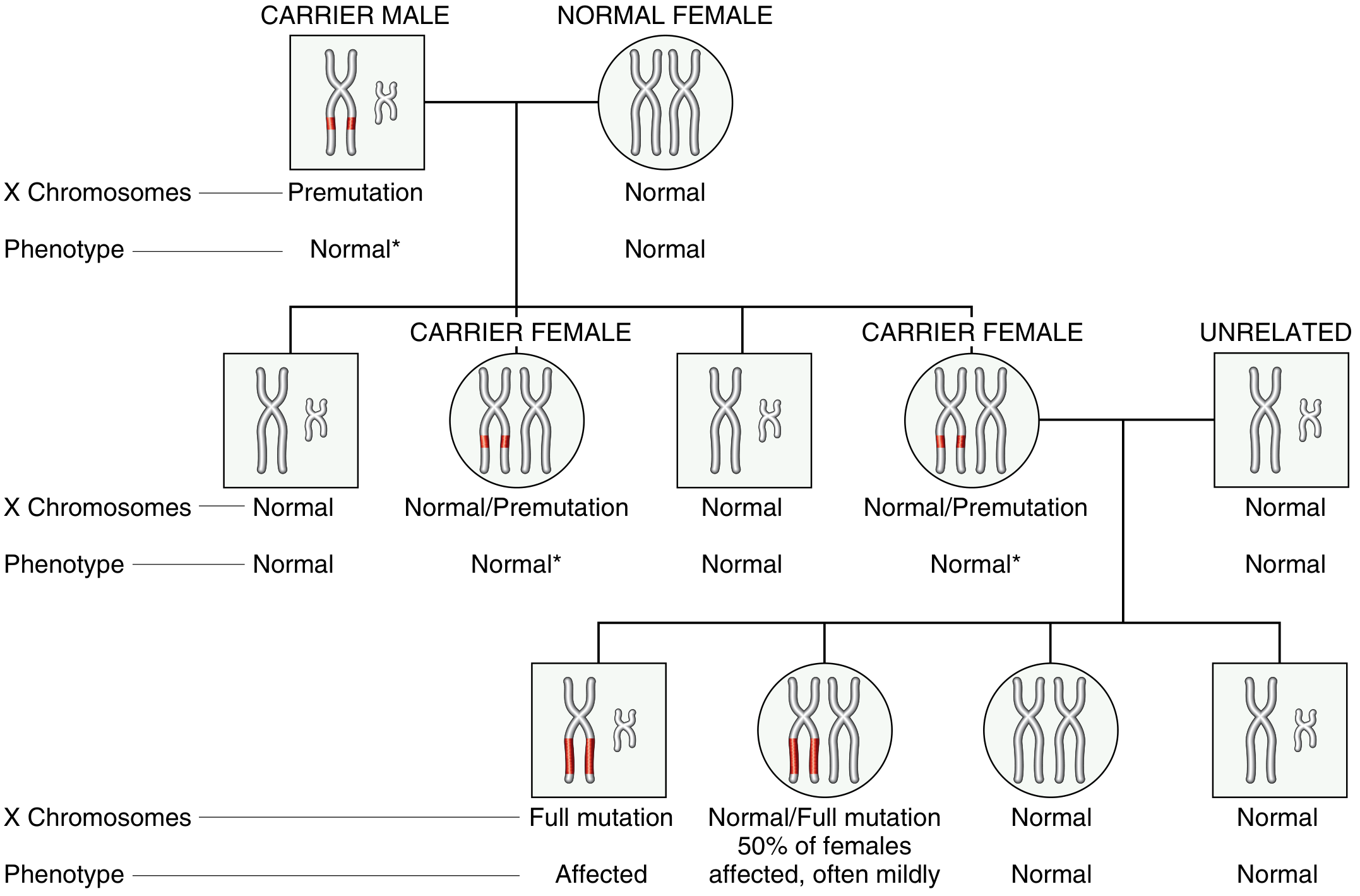
Fragile X pedigree. A carrier male (premutation) passes the allele through phenotypically normal daughters, who can then expand it to a full mutation in their offspring. - Robbins Pathologic Basis of Disease, Fig. 5.28
Key features that distinguish it from classic X-linked recessive inheritance:
- Normal transmitting males: ~20% of males carrying a premutation are phenotypically normal. They transmit through carrier daughters to affected grandchildren.
- Affected females: 30–50% of carrier (heterozygous) females have intellectual disability - far higher than most X-linked recessive conditions. This is partly because random X-inactivation is not fully protective.
- Anticipation: Symptoms worsen in successive generations as the premutation expands further.
- Risk depends on position in pedigree: Brothers of transmitting males have a 9% risk of intellectual disability; grandsons of transmitting males face a 40% risk.
- The premutation almost always expands to full mutation during maternal (female) oogenesis, not during spermatogenesis. Larger premutations carry higher expansion risk.
Clinical Features
Males (Full Mutation)
Physical features:
- Long face, prominent forehead and jaw
- Large ears
- Macroorchidism (large testes) - becomes more prominent after puberty
- Flat feet, joint hypermobility
Neurodevelopmental and behavioral features:
- Moderate intellectual disability (IQ typically 40–70)
- Delayed motor and speech development
- Autism spectrum disorder in 50–75% of males
- Anxiety and hyperactivity (50–75%)
- Epilepsy in ~30%
- Aggressive behavior in ~90%
- Hand flapping, poor eye contact, perseverative speech, temper tantrums
Females (Full Mutation)
- Features are typically milder due to random X-inactivation (the normal X is expressed in ~half of cells)
- ~30–50% have some degree of intellectual disability or learning difficulties
- Behavioral and social difficulties may still be present
Associated Premutation Disorders
Carriers of the premutation (55–200 CGG repeats) are not affected by FXS, but are at risk for two other distinct conditions caused by a toxic gain-of-function mechanism (elevated FMR1 mRNA is toxic rather than protein loss):
| Condition | Who | Features |
|---|---|---|
| Fragile X-associated Tremor/Ataxia Syndrome (FXTAS) | ~50% of premutation males >50 years | Progressive intention tremor, cerebellar ataxia, dementia, starts 6th decade |
| Fragile X-associated Primary Ovarian Insufficiency (FXPOI) | ~20% of female premutation carriers | Premature ovarian failure before age 40, elevated FSH, low anti-Müllerian hormone, early menopause |
Diagnosis
- Historically: Cytogenetic "fragile site" at Xq27.3 visible on karyotype in folate-deficient media (now obsolete).
- Current gold standard: PCR-based analysis of CGG repeat size in FMR1. This quantifies repeat number precisely and identifies methylation status.
- Methylation analysis: Full mutations show hypermethylation at the FMR1 promoter CpG island. Methylation mosaics (partial methylation) have some FMRP and milder phenotype.
- FXS caused by point mutations or large deletions in FMR1 (rare, ~few percent of cases) requires sequencing.
Management
There is no cure. Management is symptomatic and supportive:
- Intellectual disability: Special education, speech and occupational therapy
- ASD/behavioral: Applied behavioral analysis, social skills training
- Anxiety/ADHD: Stimulants (methylphenidate), SSRIs, guanfacine
- Epilepsy: Standard anticonvulsants
- Genetic counseling: Essential for carrier females regarding expansion risk and associated conditions (FXPOI)
- Research is ongoing into mGluR5 antagonists to restore synaptic plasticity, though clinical trials have not yet yielded approved therapies
Recent Evidence
Recent systematic reviews (2025) confirm the lifespan complexity of FXS:
- A 2025 systematic review by Genovese & Butler highlights the broad genetic, neurodevelopmental, behavioral, and psychiatric associations across the lifespan (PMID: 40004478).
- A 2025 review by Souski et al. details the clinical and molecular divergences between full mutation and premutation carriers (PMID: 40985503).
Summary mnemonic - "FRAGILE X":
- F - FMR1 gene (Xq27.3)
- R - Repeat CGG expansion (>200 = full mutation)
- A - Autism spectrum disorder (50–75%)
- G - Gene silencing via methylation
- I - Intellectual disability (most common heritable cause)
- L - Large ears, Long face, Large testes (macroorchidism)
- E - Epilepsy (~30%), Epilepsy, EpilEpsy
- X - X-linked with anticipation; premutation expands in female oogenesis
Aicardi syndrome
Aicardi syndrome is a rare X-linked dominant neurodevelopmental disorder characterized by the classic triad of agenesis of the corpus callosum, infantile spasms, and chorioretinal lacunae. It occurs almost exclusively in females (or 47,XXY males). Let me look this up properly.
Searching sources...
Reading File
Finding Sources
Searching PubMed
"Aicardi syndrome"[MeSH Terms]
The textbook headings results are sparse. Let me do a paragraph-level search and also search for broader paragraph content across multiple books, plus web search in parallel.
Finding Sources
Searching the Web
Aicardi syndrome clinical features diagnosis management neurology
Good. Now let me read the textbook content from Bradley & Daroff's Neurology and Kanski's Ophthalmology, and extract the GeneReviews content in parallel.
Reading File
Reading File
Reading Web Page
https://www.ncbi.nlm.nih.gov/books/NBK1381
I have good content from textbooks and GeneReviews. Let me fetch the ophthalmology image and get more detailed management/prognosis content from GeneReviews.
Loading Image
Reading Web Page
https://www.ncbi.nlm.nih.gov/books/NBK1381
I now have comprehensive information from textbooks, GeneReviews, and clinical sources. Here is the full structured answer:
Aicardi Syndrome
Overview
Aicardi syndrome (AIC) is a rare X-linked dominant neurodevelopmental disorder that occurs almost exclusively in females (or rarely in 47,XXY males). It was first described by Jean Aicardi in 1965. The estimated prevalence is 1 in 100,000–167,000 live female births. Nearly all cases are sporadic, arising from de novo mutations - familial transmission is exceptionally rare.
Note: Aicardi syndrome is completely distinct from Aicardi-Goutieres syndrome, an unrelated early-onset encephalopathy caused by interferonopathy.
Genetics
- Inheritance: X-linked dominant, presumed lethal in hemizygous males (XY karyotype) in utero - explaining the near-exclusive occurrence in females
- Rare male cases have a 47,XXY (Klinefelter) karyotype
- No causative gene has been identified to date - this remains an unsolved mystery in medical genetics
- The current hypothesis is a de novo pathogenic variant in a gene on the X chromosome subject to X-inactivation
- Genetic testing (chromosomal microarray, whole exome sequencing) is part of evaluation to exclude differential diagnoses, but will not confirm Aicardi syndrome
- Recurrence risk for parents of an affected child is very low (essentially that of a new de novo mutation)
Classic Diagnostic Triad
Aicardi syndrome was originally defined by three cardinal features, present in over 90% of classical cases:
| Feature | Details |
|---|---|
| 1. Agenesis of the corpus callosum | Complete or partial; often accompanied by other brain malformations |
| 2. Chorioretinal lacunae | Bilateral, depigmented "punched-out" lesions clustered around the optic disc; pathognomonic |
| 3. Infantile spasms | Onset typically 3-5 months of age; evolve into intractable epilepsy |
Diagnostic Criteria (Modified, Sutton et al. 2005)
The diagnosis requires either:
- All 3 features of the classic triad, or
- Any 2 features of the classic triad plus at least 2 of the following major/supporting features
Major Features
- Cortical malformations (polymicrogyria, pachygyria, lissencephaly)
- Periventricular and subcortical heterotopia
- Cysts around the third ventricle and/or choroid plexus
- Choroid plexus papilloma
- Optic disc/nerve coloboma
Supporting Features
- Vertebral and costal (rib) abnormalities
- Microphthalmia or other eye malformations
- "Split-brain" EEG pattern (asymmetric, asynchronous burst-suppression - see below)
- Gross hemispheric asymmetry
The diagnosis is clinical - there is no confirmatory genetic or molecular test.
Clinical Features in Detail
Neurological
- Seizures: Infantile spasms begin at 3-5 months; nearly all patients develop medically refractory epilepsy with multiple seizure types (spasms, tonic, clonic, myoclonic, absence, focal). Very few achieve sustained seizure freedom.
- Intellectual disability: Severe in the majority; a small minority have mild or moderate disability
- Developmental delay: Profound delays in motor, speech, and cognitive milestones
- Brain malformations: In addition to corpus callosum agenesis, polymicrogyria, heterotopia, cortical dysplasia, and interhemispheric cysts are common
- Hypotonia, progressing to spasticity in many cases
Ophthalmological
The hallmark ocular finding is chorioretinal lacunae - bilateral, creamy-white to yellow, punched-out depigmented lesions in the retina, typically clustered around the optic disc:
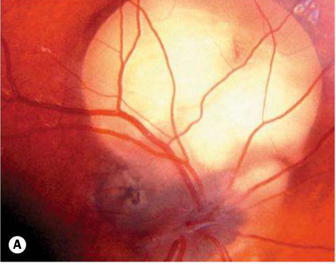
Fundus photograph showing chorioretinal lacunae in Aicardi syndrome. Note the large depigmented lesion adjacent to the optic disc. - Kanski's Clinical Ophthalmology, Fig. 19.31
Additional ocular features include:
- Optic disc hypoplasia or coloboma
- Cataract
- Microphthalmia
- Nystagmus
- Visual impairment of varying severity
Musculoskeletal
- Scoliosis (common, can be severe and progressive)
- Vertebral anomalies (hemivertebrae, butterfly vertebrae, fused vertebrae)
- Rib abnormalities (missing or fused ribs)
- Small or malformed hands
Other Systemic Features
- Microcephaly
- Prominent premaxilla (distinctive facial feature)
- Cleft lip or palate (occasional)
- Gastroesophageal reflux and feeding problems
- Precocious or delayed puberty
- Choroid plexus papillomas (associated with hydrocephalus)
- Increased risk of certain tumors (hepatoblastoma, angiosarcoma reported)
Investigations
EEG
The characteristic EEG pattern is asymmetric, asynchronous burst-suppression ("split-brain" pattern) - each hemisphere fires independently in a disorganized pattern. This is highly suggestive of Aicardi syndrome. Hypsarrhythmia may also be seen.
- Bradley and Daroff's Neurology in Clinical Practice
Brain MRI
Findings include:
- Agenesis or dysgenesis of corpus callosum (and often anterior commissure)
- Periventricular and cortical heterotopias
- Polymicrogyria
- Interhemispheric cysts
- Colpocephaly (enlarged occipital horns)
- Choroid plexus papillomas
- Marked hemispheric asymmetry
Ophthalmology
- Dilated fundus examination under anesthesia to identify and document chorioretinal lacunae
- The lacunae may increase in number and size over time
Other
- Spinal radiographs (vertebral/rib anomalies, scoliosis)
- Chromosomal microarray and/or whole exome sequencing (to exclude mimics)
Management
There are no clinical practice guidelines and no curative therapy. Management is symptomatic and requires a multidisciplinary team:
| Domain | Intervention |
|---|---|
| Epilepsy | ACTH + vigabatrin (first-line for infantile spasms); multiple AEDs for refractory epilepsy; Vagus Nerve Stimulator (VNS) for refractory cases; ketogenic diet |
| Development | Physical therapy, occupational therapy, speech/language therapy - started at diagnosis |
| Scoliosis | Orthopedic surveillance; bracing or surgical correction for severe cases |
| Ophthalmology | Regular follow-up; treatment for amblyopia if applicable |
| Nutrition | Gastrostomy tube placement for severe feeding difficulties and aspiration risk |
| Neurosurgery | Shunting for hydrocephalus from choroid plexus papilloma; resective surgery rarely feasible due to multifocal epileptogenesis |
Prognosis
- Prognosis is poor overall, with severe intellectual disability and refractory epilepsy in most patients
- Seizures are almost never fully controlled
- Mean age at death has been reported as 8.3 years, but median survival is ~18.5 years with the oldest reported survivor at 32 years (Glasmacher et al., GeneReviews)
- Probability of survival to age 27 is approximately 62% in some series
- A subset of more mildly affected girls survive longer with better functional outcomes
- Main causes of death include respiratory complications (aspiration pneumonia), status epilepticus, and complications of associated tumors
Key Points Summary
| Feature | Detail |
|---|---|
| Inheritance | X-linked dominant; de novo; lethal in XY males |
| Gene | Unknown - no gene identified |
| Classic triad | Corpus callosum agenesis + chorioretinal lacunae + infantile spasms |
| Pathognomonic finding | Bilateral chorioretinal lacunae around optic disc |
| EEG | Asymmetric asynchronous burst-suppression ("split-brain") |
| Onset | Infantile spasms at 3-5 months |
| Treatment | Symptomatic; no cure |
| Prognosis | Severe disability; median survival ~18.5 years |
- Bradley and Daroff's Neurology in Clinical Practice
- Kanski's Clinical Ophthalmology, 10th edition, p. 803
- GeneReviews: Aicardi Syndrome (NCBI)
This is a shared conversation. Sign in to Orris to start your own chat.