
Ligamentum arteriosum
ligamentum arteriosum ductus arteriosus anatomy diagram
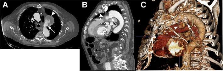
This composite of contrast-enhanced computed tomography angiography (CTA) images of the thoracic aorta illustrates a ductus arteriosus diverticulum (DAD) and an associated pseudoaneurysm. Panel A, an axial image, shows the DAD (arrow) as a contrast-filled outpouching from the aorta, connecting to a larger, more peripheral pseudoaneurysm (arrowhead) that displays heterogeneous density, indicating a mix of opacified blood and potential thrombus. Panel B is a two-dimensional sagittal reformatted image showing the DAD (arrow) and pseudoaneurysm (arrowhead) arising from the inferior aspect of the aortic arch, near the site of the ligamentum arteriosum. Panel C presents a three-dimensional volume-rendered reconstruction, providing a spatial overview of the aortic arch anatomy for surgical planning. The reconstruction clearly delineates the wide neck of the DAD (arrow) and the orientation of the pseudoaneurysm sac (arrowhead) toward the pulmonary artery. These images demonstrate key diagnostic features used to differentiate a benign ductus diverticulum from a complex vascular pathology requiring intervention.
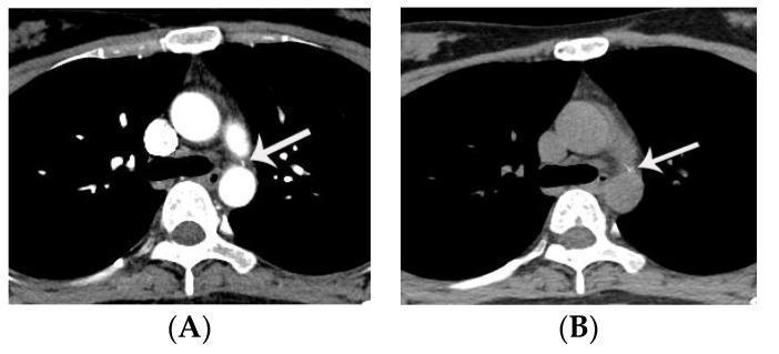
This diagnostic image consists of two axial chest CT scans—contrast-enhanced (A) and non-contrast (B)—demonstrating a calcified ligamentum arteriosum in a 32-year-old female. Both images show a small, well-defined, hyperdense focal point (indicated by white arrows) located at the level of the tracheal bifurcation. This calcification is positioned within the aortopulmonary window, specifically near the 12 o'clock position relative to the descending thoracic aorta. Anatomical landmarks include the descending thoracic aorta posteriorly, the bifurcation of the trachea (carina) centrally, and the pulmonary artery adjacent to the aorta. The primary educational focus is to distinguish this benign, physiological calcification of the ligamentum arteriosum from a small patent ductus arteriosus (PDA). On enhanced CT (A), the high density of the calcification can mimic vascular enhancement; however, the non-contrast image (B) confirms the pre-existing high density characteristic of calcification rather than contrast-filled lumen. This distinction is critical for radiologists to avoid misdiagnosing a congenital vascular shunt.
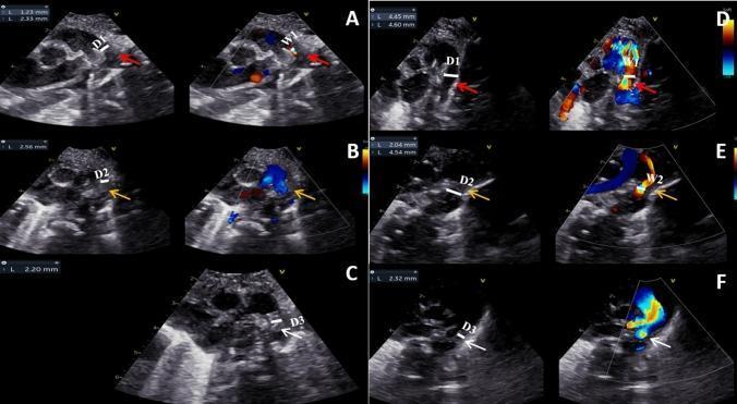
This set of diagnostic images features longitudinal two-dimensional (2D) and color Doppler echocardiography of the ductus arteriosus (DA) in neonates. Panels A-C illustrate the 'PDA-closure group,' demonstrating the progressive stages of spontaneous closure. (A) shows a thin, hypoechoic, uneven intima with a patent lumen (D1) and wide transcatheter flow (W1) within 24 hours of birth. (B) displays intimal thickening and lumen occlusion by 48-72 hours. (C) shows the final formation of the hyperechoic ligamentum arteriosum with total cessation of flow at 26 days. Panels D-F depict the 'PDA-open group,' where the intima remains thinner and the lumen (D1-D3) stays more patent over time despite some contraction. Color Doppler confirms persistent shunting (blue and red flow signals) across the DA in the open group, while showing diminishing flow in the closure group. The series highlights the pathophysiology of neonatal DA remodeling, specifically the role of intimal cushions and contraction in transition from a patent ductus arteriosus to an anatomical ligament.
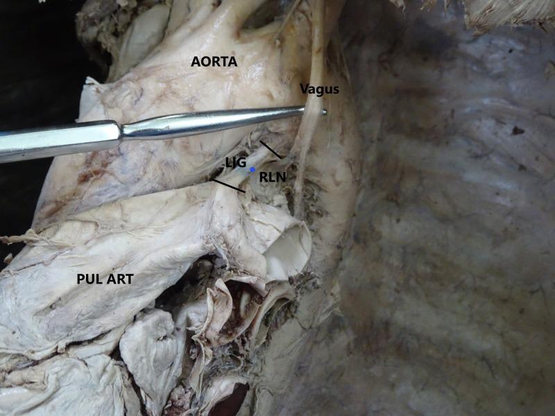
A clinical photograph of a cadaveric dissection focused on the superior mediastinum, specifically demonstrating the relationship between major cardiovascular and neurovascular structures. The aortic arch is positioned superiorly, with the pulmonary artery (PUL ART) located inferior to it. Connecting these two vessels is the ligamentum arteriosum (LIG), a fibrous remnant of the ductus arteriosus, shown as a short, taut band. The left vagus nerve descends vertically, crossing the lateral aspect of the aortic arch. The left recurrent laryngeal nerve (RLN) is clearly visible branching from the vagus nerve and looping medially and superiorly beneath the aortic arch, just posterior to the ligamentum arteriosum. A metal surgical probe is used to retract the vagus nerve for better visualization of the RLN's origin. This anatomical illustration highlights the clinical importance of the RLN's course, particularly in thoracic surgery or cases of aortic pathology where the nerve may be compressed or injured, leading to vocal cord paralysis.
Ligamentum Arteriosum
What It Is
- Gray's Anatomy for Students, p. 258
- Fischer's Mastery of Surgery, 8th ed., p. 2512
Embryological Origin
-
Functional closure occurs within the first few days as rising arterial oxygen tension causes smooth muscle contraction of the ductus wall.
-
Anatomical closure (fibrous obliteration into the ligamentum arteriosum) is normally complete by the 12th postnatal week.
-
The Developing Human: Clinically Oriented Embryology, p. 879-880
Adult Anatomy
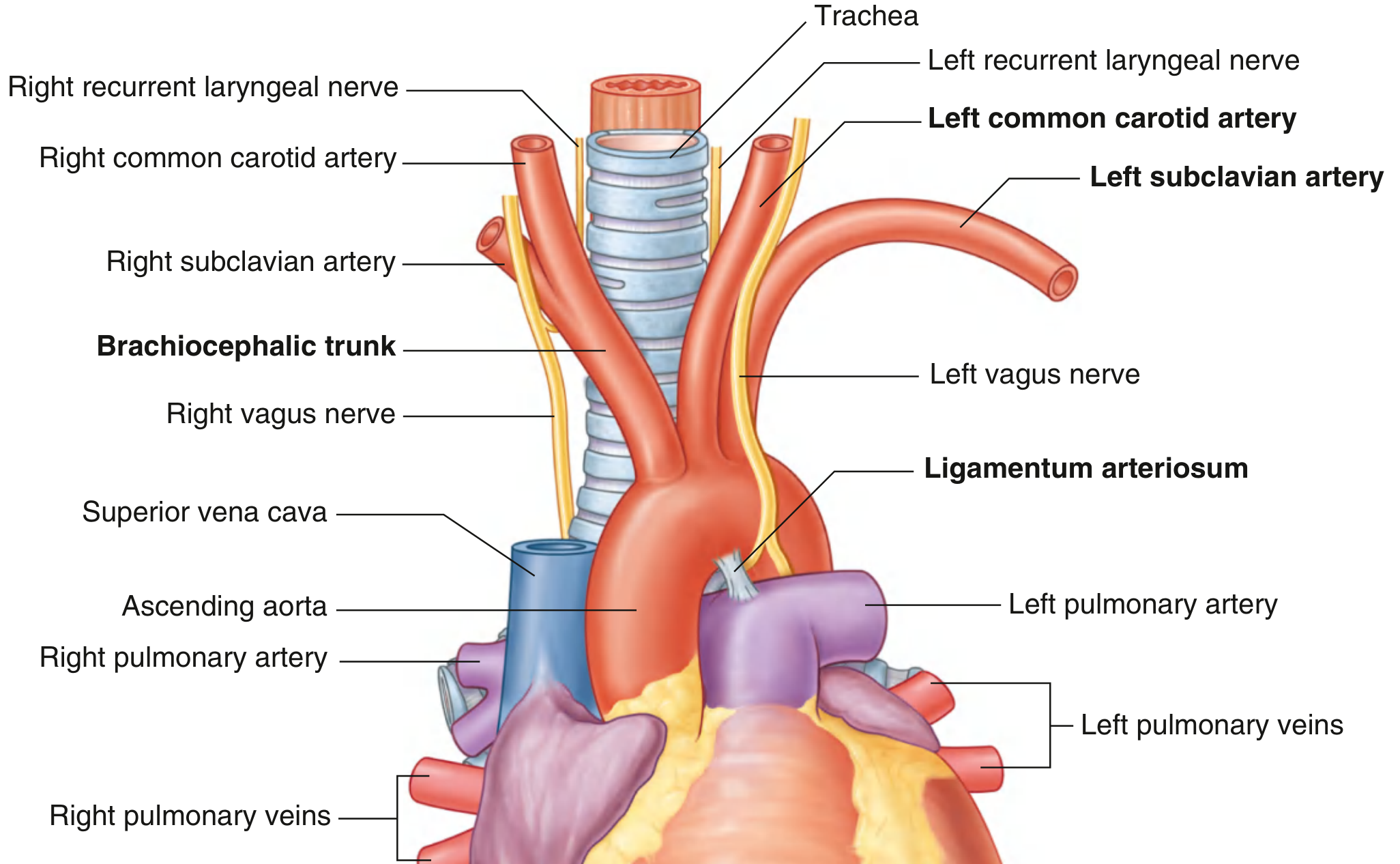
| Structure | Relationship |
|---|---|
| Aortic arch | Superior attachment of the ligament |
| Left pulmonary artery | Inferior attachment |
| Left recurrent laryngeal nerve | Loops around the aortic arch just to the right of the ligamentum arteriosum |
| Left vagus nerve | Descends anterior to the aortic arch, gives off the recurrent laryngeal at the level of the ligament |
Clinical Significance
1. Landmark for the Left Recurrent Laryngeal Nerve
- Fischer's Mastery of Surgery, p. 2512-2513
2. Aortic Trauma - Fixed Point
- Gray's Anatomy for Students, p. 258
3. Division of the Ligament in Surgery
- Fischer's Mastery of Surgery, p. 2059
4. Aortic Aneurysms - Anatomical Dividing Line
- Fuster and Hurst's The Heart, 15th ed., p. 3116
5. Patent Ductus Arteriosus (PDA) - Failure to Close
- 2-3x more common in females
- Associated with maternal rubella infection in early pregnancy
- Common in prematurity (<28 weeks), high-altitude birth, and hypoxia
- Treated medically with indomethacin (COX inhibitor, reduces prostaglandin E2 which normally keeps the ductus open) or surgical/catheter-based ligation
- The Developing Human: Clinically Oriented Embryology, p. 880
6. Vascular Rings
- Grainger & Allison's Diagnostic Radiology
7. Calcified Ligamentum Arteriosum on CT
Summary Table
| Feature | Detail |
|---|---|
| What it is | Fibrous remnant of the ductus arteriosus |
| Location | Superior mediastinum |
| Connects | Left pulmonary artery → undersurface of aortic arch |
| Functional closure | First few days of life |
| Anatomical closure | By 12th postnatal week |
| Key nerve nearby | Left recurrent laryngeal nerve loops just to its right |
| Trauma significance | Fixed point of aorta; rupture site in deceleration injury |
| Failure to close | Patent ductus arteriosus (PDA) |
Vocal cords
vocal cords larynx anatomy diagram true false vocal folds
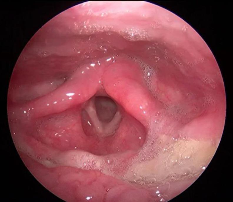
This clinical image is a frame from a video laryngoscopy showing a post-treatment view of a normal larynx in a pediatric patient. The true vocal cords (vocal folds) are clearly visualized, appearing as symmetrical, smooth, pearly-white bands that form a V-shape. The surrounding supraglottic structures, including the epiglottis, arytenoids, and false vocal cords, appear healthy with smooth, moist, pink mucosa and no evidence of edema or inflammation. The glottic opening is patent, and the vallecula and pyriform sinuses are clear of masses or lesions. This image demonstrates the resolution of a previously documented laryngeal B-cell lymphoma following chemotherapy and surgical intervention, serving as a baseline for normal laryngeal anatomy during follow-up endoscopic assessment.
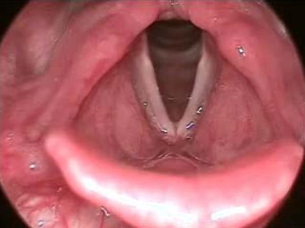
An endoscopic view of a normal human larynx, representing a baseline for clinical comparison with various voicing disorders. The diagnostic image displays the true vocal folds (vocal cords) as smooth, whitish-pink, and symmetrical structures. They are positioned in a V-shaped configuration, creating a clear triangular glottic opening that indicates the larynx is in a state of abduction (opening for respiration). The surrounding laryngeal mucosa and vestibular folds (false vocal folds) appear healthy, pink, and free of visible lesions, nodules, cysts, or inflammation. Key landmarks visible include the posterior commissure at the base of the 'V' and the surrounding subglottic space appearing dark in the background. This clinical photograph serves as an educational reference for normal laryngeal anatomy in the context of otolaryngology and speech-language pathology.
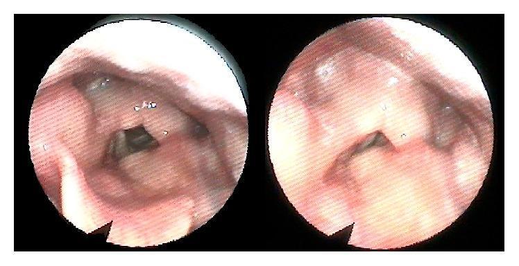
Two circular panels show a diagnostic endoscopy of the human larynx, specifically a postoperative view following surgical repair of laryngeal trauma. The clinical photographs depict the internal laryngeal anatomy, including the epiglottis, false vocal folds, and true vocal cords. The mucosal surfaces appear pinkish-red and healthy, with no visible signs of acute inflammation, significant granulation tissue, or ulceration. The true vocal folds are centrally positioned and form a dark triangular glottic opening, suggesting patency of the airway. The left panel shows the larynx in a more open (abducted) state, while the right panel shows a slightly different phase of mobilization or viewing angle. The anatomical structures, including the arytenoids and surrounding vestibular folds, appear symmetrical and well-defined. This endoscopic evaluation is used to assess postoperative healing, laryngeal lumen patency, and vocal cord mobility after complex laryngeal reconstruction, such as thyroid and cricoid cartilage fracture repair.
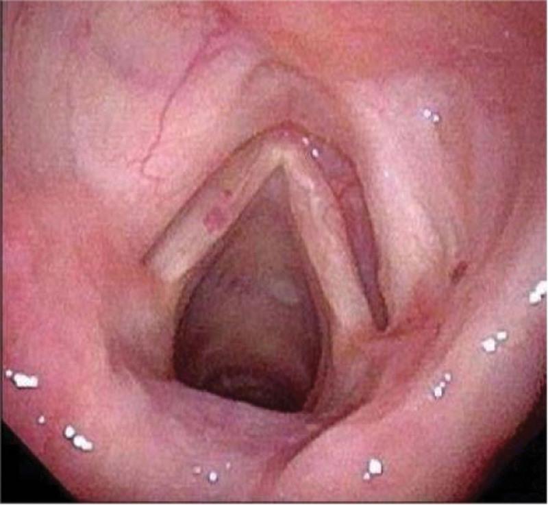
This endoscopic clinical photograph depicts the internal anatomy of the human larynx as captured via electronic video laryngoscopy. The visual focuses on the glottis during inspiration, showing the true vocal cords in an abducted position, forming a V-shaped aperture that reveals the subglottic space. The vocal cords exhibit a normal pearly-white to pale-pink mucosal color with a smooth surface, free of visible nodules, polyps, or leukoplakia. Surrounding supraglottic structures, including the aryepiglottic folds and the ventricular zone (false vocal cords), appear symmetrical with healthy pink mucosa. There is no evidence of edema, erythema, or residual tumor mass in this 9-month postoperative view. The image serves as a representative example of a healthy laryngeal recovery following the surgical resection of an extramedullary plasmacytoma (EMP), demonstrating restored anatomical integrity and no signs of clinical recurrence.
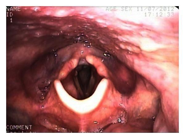
This clinical photograph displays an endoscopic view of the human larynx, specifically a fiberoptic nasoendoscopy. The image highlights the glottis and surrounding supraglottic structures. Centrally, the true vocal folds (vocal cords) appear as bright, pearly-white, crescent-shaped bands. The edges of the folds are smooth and symmetrical, framing the dark, patent rima glottidis (airway opening). Above the vocal folds, the vestibular folds (false vocal cords) and the laryngeal vestibule are visible, exhibiting a healthy, reddish-pink mucosal lining with normal vascularity. The epiglottis is partially visible at the top of the frame. Small droplets of clear mucus are scattered across the mucosal surface, which is a normal finding. This image serves as an educational example of normal post-traumatic recovery, demonstrating preserved laryngeal anatomy and unobstructed airway patency following a penetrating neck injury.
Vocal Cords (Vocal Folds)
Overview
Anatomy of the Larynx and Vocal Fold Position
- The space between the true vocal folds is the rima glottidis (glottic opening)
- The area above is the supraglottis; below is the subglottis
- Scott-Brown's Otorhinolaryngology, Vol. 1, p. 2466-2470
Endoscopic view - normal larynx:
Microarchitecture - The Layered Structure
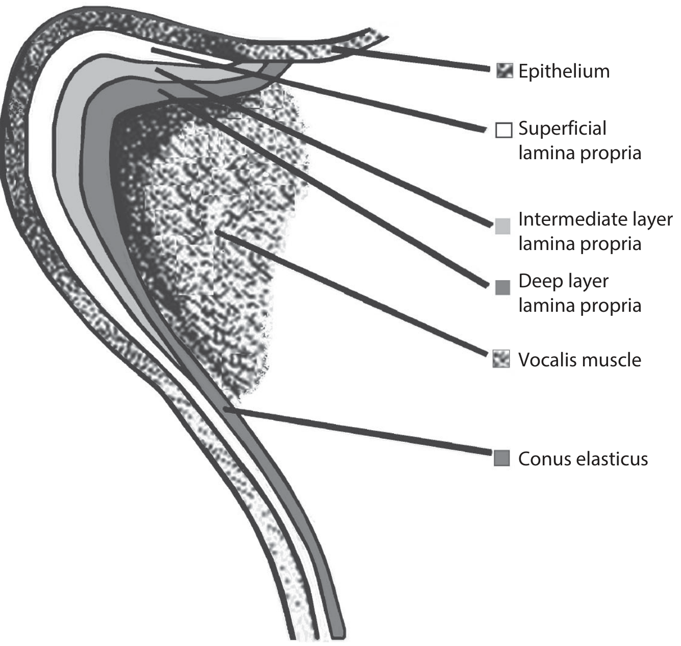
| Layer | Composition | Notes |
|---|---|---|
| Epithelium | Stratified squamous (~50 µm thick) | Transitions to pseudocolumnar toward the ventricle and subglottis |
| Superficial lamina propria | Loose areolar tissue (Reinke's space) | Gelatin-like; allows mucosal wave during phonation |
| Intermediate lamina propria | Elastic fibres | Together with deep layer forms the vocal ligament |
| Deep lamina propria | Collagen fibres (denser) | |
| Vocalis muscle (TAv) | Medial portion of thyroarytenoid | Forms the bulk of the vocal fold body; controls length, tension, stiffness |
| Conus elasticus | Elastic membrane | Underlies and supports the fold |
- Scott-Brown's, p. 2441-2448; KJ Lee's Essential Otolaryngology, p. 1180
Macula Flava
- Scott-Brown's, p. 2462
Intrinsic Laryngeal Muscles
| Muscle | Origin → Insertion | Action | Nerve |
|---|---|---|---|
| Posterior cricoarytenoid (PCA) | Posterior cricoid lamina → muscular process of arytenoid | Abduction (only abductor) | RLN |
| Lateral cricoarytenoid (LCA) | Lateral cricoid arch → muscular process | Adduction (primary adductor) | RLN |
| Thyroarytenoid (TA) | Anterior thyroid → vocal process + arytenoid | Adduction; shortens/thickens fold | RLN |
| - Vocalis (TAv, medial part) | Controls tension, length, stiffness | RLN | |
| Interarytenoid (IA) | Connects the two arytenoids | Closes posterior glottis; only unpaired muscle | RLN (bilateral) |
| Cricothyroid (CT) | Anterior thyroid-cricoid | Lengthens/tenses fold (raises pitch) | External SLN |
- The PCA is the sole abductor - its bilateral paralysis causes life-threatening airway obstruction
- The LCA is the primary adductor (strongest), but complete glottic closure requires all adductors together
- Cummings Otolaryngology, p. 1019; KJ Lee's, p. 1021-1068
Innervation
Superior Laryngeal Nerve (SLN)
- Internal branch (sensory): pierces the thyrohyoid membrane; carries sensation from the supraglottic and glottic larynx
- External branch (motor): supplies the cricothyroid muscle → controls pitch
Recurrent Laryngeal Nerve (RLN)
- Motor nucleus: nucleus ambiguus
- Supplies all intrinsic laryngeal muscles except cricothyroid (ipsilateral)
- The interarytenoid muscle receives bilateral innervation
- Carries sensory fibres from the subglottis and trachea to nucleus solitarius
- Left RLN: loops around the ligamentum arteriosum (arch of aorta), ascends in the tracheoesophageal groove - hence its vulnerability in thoracic and thyroid disease
- Right RLN: loops around the right subclavian artery, shorter course
- Both enter the larynx near the cricothyroid joint
- A non-recurrent RLN occurs with anomalous retroesophageal subclavian artery
- KJ Lee's Essential Otolaryngology, p. 1163-1213
Phonation - How the Vocal Folds Produce Sound
- Adductors close the glottis
- Subglottic air pressure builds until it forces the folds apart
- The Bernoulli effect and tissue elasticity snap the folds back together
- This cycle repeats ~100-300 times/second (fundamental frequency)
- Cricothyroid muscle (lengthens/tenses fold → higher pitch)
- Vocalis muscle (shortens/thickens fold → lower pitch)
- Cummings Otolaryngology, p. 2203-2216
Respiratory Role
- Cummings Otolaryngology, p. 2266-2268
Clinical Conditions
Vocal Cord Paralysis
- Unilateral: usually presents as hoarseness/dysphonia, breathy voice; rarely dyspnea
- Bilateral: presents with biphasic stridor, respiratory distress, and a near-normal voice
- Common causes: thyroid surgery (most common for bilateral), lung/mediastinal malignancy (left RLN), neck surgery, aortic arch aneurysm, skull base tumors, idiopathic (viral)
- Treated with vocal cord medialization (injection laryngoplasty with CMC, gel foam, calcium hydroxyapatite for temporary; fat or Teflon for permanent), medialization thyroplasty, or arytenoid adduction
- KJ Lee's, p. 6141-7178; Textbook of Family Medicine 9e
Reinke's Edema
Vocal Cord Nodules
Anterior Commissure Involvement in Laryngeal Cancer
- Scott-Brown's, p. 2483-2487
Summary
| Feature | True Vocal Fold | Vestibular Fold (False Cord) |
|---|---|---|
| Position | Inferior | Superior |
| Role | Phonation, airway protection | No phonation role; protects true cords |
| Color | Pearly-white | Pinkish-red |
| Epithelium | Stratified squamous | Respiratory (pseudostratified columnar) |
| Key muscle | Vocalis (thyroarytenoid) | None |