

For my dermatology practical exam please give me a proper case description of “neurofibromatoses” taking this as reference patient and also prepare me extensively for the questions that examiner can ask from medium to hard to very hard
neurofibromatosis type 1 AND dermatology
Neurofibromatosis type 1 diagnostic criteria management surveillance GeneReviews 2025
neurofibromatosis type 1 clinical cutaneous neurofibromas café au lait macules plexiform neurofibroma
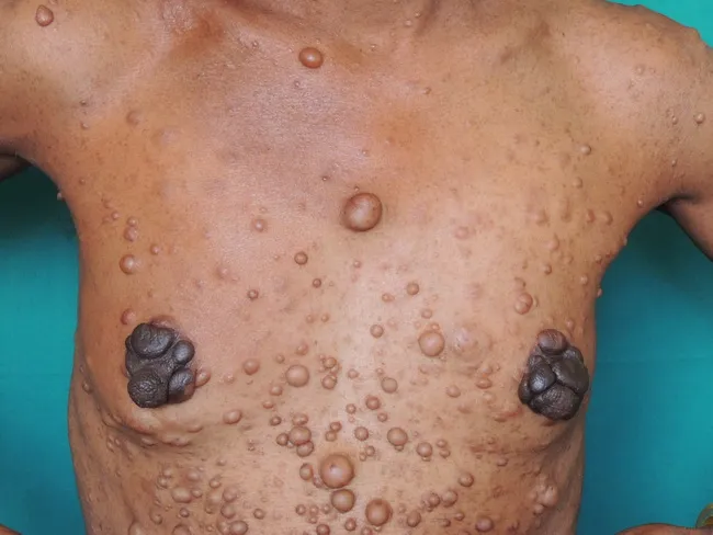
Imaging modality: Clinical photography of a living patient. Anterior trunk and upper torso view showing numerous cutaneous neurofibromas characteristic of Neurofibromatosis type 1 (NF1, von Recklinghausen disease). Primary subject: skin and subcutaneous tissue involvement with widespread, discrete nodules of varying size on the chest, abdomen, face, and limbs. Lesion morphology includes sessile and pedunculated, dome-shaped nodules that are firm, non-tender, and skin-colored to brown. Two prominent bilateral nipple-areola complex neurofibromas are evident, a finding described as uncommon but highly associated with NF1. The size range observed in this field image is approximately 0.2–2.5 cm. Additional cutaneous manifestations may include cafe-au-lait macules (not clearly visible here) and other soft tissue lesions. The clinical diagnosis is supported by family history and biopsy confirming NF1. Plexiform neurofibromas, when present, are virtually diagnostic of NF1 and warrant ongoing oncologic and neurosurgical surveillance due to potential malignant transformation to malignant peripheral nerve sheath tumors (MPNST). This photograph is valuable for dermatology education and for illustrating characteristic lesion distribution, nomenclature (neurofibroma, plexiform neurofibroma, café-au-lait spots, Lisch nodules), and differential considerations (schwannomatosis, multiple benign cutaneous tumors). Imaging aids in monitoring lesion burden and guiding genetic counseling and clinical management.
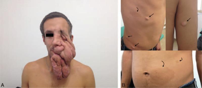
Clinical photographs demonstrating the cutaneous manifestations of Neurofibromatosis Type 1 (NF1). Figure A presents a frontal view of a male patient with a massive plexiform neurofibroma on the left side of the face. The soft tissue tumor causes severe facial asymmetry and disfigurement, characterized by overhanging skin folds, complete eyelid ptosis, and inferior displacement of the left eye, involving the temporal, orbital, cheek, nose, and chin regions. Figure B provides a detailed view of the patient's trunk and arms, illustrating characteristic dermatological findings. These include multiple pedunculated and sessile neurofibromas (indicated by straight arrows), which appear as skin-colored, raised nodules, and several café-au-lait macules (indicated by curved arrows), which are flat, hyperpigmented, light-brown patches with smooth borders. This composite image serves as a visual reference for the range of severity in NF1, from common cutaneous lesions to debilitating plexiform neurofibromas.
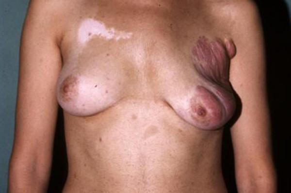
Clinical photograph of a patient's torso displaying characteristic cutaneous manifestations of Neurofibromatosis type 1 (NF1). The right breast exhibits severe deformity and soft tissue overgrowth caused by an extensive plexiform neurofibroma. The lesion involves the outer quadrants, extending to the nipple-areolar complex and toward the homolateral axilla, resulting in a pendulous, bag-like enlargement with a reddish-purple hue and a striated, thickened skin texture (pachydermatous changes). In contrast, the left breast appears normal in morphology. Adjacent to the left breast on the upper chest is a large, irregular depigmented patch consistent with vitiligo. Additionally, the trunk displays multiple café-au-lait macules—well-circumscribed, light-brown pigmented spots—and smaller, scattered cutaneous neurofibromas. The image illustrates the significant aesthetic and structural disfigurement associated with plexiform neurofibromas, which are pathognomonic for NF1 and involve diffuse nerve sheath thickening that can infiltrate surrounding dermal and subcutaneous layers.
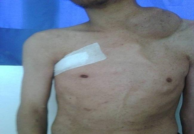
Clinical photograph of a 26-year-old male showing cutaneous manifestations of Neurofibromatosis type 1 (Von Recklinghausen disease). The most striking feature is a large, firm, and irregular plexiform neurofibroma located in the left latero-cervical and supraclavicular region, measuring approximately 10 cm and significantly distorting the neck contour. The patient's torso exhibits classic stigmata of the condition, including multiple hyperpigmented macules (café-au-lait spots) and small, disseminated cutaneous neurofibromas. A surgical dressing is visible on the upper right thoracic wall, corresponding to the site of a synovial sarcoma resection. This image illustrates the multi-system involvement of NF1 and the increased risk for associated malignancies, such as soft tissue sarcomas. The photograph is an educational resource for medical students and clinicians to recognize both the benign neurofibromas and the complex clinical presentation of syndromic oncology patients.
https://pmc.ncbi.nlm.nih.gov/articles/PMC8354850
Which viva-preparation module should I expand first after the case presentation?
Spoken long-case format
“This is a middle-aged female patient with multiple asymptomatic, gradually progressive, skin-colored papules and nodules over the face. The lesions are discrete, smooth-surfaced, skin-colored, soft-looking, and non-tender, without ulceration or discharge. The facial lesions are clinically suggestive of cutaneous neurofibromas.I would examine the whole skin for café-au-lait macules, axillary and inguinal freckling, additional cutaneous or subcutaneous neurofibromas, and any plexiform neurofibroma. I would perform ophthalmic examination for Lisch nodules and optic pathway involvement, assess blood pressure and cardiovascular status, examine the spine and long bones for skeletal deformity, and assess neurologic and developmental status.I would also ask for family history of similar lesions, visual complaints, headache, seizures, limb weakness, pain or rapid enlargement of any swelling, and learning difficulties in childhood.My provisional diagnosis is Neurofibromatosis type 1 with multiple cutaneous neurofibromas. The important concern is to identify a plexiform neurofibroma or a malignant peripheral nerve sheath tumour.”
Points to demonstrate on examining this patient
- Describe lesion site, number, morphology, surface, color, consistency, and symptoms.
- Palpate lesions. Cutaneous neurofibromas may be soft and compressible. The classic buttonhole sign is invagination of a soft neurofibroma into the skin on pressure.
- Search for:
- Café-au-lait macules: uniformly light-to-dark brown, well-defined, usually smooth-bordered ("coast of California") macules.
- Axillary or inguinal freckling: Crowe sign.
- Plexiform neurofibroma: diffuse, ill-defined, tortuous thickening along nerves, often described as a "bag of worms."
- Skeletal signs: scoliosis, tibial anterolateral bowing/pseudarthrosis, sphenoid wing dysplasia.
- Ophthalmic signs: Lisch nodules and choroidal abnormalities.
- Hypertension and symptoms suggesting pheochromocytoma.
- Neurological deficits or signs of intracranial lesion.
The key diagnostic criteria you must know
- Six or more café-au-lait macules
-
5 mm in greatest diameter before puberty
-
15 mm after puberty
-
- Axillary or inguinal freckling
- Two or more neurofibromas of any type, or one plexiform neurofibroma
- Optic pathway glioma
- Two or more Lisch nodules on slit-lamp examination, or two or more choroidal abnormalities on OCT/near-infrared imaging
- A characteristic bony lesion:
- Sphenoid dysplasia
- Anterolateral bowing of tibia
- Pseudarthrosis of a long bone
- A heterozygous pathogenic NF1 variant in normal tissue.
Examiner-viva roadmap
-
Clinical case presentation and morphology
- How to describe neurofibromas correctly
- Buttonhole sign
- Cutaneous versus subcutaneous versus plexiform neurofibroma
-
NF1 diagnosis and differentials
- Revised diagnostic criteria
- Legius syndrome
- Segmental or mosaic NF1
- NF2-related schwannomatosis and other schwannomatoses
- Café-au-lait macules in other disorders
-
Genetics and pathogenesis
- NF1 gene, chromosome 17q11.2
- Neurofibromin and RAS-MAPK pathway
- Autosomal-dominant inheritance, variable expressivity, de novo disease
-
Systemic manifestations and complications
- Optic pathway glioma
- Plexiform neurofibroma
- MPNST
- Skeletal disease, learning issues, vasculopathy, pheochromocytoma, breast cancer risk
-
Investigations, counselling, surveillance, and treatment
- When MRI or PET imaging is appropriate
- Red flags for MPNST
- Surgical and medical management
- MEK inhibitors, particularly selumetinib for selected inoperable symptomatic plexiform neurofibromas
- Genetic counselling and family screening
Describe the location of the lesion near eyebrow
facial anatomical terminology forehead supraorbital eyebrow region dermatology lesion description
“A solitary, skin-colored, dome-shaped nodule is present over the left supraorbital region of the forehead, approximately 1-2 cm above the lateral aspect of the left eyebrow.”
One near the eye at right side
“Multiple small skin-colored papules are present over the left infraorbital and malar region, located just below and lateral to the left eye.”
Prepare me for questions examiner can ask on this topic from moderate to hard to very hard questions with answers
neurofibromatosis type 1 AND management
2025 neurofibromatosis type 1 clinical management surveillance selumetinib guideline
Neurofibromatosis type 1: examiner-viva map
-
Recognition and clinical description
- Morphology of cutaneous neurofibromas
- Buttonhole sign
- Café-au-lait macules, Crowe sign, plexiform neurofibroma
-
Diagnosis and differentials
- Revised NF1 diagnostic criteria
- Legius syndrome, segmental/mosaic NF1
- NF2-related schwannomatosis and other causes of café-au-lait macules
-
Genetics and pathology
- NF1 gene, neurofibromin, RAS-MAPK pathway
- Autosomal-dominant inheritance, penetrance, variable expression
- Histopathology and immunohistochemistry
-
Complications and surveillance
- Optic pathway glioma, skeletal disease, learning problems, hypertension
- Plexiform neurofibroma and MPNST
- Pheochromocytoma/paraganglioma, GIST, breast cancer risk
-
Management
- Counselling and family screening
- Indications for imaging and biopsy
- Surgery and selumetinib for selected symptomatic inoperable plexiform neurofibromas
- A. Diagnosis and differentials
- B. Genetics, pathogenesis and histopathology
- C. Complications and surveillance
- D. Management and treatment
- E. Full mixed mock viva
Just give me all the questions you gave earlier for other spotters
Neurofibromatosis type 1 (NF1): Spotter Viva Questions with Answers
Moderate-level questions
1. What is your diagnosis?
2. What are the lesions seen in this patient?
3. Describe a cutaneous neurofibroma.
4. What is the buttonhole sign?
5. What are the main types of neurofibroma?
- Localized cutaneous neurofibroma
- Subcutaneous neurofibroma
- Diffuse neurofibroma
- Plexiform neurofibroma
6. What is a plexiform neurofibroma?
7. What is the importance of plexiform neurofibroma?
8. What are café-au-lait macules?
9. How many café-au-lait macules are significant in NF1?
- More than 5 mm in greatest diameter in a prepubertal child
- More than 15 mm in a postpubertal person
10. What is Crowe sign?
11. What are Lisch nodules?
12. Are Lisch nodules present at birth?
13. What should you ask in history?
- Age at onset and progression of lesions
- Family history of similar lesions, pigmentary lesions, or known NF1
- Pain, rapid growth, hardening, or neurological symptoms in any tumour
- Visual symptoms, proptosis, squint, headache, seizures
- Learning difficulty, poor school performance, ADHD
- Bone pain, limb deformity, scoliosis
- Hypertension symptoms, palpitations, sweating, headache
- Menstrual/sexual precocity in children, suggesting optic/hypothalamic involvement
14. What should you examine apart from the visible lesions?
- Full skin examination for café-au-lait macules and neurofibromas
- Axillae and groins for freckling
- Eyes for Lisch nodules and visual impairment
- Spine for scoliosis
- Long bones for tibial bowing/pseudarthrosis
- Blood pressure
- Neurological examination
- Development, cognition, and school performance in children
- Breast examination and appropriate screening discussion in adult women
Diagnostic criteria
15. State the revised diagnostic criteria for NF1.
- Six or more café-au-lait macules of appropriate size
- Axillary or inguinal freckling
- Two or more neurofibromas of any type, or one plexiform neurofibroma
- Optic pathway glioma
- Two or more Lisch nodules, or two or more choroidal abnormalities on appropriate imaging
- Distinctive osseous lesion: sphenoid dysplasia, anterolateral tibial bowing, or pseudarthrosis of a long bone
- A heterozygous pathogenic NF1 variant in normal tissue
16. Is genetic testing mandatory for diagnosis?
17. Which gene is mutated in NF1?
18. What does the NF1 gene encode?
19. Explain the role of neurofibromin.
20. What is the inheritance pattern?
21. What is the recurrence risk for offspring of an affected person?
22. How often is NF1 a new mutation?
23. Can severity be predicted from the severity in the parent?
Differential diagnosis
24. What is the differential diagnosis of multiple skin-colored papules/nodules on the face?
- Multiple cutaneous neurofibromas in NF1
- Multiple schwannomas
- Multiple trichoepitheliomas
- Fibrofolliculomas, such as in Birt-Hogg-Dubé syndrome
- Angiofibromas of tuberous sclerosis
- Syringomas
- Skin tags, if pedunculated
- Leiomyomas, usually firmer and may be painful
25. What is the differential diagnosis of multiple café-au-lait macules?
- NF1
- Legius syndrome
- McCune-Albright syndrome
- Constitutional mismatch repair deficiency syndrome
- Noonan syndrome with multiple lentigines
- Watson syndrome
- Isolated familial café-au-lait macules
26. Differentiate NF1 from Legius syndrome.
| Feature | NF1 | Legius syndrome |
|---|---|---|
| Gene | NF1 | SPRED1 |
| Café-au-lait macules | Present | Present |
| Axillary freckling | Present | May occur |
| Neurofibromas | Present | Absent |
| Lisch nodules | May occur | Absent |
| Optic pathway glioma | May occur | Absent |
| Major tumour predisposition | Present | Not characteristic |
27. What is segmental or mosaic NF1?
28. Can a patient with segmental NF1 transmit generalized NF1 to their children?
29. Differentiate NF1 from NF2-related schwannomatosis.
Hard questions: complications
30. Name the important complications of NF1.
- Plexiform neurofibroma
- MPNST
- Optic pathway glioma
- Other low-grade gliomas
- Learning disability, ADHD, developmental delay
- Skeletal abnormalities: scoliosis, tibial pseudarthrosis, sphenoid wing dysplasia
- Hypertension due to renal artery stenosis, coarctation, or pheochromocytoma
- Pheochromocytoma/paraganglioma
- Gastrointestinal stromal tumour
- Glomus tumours
- Juvenile myelomonocytic leukaemia, especially in children
- Increased breast cancer risk in women
- Cerebrovascular abnormalities, including moyamoya vasculopathy
31. Which tumour is most feared in NF1?
32. What features suggest malignant transformation of a plexiform neurofibroma?
- Rapid increase in size or change in growth rate
- New, persistent, severe, or nocturnal pain
- New hard area within a previously soft lesion
- New motor weakness, sensory loss, or neuropathic symptoms
- Bladder/bowel dysfunction, dysphagia, or respiratory compromise depending on location
33. What is the work-up if you suspect MPNST?
- Urgent assessment by an NF1/sarcoma multidisciplinary team
- Regional MRI to define the lesion
- FDG-PET/CT or FDG-PET/MRI to identify metabolically active suspicious areas
- Image-guided core biopsy targeting the suspicious region, planned with the sarcoma team
- Staging imaging if malignancy is confirmed
34. Why should an incisional biopsy not be done casually in suspected MPNST?
35. What is ANNUBP?
36. What is an optic pathway glioma?
37. Does every child with NF1 need screening MRI of brain and orbits?
38. Which child with NF1 is at risk of precocious puberty?
39. What are the skeletal manifestations of NF1?
- Scoliosis, both dystrophic and non-dystrophic
- Anterolateral bowing of tibia
- Tibial pseudarthrosis
- Sphenoid wing dysplasia
- Vertebral scalloping
- Short stature and reduced bone mineral density in some patients
40. Why should blood pressure be checked at every visit?
- Essential hypertension
- Renal artery stenosis
- Coarctation of the aorta
- Pheochromocytoma or paraganglioma
- Other NF1-associated vasculopathies
41. When should you suspect pheochromocytoma in NF1?
42. What malignancy screening issue is important in adult women with NF1?
Histopathology and very hard questions
43. What is the histopathology of a neurofibroma?
44. Why is a neurofibroma usually non-encapsulated?
45. How is neurofibroma differentiated from schwannoma histologically?
| Neurofibroma | Schwannoma |
|---|---|
| Usually non-encapsulated | Usually encapsulated |
| Entraps axons | Typically displaces axons outside the tumour |
| Mixed cell population | Predominantly Schwann cells |
| Wavy spindle cells in myxoid background | Antoni A and Antoni B areas, Verocay bodies |
| Can be plexiform in NF1 | Plexiform schwannoma is distinct and not typical of NF1 |
46. Which immunohistochemical markers are useful?
- S100 and SOX10: positive in Schwann-cell component, often patchy in neurofibroma
- CD34: highlights fibroblastic component
- Neurofilament protein: highlights entrapped axons
- EMA: may highlight perineurial cells
- Ki-67: helps evaluate proliferative activity in atypical or malignant lesions
- H3K27me3 loss can support MPNST in the appropriate pathological context
47. Why do neurofibromas develop in NF1 although every cell carries one altered NF1 allele?
48. What is the role of mast cells in neurofibroma formation?
49. Why are cutaneous neurofibromas common in adolescence and pregnancy?
50. Are cutaneous neurofibromas premalignant?
Management questions
51. How do you manage a patient with NF1?
- Clinical diagnosis and genetic counselling
- Education about red-flag symptoms
- Annual or periodic clinical review depending on age and complications
- Full skin, neurologic, skeletal, visual, and blood-pressure assessment
- Ophthalmology surveillance in children
- Developmental, behavioural, and educational support
- Targeted imaging only when clinically indicated
- Referral to relevant specialties for tumours or complications
52. How are asymptomatic cutaneous neurofibromas managed?
53. What treatment options exist for cutaneous neurofibromas?
- Surgical excision
- Shave excision
- Electrosurgery/electrodessication
- Laser treatment in selected cases
- Radiofrequency ablation in selected settings
54. How is a plexiform neurofibroma managed?
- Assess by an NF1 multidisciplinary team
- MRI for anatomy, extent, and serial follow-up when indicated
- Surgery if complete resection is feasible and morbidity is acceptable
- Observe stable asymptomatic lesions
- Consider a MEK inhibitor, such as selumetinib, for selected symptomatic, inoperable plexiform neurofibromas, especially in paediatric patients where locally approved
55. What is selumetinib and how does it work?
56. What adverse effects should be monitored with selumetinib?
- Acneiform or other rash
- Diarrhoea
- Nausea/vomiting
- Elevated creatine kinase
- Paronychia
- Ocular toxicity, including retinal changes
- Reduced left ventricular ejection fraction
57. What counselling should be provided?
- NF1 is autosomal dominant
- Each child has a 50% transmission risk if the parent has a germline pathogenic variant
- Severity cannot be reliably predicted in offspring
- Offer genetic counselling before conception
- Discuss testing options according to local laws and services
- Counsel on warning symptoms of MPNST and need for lifelong follow-up
Rapid-fire very hard viva
58. Why are café-au-lait macules not specific for NF1?
59. Why are the borders of NF1 café-au-lait macules described as coast of California?
60. What is the difference between generalized NF1 and mosaic NF1 genetically?
61. Why can a blood NF1 genetic test be negative in mosaic NF1?
62. Is NF1 the same as “elephant man disease”?
63. Which tumour is more associated with NF1: schwannoma or neurofibroma?
64. What is the significance of a solitary neurofibroma?
65. Is every patient with NF1 equally likely to develop MPNST?
66. Why should radiotherapy be avoided where possible in NF1?
67. What is the most appropriate single sentence to end your spotter presentation?
“This patient has multiple facial cutaneous neurofibromas, and my provisional diagnosis is neurofibromatosis type 1; I would complete the diagnostic assessment by looking for pigmentary, ocular, skeletal, neurological, and familial features, while specifically excluding a plexiform neurofibroma or features of malignant peripheral nerve sheath tumour.”
What is the prognosis of neurofibromatosis
neurofibromatosis type 1 AND mortality
GeneReviews neurofibromatosis 1 prognosis life expectancy complications
Prognosis of Neurofibromatosis Type 1
Good prognostic points
- Isolated or multiple cutaneous neurofibromas generally do not become malignant.
- Most optic pathway gliomas are indolent, particularly when detected in childhood.
- Early recognition of learning, visual, skeletal, and blood-pressure problems improves outcomes.
- Regular surveillance helps identify complications early.
Poor prognostic factors
- Large or deep plexiform neurofibromas
- Rapidly enlarging, painful, hard, or neurologically symptomatic lesion
- Development of malignant peripheral nerve sheath tumour (MPNST)
- Central nervous system tumours or significant vasculopathy
- Severe scoliosis, tibial pseudarthrosis, or major skeletal deformity
- Significant cognitive, behavioural, or psychosocial problems
- NF1-associated breast cancer in women
Important figures for viva
- Plexiform neurofibromas occur clinically in about 30% of patients.
- Lifetime risk of MPNST is roughly 8%-13%.
- Learning difficulties occur in around 50%-60% of affected children.
- Median life expectancy is reported to be about 8 years lower than the general population, mainly due to malignant tumours and vascular complications. This is a population figure, not a prediction for an individual patient. See GeneReviews prognosis data.
Best viva answer
“The prognosis of NF1 is highly variable. Most patients with only cutaneous neurofibromas have a good life expectancy, although cosmetic and psychosocial morbidity may be substantial. Prognosis worsens in patients with plexiform neurofibromas, MPNST, CNS tumours, vasculopathy, severe skeletal disease, and significant learning disability. Lifelong multidisciplinary surveillance is required, particularly for red flags of malignant transformation: rapid growth, persistent or nocturnal pain, hardening of a lesion, or new neurological deficit.”