I now have comprehensive information to compile the full case discussion. Let me compile the complete presentation.
DMSF IMD 27 - Anal Mass Case Discussion
Patient: 50-year-old Male | OPD Consult
👤 Presenter: Pranav — Intro, History & Physical Exam
History of Present Illness
- 50/M presenting with an anal mass
- 5 years prior to consultation (PTC): Intermittent, painless bright-red rectal bleeding - minimal (~2-3 drops), occurring once every 1-3 months
- 1 year PTC: Noticed a prolapsing anal mass upon straining - manually reducible
- 4 days ago: Sudden onset perianal pain (NRS 7/10) associated with the anal mass, worsening with defecation. Took pain medications
- Current (in-interim): Pain decreased to NRS 1/10
Associated Symptoms
- (+) Constipation - BM every 2-3 days, hard stools
- (+) Straining during defecation
- (-) Weight loss, change in stool caliber, abdominal pain, melena
Past Medical History
- Hypertension - on Amlodipine 5 mg OD
- No prior surgeries
Family History
- Father: Hypertension
- Mother: Type 2 Diabetes Mellitus
Personal & Social History
- Low fiber/fruit/vegetable intake
- Drinks < 2L water/day
- Virtual assistant (sedentary occupation)
- Non-smoker, occasional alcohol drinker
Physical Examination
| Parameter | Finding |
|---|
| BP | 132/82 mmHg |
| HR | 78 bpm |
| RR | 14/min |
| Temp | 37°C |
| BMI | 29 (overweight) |
- General: Alert, oriented
- HEENT: Pink palpebral conjunctivae, anicteric sclerae
- Chest: Distinct heart sounds, clear breath sounds
- Abdomen: Soft, nontender, nondistended, normoactive bowel sounds, no palpable mass
Anorectal Exam
- Visual Inspection (Relaxed): Firm, slightly tender, bluish-purple nodule (~1.5 cm) at the anal verge - right posterolateral area
- Valsalva Maneuver: Bearing down reveals prolapse of pink, engorged mucosal tissue
- DRE: Normal resting sphincter tone; no palpable rectal masses, strictures, or fluctuance; prostate smooth and mildly enlarged
🔍 Presenter: Jeni — Primary Impression with Rationale
Primary Impression: Mixed Hemorrhoidal Disease (Thrombosed External Hemorrhoid + Prolapsed Internal Hemorrhoid)
Rationale:
Two concurrent findings explain the clinical picture:
| Finding | Interpretation |
|---|
| Firm, tender, bluish-purple nodule at anal verge | Thrombosed external hemorrhoid - clot formation in external hemorrhoidal plexus (below dentate line) |
| Prolapsing pink mucosal tissue on Valsalva | Internal hemorrhoid (at least Grade II-III) prolapsing through the anal canal |
| Painless rectal bleeding for 5 years | Classic for internal hemorrhoids (above dentate line = no somatic sensory innervation) |
| Sudden perianal pain 4 days ago | Thrombosis of the external component - characteristically acute, constant, and severe |
| Constipation + straining + low fiber diet | Well-established risk factors for hemorrhoidal disease |
| Pain now improving (NRS 7 → 1) | Consistent with natural history: peak pain at 48-72 hours, then spontaneous clot resolution begins |
The combination of a prolapsing internal component (mucosal prolapse on Valsalva) and a thrombosed external nodule constitutes mixed hemorrhoidal disease.
📋 Presenter: Vignesh — Differential Diagnoses
| Diagnosis | Supporting Features | Against |
|---|
| Mixed hemorrhoids (thrombosed external + prolapsed internal) (Primary) | Bluish nodule at verge, mucosal prolapse, 5-yr bleeding history, straining, constipation | - |
| Perianal abscess/fistula | Perianal pain, lump | No fever, no fluctuance on DRE, no purulent discharge, no surrounding induration |
| Rectal prolapse | Tissue protruding on straining | Prolapse here involves mucosal tissue, not full-thickness rectal wall; reducible; no circumferential protrusion |
| Anal fissure | Perianal pain worsening with defecation | No sentinel pile/posterior midline tear described; mass is nodular not linear |
| Condyloma acuminata (anal warts) | Perianal mass | No sexual history suggestive; mass is bluish-purple and solitary, not verrucous |
| Anal/rectal carcinoma | Rectal bleeding in a 50-yr-old male | No weight loss, change in stool caliber, or palpable rectal mass; bleeding is painless bright red - not consistent with cancer pattern |
| Skin tag | Visible perianal lump | Not tender, not bluish-purple, not acutely symptomatic |
Note: At age 50 with rectal bleeding, colorectal malignancy must be excluded. Colonoscopy is indicated.
🔬 Presenter: Kashmira — Diagnostic Plan
Bedside/Immediate
| Test | Rationale |
|---|
| Anoscopy | Gold standard to classify internal hemorrhoids by grade; visualize the prolapsing tissue and identify bleeding source |
| Proctoscopy | Examine rectal mucosa, rule out rectal polyp or mass |
| Valsalva maneuver (done) | Demonstrate prolapse extent |
Laboratory (Already Available)
| Lab | Result | Interpretation |
|---|
| Hemoglobin | 13.2 g/dL (Low normal) | Mild - consistent with chronic minimal blood loss |
| Hematocrit | 40% | Mildly reduced |
| WBC | 6.5 x 10³/µL | Normal - no infection/abscess |
| Platelets | 250 x 10³/µL | Normal |
Additional labs: Coagulation profile (PT/INR, aPTT) if surgery is anticipated; Fasting blood glucose (family Hx T2DM, BMI 29); Creatinine (pre-operative baseline)
Endoscopy
| Procedure | Indication |
|---|
| Colonoscopy | Patient is 50 years old with rectal bleeding - colorectal cancer screening is indicated per guidelines; symptoms alone insufficient to attribute bleeding solely to hemorrhoids without ruling out proximal pathology |
✅ Presenter: Avadhat — Final Diagnosis
Mixed Hemorrhoidal Disease
- Thrombosed External Hemorrhoid (right posterolateral, ~1.5 cm, acutely thrombosed with resolving symptoms)
- Internal Hemorrhoid - Grade III (prolapsing on straining, manually reducible, with history of painless rectal bleeding)
Basis:
- Painless bleeding (5 yrs) = internal hemorrhoid component
- Prolapse requiring manual reduction = Grade III internal hemorrhoid
- Acute bluish-purple tender nodule at anal verge = thrombosed external hemorrhoid
- Current improvement in pain = thrombosis past its peak (>72 hrs), now resolving
- Normal WBC, no fever, no fluctuance = no abscess/sepsis
🫀 Presenter: Avinash — Anatomy
The hemorrhoidal system is made up of submucosal vascular cushions composed of arteriovenous communications, smooth muscle (Treitz muscle), and connective tissue that contribute to anal continence by providing a compressible "seal."
Key Anatomical Landmarks
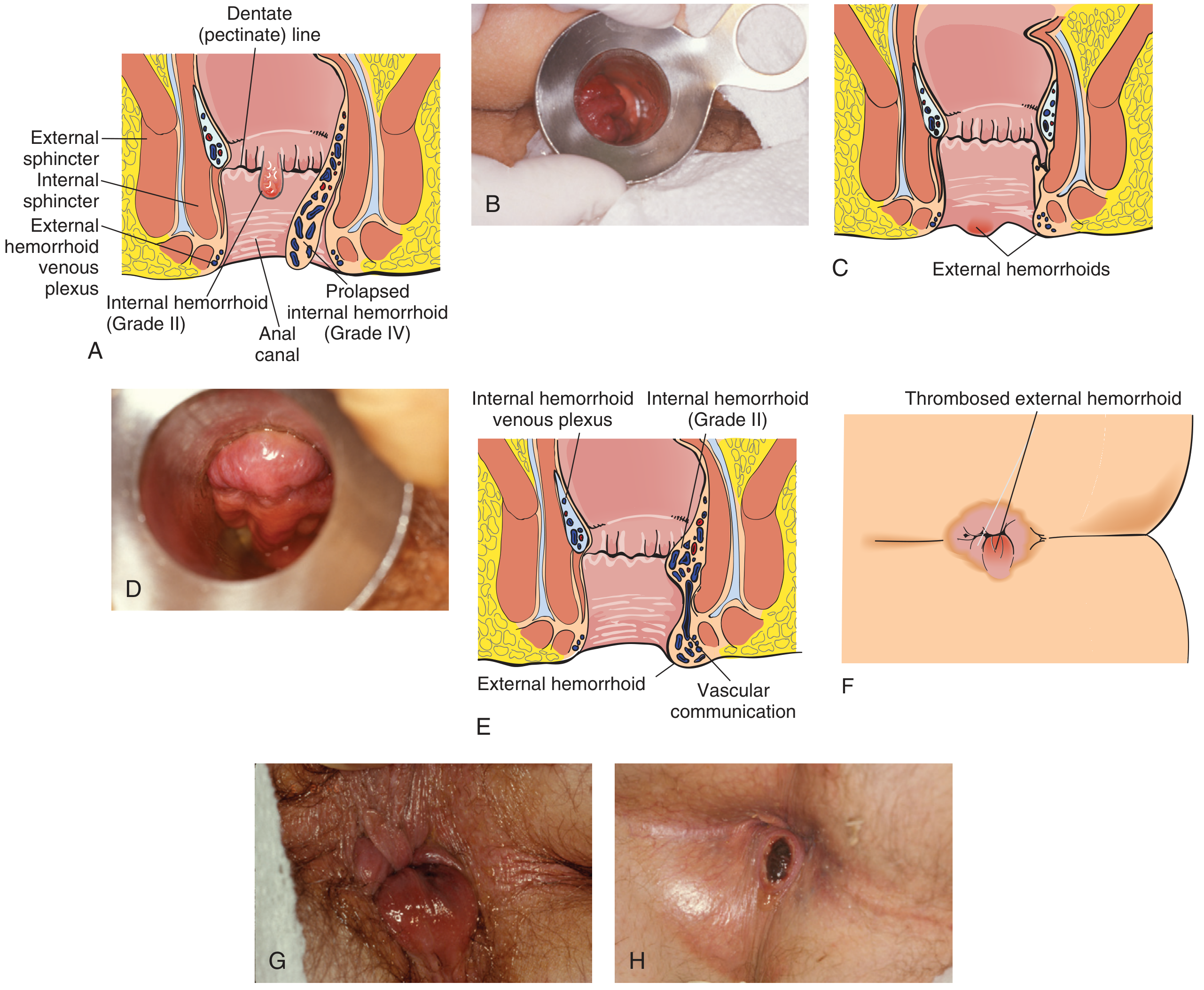
Panels A-H showing: internal hemorrhoid Grade II, Grade IV prolapsed internal hemorrhoid, external hemorrhoids, thrombosed external hemorrhoid, and mixed hemorrhoid with vascular communication.
| Structure | Location | Clinical Relevance |
|---|
| Dentate (Pectinate) Line | 2 cm from anal verge | Divides internal from external hemorrhoids; marks transition from columnar to squamous epithelium |
| Internal hemorrhoidal plexus | Above dentate line; covered by mucosa | No somatic sensation → painless bleeding |
| External hemorrhoidal plexus | Below dentate line; covered by anoderm | Rich somatic innervation (pudendal nerve) → painful when thrombosed |
| Columns of Morgagni | Proximal to dentate line | Anal crypts with mucous glands for lubrication |
| Anal sphincter complex | Surrounds anal canal | Internal (involuntary, smooth muscle) + External (voluntary, skeletal muscle) |
Hemorrhoidal Blood Supply
- Superior hemorrhoidal artery - branch of inferior mesenteric artery (IMA) → internal hemorrhoids
- Middle hemorrhoidal artery - from internal iliac artery
- Inferior hemorrhoidal artery - from internal pudendal artery → external hemorrhoids
Venous Drainage
- Superior hemorrhoidal veins → portal system (clinically important in portal hypertension)
- Inferior hemorrhoidal veins → caval system
- This creates a portosystemic anastomosis at the anorectal junction
Innervation
- Above dentate line: Autonomic (visceral) - no pain fibers → internal hemorrhoids painless
- Below dentate line: Somatic (pudendal nerve S2-S4) → external hemorrhoids exquisitely painful
Classic Hemorrhoid Positions
Internal hemorrhoids classically occur at the 3 o'clock (left lateral), 7 o'clock (right posterior), and 11 o'clock (right anterior) positions - corresponding to the three major vascular pedicles.
(Source: Pfenninger and Fowler's Procedures for Primary Care, 3rd ed. | Rosen's Emergency Medicine)
⚙️ Presenter: Keshav — Pathophysiology
Normal Function
Hemorrhoidal cushions are normal anatomical structures - not inherently pathological. They consist of submucosal arteriovenous sinusoids supported by smooth muscle (muscularis submucosae ani) and connective tissue. They contribute approximately 15-20% of resting anal canal pressure and help with fine continence discrimination.
How Hemorrhoids Become Symptomatic
Two main theories:
1. Vascular Engorgement Theory
Chronic straining and constipation → increased intra-abdominal pressure → venous congestion within hemorrhoidal plexuses → progressive engorgement and enlargement of the vascular cushions.
2. Sliding Anal Lining Theory (more accepted)
Chronic straining → repetitive downward force → progressive degeneration of the supporting connective tissue (Parks ligament) and Treitz muscle → downward displacement of the anal cushions → prolapse.
Thrombosis of External Hemorrhoid (This Patient)
- Acute increase in venous pressure (straining/defecation) → rupture of a vein in the external hemorrhoidal plexus → blood clot forms within the subcutaneous tissue below the dentate line
- Result: Acute, painful, firm, bluish-purple nodule
- Covered by anoderm (somatic pain fibers) → severe pain at onset (NRS 7/10)
- Natural history: Peak pain at 48-72 hours → clot begins to organize and reabsorb → pain diminishes over 7-10 days (explains NRS 1/10 now)
Risk Factors in This Patient
| Risk Factor | Mechanism |
|---|
| Low fiber diet | Hard stools → increased straining → higher intra-abdominal pressure |
| Low water intake | Worsens constipation |
| Constipation | Prolonged straining → sustained increase in venous pressure |
| Sedentary occupation (virtual assistant) | Prolonged sitting → reduced perineal muscle tone, venous pooling |
| BMI 29 (overweight) | Increased intra-abdominal pressure |
| Age 50 | Progressive connective tissue degeneration |
(Source: Textbook of Family Medicine 9e | Rosen's Emergency Medicine)
📊 Presenter: Pragati — Grading
Internal Hemorrhoids: Goligher Classification
| Grade | Description | Example in This Patient |
|---|
| Grade I | Bleed but do not prolapse | Early phase (5 yrs ago - bleeding only) |
| Grade II | Prolapse on straining but reduce spontaneously | Intermediate phase |
| Grade III | Prolapse on straining; require manual reduction | Current internal component |
| Grade IV | Permanently prolapsed; cannot be manually reduced | Not present |
This patient has Grade III internal hemorrhoids - the prolapsed mucosal tissue reduced manually.
External Hemorrhoids: No Formal Grading Scale
External hemorrhoids are classified by their clinical state:
- Non-thrombosed: Soft, asymptomatic or mildly symptomatic folds
- Thrombosed: Acute, firm, tender, bluish-purple nodule (present in this case)
- Gangrenous: Rare, associated with severe edema and necrosis (not present)
Mixed Hemorrhoids
When both internal and external components are present with a vascular communication, it is termed mixed hemorrhoidal disease - as in this patient.
(Source: Pfenninger and Fowler's Procedures for Primary Care, 3rd ed. | Textbook of Family Medicine 9e)
💊 Presenter: Sanya — Management
Acute Management (ED/OPD - This Presentation)
Since the patient's thrombosed external hemorrhoid is now > 72 hours and pain is declining (NRS 7 → 1), conservative management is the preferred approach:
WASH Regimen (Rosen's Emergency Medicine)
| Component | Specific Recommendation |
|---|
| W - Warm water | Sitz baths 3-4x/day for 15 min; 40°C water reduces anal canal pressure and promotes comfort |
| A - Analgesics | Paracetamol/NSAIDs (ibuprofen) for pain relief; topical lidocaine for local anesthesia |
| S - Stool softeners | Docusate sodium OR lactulose to prevent hard stools and further straining |
| H - High-fiber diet | Psyllium husk 1-2 tsp in 8 oz water OD; target 20-30 g fiber/day; increase water to ≥2L/day |
Topical Agents
- Topical hydrocortisone cream for short-term (<5 days) to reduce edema and inflammation
- Topical anesthetics (lidocaine 5% ointment) for symptomatic relief
- Witch hazel astringent pads for hygiene
Lifestyle Modifications
- Increase dietary fiber and water intake
- Avoid prolonged sitting/straining
- Regular light exercise to improve perineal tone and venous return
Outpatient Follow-up Management for Grade III Internal Hemorrhoid
Once the acute thrombosis resolves, the Grade III internal hemorrhoid requires definitive treatment:
Office-Based Procedures (First-line for Grade I-III):
| Procedure | Description | Efficacy |
|---|
| Rubber Band Ligation (RBL) | Most effective office procedure; band placed at base of hemorrhoid, causing ischemic necrosis and scarring | Most effective; recommended by ASCRS 2024 as gold standard office procedure |
| Sclerotherapy | Injection of sclerosant (5% phenol in almond oil) into hemorrhoid pedicle → fibrosis | Effective for Grade I-II |
| Infrared Coagulation (IRC) | Infrared energy coagulates hemorrhoidal tissue | Useful for Grade I-II |
Per
ASCRS 2024 guidelines: Office-based procedures (especially rubber band ligation) are recommended for most Grade I-II and selected Grade III hemorrhoids.
🔪 Surgical Options (Sanya - if applicable)
Indicated if:
- Grade III hemorrhoids fail conservative therapy + office procedures
- Grade IV hemorrhoids
- Mixed internal/external hemorrhoids with large external component
- Patient preference
- Complicated disease (thrombosis, strangulation, gangrenous hemorrhoids)
Surgical Options
| Procedure | Description | When to Use |
|---|
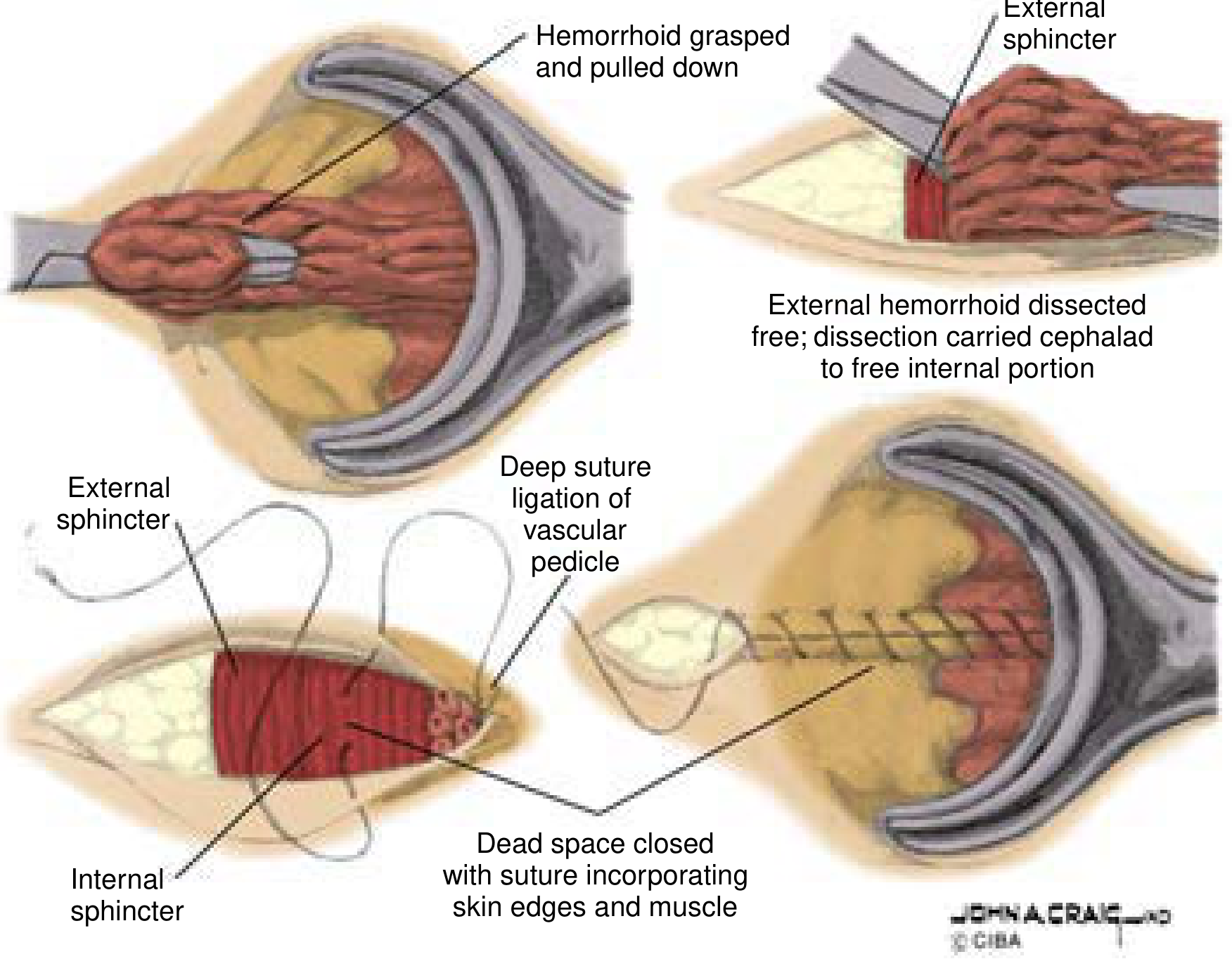
| Ferguson (Closed) Hemorrhoidectomy | V-shaped excision, pedicle ligated, wound closed with running absorbable suture (most common in USA) | Grade III-IV, mixed disease, large external hemorrhoids |
| Milligan-Morgan (Open) Hemorrhoidectomy | Same excision but wound left open (common in Europe/UK) | Same indications; longer healing time |
| Stapled Hemorrhoidopexy (PPH) | Circular stapler removes band of mucosa proximal to hemorrhoids; repositions tissue without excision; less pain but higher recurrence | Grade I-III circumferential internal hemorrhoids without large external component |
| Doppler-Guided Hemorrhoid Artery Ligation (HAL) + Mucopexy | Doppler probe identifies hemorrhoidal arteries; Z-stitch ligation to devascularize; mucopexy tacks up prolapsing tissue | Grade II-IV, less pain than excisional hemorrhoidectomy; higher recurrence rate |
| Excision of Thrombosed External Hemorrhoid | Elliptical excision under local anesthesia | If pain > 72 hrs and still severe, or if requested by patient |
Ferguson (closed) hemorrhoidectomy: dissection of external and internal components off sphincter muscles, ligation of vascular pedicle, and closure with running suture.
For THIS patient: Given Grade III disease with mixed hemorrhoid (thrombosed external now resolving), the algorithm would be:
- Conservative management now (WASH + lifestyle)
- Rubber band ligation for the internal Grade III component at follow-up visit
- If RBL fails or recurs → excisional hemorrhoidectomy (Ferguson or Milligan-Morgan)
(Source: Current Surgical Therapy 14e | ASCRS 2024 Clinical Practice Guidelines)
🏥 Presenter: Poornema — Post-Surgical Approach
Immediate Post-Operative Period (Day 0-1)
| Domain | Management |
|---|
| Pain | Multimodal analgesia: paracetamol + NSAIDs + short-course oral opioid (tramadol); topical anesthetic ointment; minimize narcotic use |
| Urinary retention | Monitor urine output; urinary retention occurs in up to 15% of patients post-hemorrhoidectomy; fluid restrict intraoperatively; catheterize if needed |
| Wound care | Keep perianal area clean and dry; sitz baths 3x/day starting Day 1 |
| Diet | Clear liquids initially; advance to soft diet |
| Activity | Ambulate early; avoid prolonged sitting |
Days 1-14
| Domain | Management |
|---|
| Bowel regimen | Stool softeners (docusate) + oral fiber supplement mandatory to prevent straining; target soft, formed stool; avoid constipation which increases tension on repair |
| Sitz baths | 15 min warm water soaks 3-4x/day and after each bowel movement |
| Wound monitoring | Spotting/minor bleeding for 8-10 days is expected; patient education critical |
| Pain | Expected to decrease by Day 3-5; prescribe adequate analgesics for 1-2 weeks |
| Infection surveillance | Instruct patient to report fever, increasing pain, urinary difficulty, swelling → sign of perineal sepsis (rare but serious) |
Discharge Instructions & Follow-up
| Item | Instruction |
|---|
| Bleeding | Small amounts expected for 7-10 days as sutures absorb; profuse bleeding (soaking through pad) → return to ER immediately - secondary hemorrhage peaks at 7-14 days |
| Fiber & Hydration | High-fiber diet (20-30 g/day), ≥2L water/day permanently |
| Activity | Light activity at 1-2 weeks; avoid heavy lifting/straining for 3-4 weeks |
| Return to Work | Sedentary work (virtual assistant): 1-2 weeks; physical labor: 3-4 weeks |
| Follow-up | 2-4 weeks post-op for wound check; colonoscopy scheduled for colorectal cancer screening |
Potential Post-Operative Complications
| Complication | Timing | Management |
|---|
| Urinary retention | Immediate | Catheterization |
| Primary bleeding | < 24 hrs | Return to OR, oversew |
| Secondary hemorrhage | 7-14 days | Return to OR; often no active bleeding seen on exam |
| Perineal/pelvic sepsis | Variable | Broad-spectrum IV antibiotics + surgical debridement; emergency |
| Anal stenosis | Late | Anal dilators; rarely requires revision |
| Fecal incontinence | Rare | Sphincter-preservation technique; pelvic floor PT |
| Recurrence | Late | Rubber band ligation for recurrence; revision surgery if needed |
(Source: Current Surgical Therapy 14e | Pfenninger & Fowler's Procedures for Primary Care, 3rd ed. | ASCRS 2024 Guidelines)
📎 Summary Table
| Section | Assigned To | Key Point |
|---|
| Intro/HPI/PE | Pranav | 50/M, 5-yr painless rectal bleeding, 1-yr prolapsing mass, 4-day acute pain, bluish-purple nodule at verge, mucosal prolapse on Valsalva |
| Primary Impression | Jeni | Mixed hemorrhoidal disease (thrombosed external + Grade III internal) |
| Differentials | Vignesh | Perianal abscess, rectal prolapse, anal fissure, condyloma, anal cancer |
| Diagnostics | Kashmira | Anoscopy, proctoscopy, CBC (mild anemia), colonoscopy for CRC screening |
| Final Diagnosis | Avadhat | Thrombosed external hemorrhoid + Grade III internal hemorrhoid (mixed disease) |
| Anatomy | Avinash | Dentate line divides internal/external; 3 vascular pedicles; somatic vs. autonomic innervation |
| Pathophysiology | Keshav | Sliding/vascular theory; connective tissue degeneration; thrombosis mechanism; risk factors |
| Grading | Pragati | Goligher Grade III (prolapse requiring manual reduction); thrombosed external component |
| Management | Sanya | WASH regimen acutely; RBL for Grade III at follow-up; hemorrhoidectomy if RBL fails |
| Post-surgical | Poornema | Multimodal analgesia, stool softeners, sitz baths, wound monitoring, secondary hemorrhage warning at 7-14 days |