
Mid brain at level of superior colliculus image
Reading File
This is a Visual query - I need to search for midbrain/superior colliculus anatomy images from the textbook library.
Finding Sources
Finding Sources
Excellent! Multiple good sources found. Let me read the sections with cross-section images - the Bradley & Daroff Neurology book and Gray's Anatomy look most promising for a cross-sectional image at the level of the superior colliculus.
Reading File
Reading File
I found two excellent images. Let me fetch both - the Bradley & Daroff clinical diagram (Fig. 21.4) and the Gray's Anatomy transverse section (eFig. 9.28).
Loading Image
Loading Image
Here are two high-quality images of the midbrain at the level of the superior colliculus from authoritative textbooks:
Midbrain at the Level of the Superior Colliculus
Image 1 - Clinical Diagram (Bradley and Daroff's Neurology in Clinical Practice)
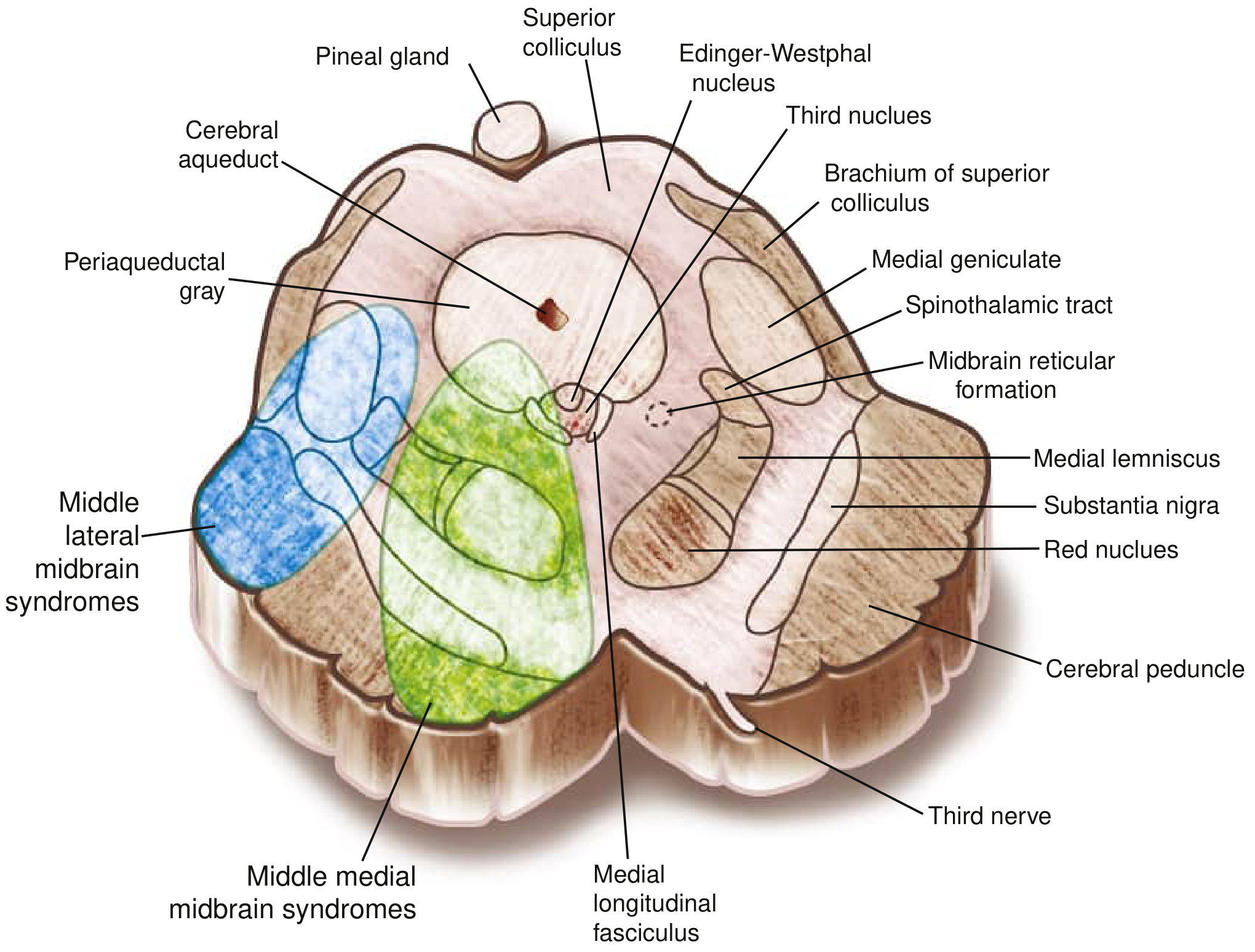
Fig. 21.4 - Midbrain at the level of the superior colliculus, showing medial and lateral vascular territories (blue = lateral midbrain syndrome zone, green = medial midbrain syndrome zone)
Key structures labeled:
- Tectum (dorsal): Superior colliculus, Pineal gland
- Around the aqueduct: Cerebral aqueduct, Periaqueductal gray, Edinger-Westphal nucleus
- Tegmentum: Red nucleus, Substantia nigra, Medial longitudinal fasciculus, Midbrain reticular formation
- Tracts: Medial lemniscus, Spinothalamic tract
- Lateral: Brachium of superior colliculus, Medial geniculate body
- Ventral (basis pedunculi): Cerebral peduncle, Third nerve (CN III)
Image 2 - Histological Section (Gray's Anatomy for Students, from Crossman's Neuroanatomy, 6th ed.)
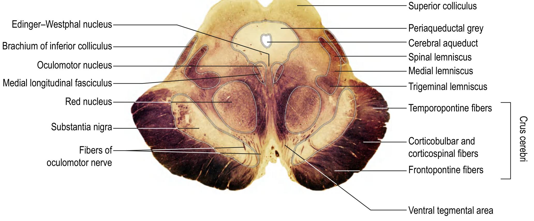
eFig. 9.28 - Transverse section of the rostral midbrain at the location of the superior colliculus
Key structures labeled:
- Tectum (dorsal): Superior colliculus, Periaqueductal grey, Cerebral aqueduct
- Tegmentum: Oculomotor nucleus (CN III), Edinger-Westphal nucleus, Red nucleus, Substantia nigra, Medial longitudinal fasciculus (MLF), Ventral tegmental area
- Sensory tracts: Medial lemniscus, Spinal lemniscus, Trigeminal lemniscus
- Crus cerebri (ventral): Corticobulbar + corticospinal fibers (middle), Frontopontine fibers (medial), Temporopontine fibers (lateral)
- Emerging nerve: Fibers of oculomotor nerve (CN III), Brachium of inferior colliculus
Anatomical summary - At the level of the superior colliculus, the midbrain has three compartments:
- Tectum (roof, dorsal) - superior colliculi for visual reflex relay
- Tegmentum (middle) - CN III nucleus, Edinger-Westphal nucleus, red nucleus, substantia nigra, MLF, periaqueductal gray
- Crus cerebri/Basis pedunculi (ventral) - descending corticospinal, corticobulbar, and pontine projection fibers
- Bradley and Daroff's Neurology in Clinical Practice, Fig. 21.4
- Gray's Anatomy for Students (Crossman's Neuroanatomy 6th ed.), eFig. 9.28
This is a shared conversation. Sign in to Orris to start your own chat.