
What is e coli
E. coli bacteria overview types infections 2024
E. coli gram negative rod bacteria microscopy
E coli gram negative rod bacteria microscopy
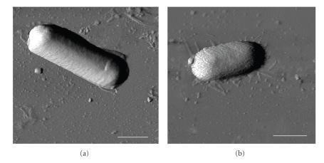
This diagnostic imaging set displays Atomic Force Microscopy (AFM) phase images of two distinct bacterial species, Bacillus cereus (a) and Escherichia coli (b), deposited on a mica surface. The images facilitate a morphological comparison between Gram-positive and Gram-negative bacteria at the nanometer scale. Panel (a) shows B. cereus as a distinctly elongated, rod-shaped cell with a relatively uniform, smooth surface texture and well-defined edges. In contrast, panel (b) depicts E. coli with a more compact, oval or capsule-shaped morphology. The E. coli cell exhibits a significantly rougher surface topography with visible granularity compared to the smoother envelope of B. cereus. The background depicts the mica substrate with faint thread-like structures visible around the E. coli cell, likely representing extracellular filaments or surface dehydration artifacts. A 1 μm scale bar is provided in both panels. These images are used in microbiology to study bacterial cell wall structural integrity, surface roughness (Rq), and biophysical responses to environmental conditions like relative humidity (RH).
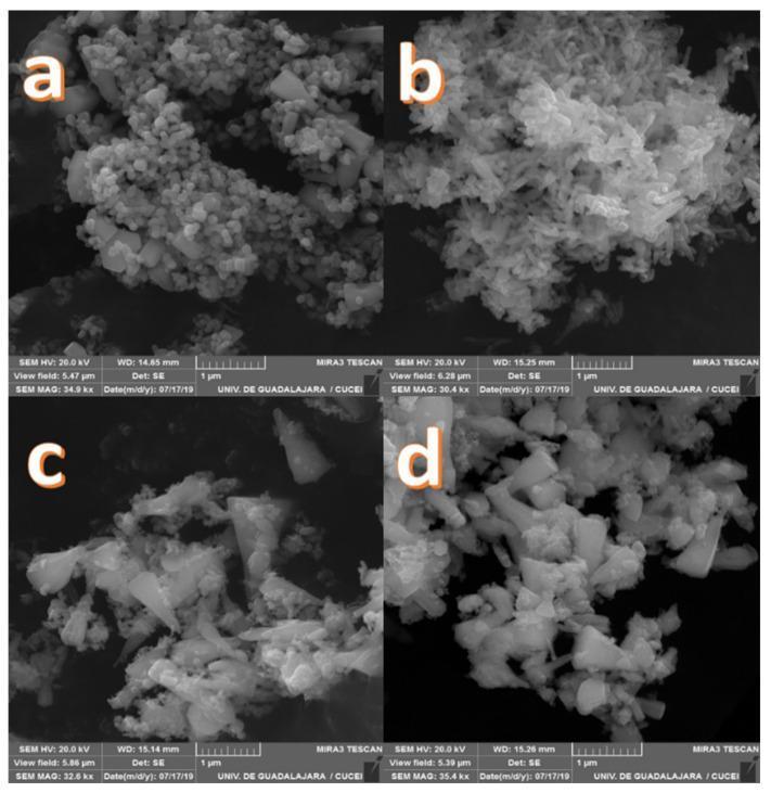
Scanning electron microscopy (SEM) images of Zinc-Calcium (Zn-Ca) nanocomposites, synthesized for biomedical applications such as antibacterial agents and dental restorative materials. The four-panel figure displays varying morphologies based on calcium dopant concentration: (a) pure ZnO, (b) Zn-Ca 1%, (c) Zn-Ca 3%, and (d) Zn-Ca 5%. The images demonstrate several characteristic nanoparticle structures, including spherical particles, conical shapes with hexagonal bases, and elongated rod-like morphologies. Magnification ranges from 30.4 kx to 35.4 kx, with scale bars of 1 μm. These nanocomposites are studied for their potential to inhibit both Gram-negative (e.g., E. coli) and Gram-positive (e.g., S. aureus) bacteria through the release of reactive oxygen species (ROS) and Zn2+ ions, which damage bacterial cell membranes and cytoplasmic contents. The visual data is essential for correlating physical particle morphology and surface area with the observed antibacterial efficacy and clinical utility in medical biomaterial engineering.
Escherichia coli (E. coli)
What Is It?
Basic Biology
| Feature | Detail |
|---|---|
| Shape | Rod (bacillus) |
| Gram stain | Gram-negative |
| Metabolism | Facultative anaerobe (can live with or without oxygen) |
| Oxidase test | Negative |
| Key structure | Lipopolysaccharide (LPS) cell wall with lipid A (endotoxin), O polysaccharide, and core polysaccharide |
Types of Pathogenic E. coli
1. Intestinal (Diarrheagenic) Strains
| Strain | Full Name | Key Features | Main Victims |
|---|---|---|---|
| STEC/EHEC | Shiga toxin-producing / Enterohemorrhagic | Bloody diarrhea, hemolytic uremic syndrome (HUS); O157:H7 is the classic strain | Children <5, elderly |
| ETEC | Enterotoxigenic | "Traveler's diarrhea"; watery, non-bloody diarrhea | Travelers to developing countries |
| EPEC | Enteropathogenic | Watery diarrhea; major cause of infant diarrhea | Infants <1 year |
| EIEC | Enteroinvasive | Watery or bloody diarrhea, fever; acts like Shigella | Children, travelers |
| EAEC | Enteroaggregative | Persistent (>2 weeks) watery diarrhea | Children, HIV patients |
2. Extraintestinal Strains (ExPEC)
- Urinary tract infections (UTIs) - E. coli is the #1 cause of UTIs (especially in women)
- Neonatal meningitis - a leading cause of meningitis in newborns
- Bacteremia / Sepsis - bloodstream infections
- Pneumonia - especially hospital-acquired
- Intra-abdominal infections - cholecystitis, peritonitis, cholangitis
How Is It Spread?
- Most infections are endogenous (from the patient's own gut flora moving to other body sites, like the bladder)
- Gastroenteritis strains are typically acquired exogenously via:
- Contaminated food (especially undercooked ground beef, leafy greens, raw milk)
- Contaminated water
- Contact with infected animals or their feces
- Person-to-person spread (fecal-oral route)
Diagnosis
- Grows rapidly on standard laboratory culture media
- Enteric multiplex nucleic acid amplification tests (NAATs) are considered the gold standard for diagnosing intestinal E. coli infections
- Urine culture for UTIs; blood cultures for bacteremia
Treatment
- Intestinal infections: Mostly supportive (fluids, rest). Antibiotics are generally avoided for STEC/O157:H7 as they may worsen HUS.
- Extraintestinal infections: Antibiotics guided by susceptibility testing. Key concern is rising antibiotic resistance, especially via extended-spectrum beta-lactamases (ESBLs) which break down penicillins and cephalosporins.
- Severe/resistant cases may require carbapenems or newer agents per IDSA 2024 guidelines.
Prevention
- Proper cooking of meat (especially ground beef)
- Good hand hygiene
- Safe water and food handling
- Avoiding unnecessary urinary catheters to prevent hospital-acquired UTIs
- Restricting antibiotic overuse to limit resistance
- Medical Microbiology 9e, block 3, lines 1867-1902
- NIH StatPearls overview
- CDC E. coli types