
Calcification.
Reading File
The topic "calcification" is broad - it covers pathological calcification (dystrophic vs. metastatic), vascular calcification, organ-specific calcification, and more. Let me search the medical library comprehensively.
Finding Sources
Finding Sources
Good. The most relevant sources are Robbins (pathologic calcification - core pathology), Harrison's (extraskeletal/ectopic), and the kidney textbook. Let me read the primary sources in parallel.
Reading File
Reading File
Reading File
Excellent content from Robbins and Harrison's. Let me get the rest of the Robbins metastatic calcification section, plus fetch the aortic valve image, and check for any additional pathogenesis details.
Reading File
Loading Image
Reading File
I have comprehensive content from Robbins and Harrison's. Let me also fetch the psammoma body image and get the pathogenesis mechanism from Robbins.
Loading Image
Finding Sources
The psammoma body image is just a placeholder graphic (concentric rings icon). I have all the core content needed. Now I'll compile the comprehensive answer.
Pathologic Calcification
Pathologic calcification is the abnormal tissue deposition of calcium salts (primarily calcium phosphate/hydroxyapatite), together with smaller amounts of iron, magnesium, and other mineral salts. There are two major forms - dystrophic and metastatic - plus a third special category of ectopic ossification.
- Robbins, Cotran & Kumar Pathologic Basis of Disease, p. 76
1. Dystrophic Calcification
Definition: Deposition of calcium in necrotic or dying tissue, occurring despite normal serum calcium levels and no systemic disturbance of calcium metabolism.
Sites
- Areas of coagulative, caseous, or liquefactive necrosis
- Enzymatic fat necrosis (e.g., pancreatitis)
- Atheromatous plaques of advanced atherosclerosis (almost invariably present)
- Aging or damaged heart valves (e.g., calcific aortic stenosis - see image below)
- Tuberculous lymph nodes (may be virtually converted to stone)
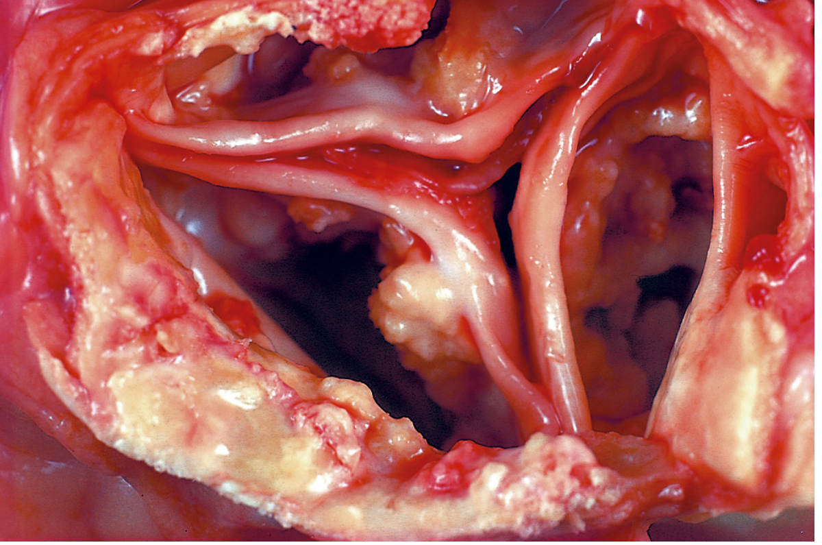
Morphology (Histology)
- On H&E stain: basophilic, amorphous granular (sometimes clumped) deposits
- Can be intracellular, extracellular, or both
- Psammoma bodies: Lamellated (concentric ring) configurations formed when single necrotic cells act as seed crystals that become progressively encrusted. Named for resemblance to grains of sand (psammos = sand). Seen in:
- Papillary thyroid carcinoma
- Meningioma
- Papillary serous ovarian carcinoma
- Asbestos bodies: In asbestosis, calcium and iron salts gather around long slender asbestos spicules in the lung, creating beaded dumbbell forms
- Heterotopic bone may eventually form at the focus of calcification
- Serum calcium is normal
Clinical significance
- May simply mark previous injury - or may cause significant organ dysfunction:
- Calcific valvular disease (aortic stenosis)
- Atherosclerotic plaque instability/stenosis
2. Metastatic Calcification
Definition: Deposition of calcium salts in otherwise normal tissues, almost always due to hypercalcemia from systemic calcium/phosphate metabolic disturbance.
Four Principal Causes of Hypercalcemia (Robbins)
- Increased PTH secretion - primary hyperparathyroidism (parathyroid tumors), or ectopic PTH-related protein (PTHrP) secretion by malignant tumors
- Bone resorption - multiple myeloma, diffuse skeletal metastases (e.g., breast cancer), Paget disease, immobilization
- Vitamin D-related disorders - vitamin D intoxication; sarcoidosis (macrophages activate vitamin D precursor); Williams syndrome (idiopathic hypercalcemia of infancy)
- Renal failure - phosphate retention leads to secondary hyperparathyroidism; also aluminum intoxication (chronic dialysis), milk-alkali syndrome (excessive calcium + absorbable antacids)
Sites Predominantly Affected
Tissues that excrete acid have an internal alkaline compartment that predisposes them:
- Gastric mucosa (parietal cells pump H⁺ → alkaline interstitium)
- Kidneys (nephrocalcinosis)
- Lungs (may cause respiratory compromise)
- Systemic arteries and pulmonary veins
Key rule from Harrison's: Calcifications in hypercalcemic states with normal or low phosphate preferentially affect kidney, lungs, and gastric mucosa. Hyperphosphatemia with normal/low calcium preferentially affects kidney and arteries.
Calcium-phosphate precipitation is especially likely when serum Ca × PO₄ concentration product >75.
Morphology
- Morphologically similar to dystrophic calcification - amorphous noncrystalline deposits or hydroxyapatite crystals
- Usually causes no clinical dysfunction, but massive pulmonary involvement can cause respiratory compromise; massive renal deposits (nephrocalcinosis) can cause renal damage
3. Mixed Calcification
A combination of metastatic and dystrophic mechanisms. Calcium dysregulation initiates the process (metastatic component), and trauma then propagates it (dystrophic component). Seen in disorders like dermatomyositis.
- Dermatology 2-Volume Set 5e
4. Ectopic Ossification / Tumoral Calcinosis
A third mechanism described in Harrison's: true bone formation in soft tissues (as opposed to calcium salt deposition). Examples:
- Myositis ossificans - heterotopic bone in muscle after trauma
- Fibrodysplasia ossificans progressiva (FOP) - rare genetic disorder
- Tumoral calcinosis - rare, caused by FGF23/GALNT3/α-Klotho gene mutations → reduced FGF23 activity → increased renal phosphate reabsorption → hyperphosphatemia → periarticular calcium phosphate masses (shoulders, hips, ankles). Labs show elevated serum phosphate and 1,25-(OH)₂ vitamin D; PTH and calcium are normal.
Summary Table
| Feature | Dystrophic | Metastatic |
|---|---|---|
| Site | Necrotic/injured tissue | Normal tissue |
| Serum calcium | Normal | Elevated (usually) |
| Serum phosphate | Normal | Often elevated |
| Mechanism | Local cell death → phosphatase release → nucleation | Systemic hypercalcemia/hyperphosphatemia |
| Examples | Atherosclerosis, calcific aortic stenosis, TB node, fat necrosis | Hyperparathyroidism, renal failure, sarcoidosis, vitamin D toxicity |
| Preferred sites | Anywhere necrosis occurs | Gastric mucosa, lungs, kidneys, arteries |
| Clinical significance | May cause valve dysfunction, atherosclerosis | Usually asymptomatic; nephrocalcinosis, pulmonary calcification if severe |
Sources:
- Robbins, Cotran & Kumar Pathologic Basis of Disease, pp. 76-77 (Pathologic Calcification)
- Harrison's Principles of Internal Medicine, 22E (2025), p. 3362 (Extraskeletal Calcification and Ossification)
- Dermatology 2-Volume Set 5e (Mixed Calcification)
This is a shared conversation. Sign in to Orris to start your own chat.