
provide a brief overview on the structural organization of male reproductive system with figures
Reading File
Finding Sources
Searching Images
male reproductive system anatomy diagram testis epididymis vas deferens prostate
Reading File
query_images_db
Structural Organization of the Male Reproductive System
The male reproductive system is organized into four functional components: the testes, the genital (excretory) ducts, the accessory glands, and the penis. Together these structures produce sperm, synthesize androgens, and deliver semen into the female reproductive tract.
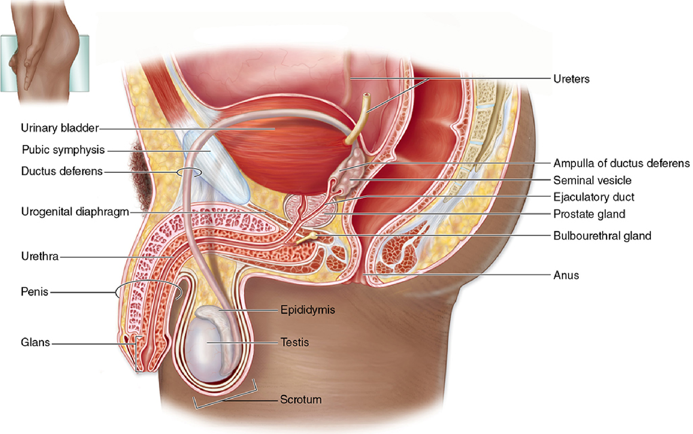
The diagram shows the relationships between the testes, epididymis, ductus deferens, and accessory glands as the duct system courses from the scrotum, along the posterior bladder, to the urethra - Junqueira's Basic Histology, Figure 21-1.
1. Testes
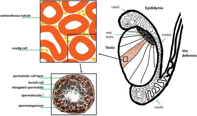
A pair of ovoid organs suspended in the scrotum, each testis is enclosed by a dense connective tissue capsule (tunica albuginea) that sends septa inward, dividing the gland into lobules.
- Seminiferous tubules - highly coiled tubules within each lobule where spermatogenesis occurs. Their wall (germinal/seminiferous epithelium) contains Sertoli cells (support, blood-testis barrier, nourishment of germ cells) and developing germ cells (spermatogonia -> spermatocytes -> spermatids -> spermatozoa).
- Interstitial (Leydig) cells - lie in the loose connective tissue between tubules and secrete testosterone.
- Tubules converge into short tubuli recti, which lead into an anastomosing network, the rete testis.
2. Intratesticular and Excretory Genital Ducts
This is the conducting system carrying sperm out of the testis:
- Rete testis -> efferent ductules (10-20 ducts leaving the testis) -> epididymis (a single highly coiled duct divided into head/caput, body/corpus, and tail/cauda; site of sperm maturation and storage) -> ductus (vas) deferens, a thick-walled muscular tube that ascends in the spermatic cord, passes through the inguinal canal, and enters the pelvis to join the ejaculatory duct near the prostate.
3. Accessory Glands
These add secretions that make up the bulk of semen and support sperm viability:
- Seminal vesicles - paired, highly folded glandular pouches posterior to the bladder; secrete a fructose-rich fluid that nourishes sperm and forms most of the ejaculate volume.
- Prostate gland - a single gland encircling the urethra just below the bladder, made of 30-50 tubuloalveolar glands arranged in three concentric zones (central, peripheral, transition), embedded in fibromuscular stroma; secretes a thin, slightly acidic fluid rich in citric acid and prostatic acid phosphatase.
- Bulbourethral (Cowper's) glands - small paired glands at the base of the penis that secrete a lubricating mucus into the urethra before ejaculation.
4. Penis
The copulatory organ, composed of three cylindrical erectile bodies: two dorsal corpora cavernosa and one ventral corpus spongiosum, which surrounds the penile (spongy) urethra and expands distally to form the glans penis. Erection results from engorgement of vascular spaces (sinusoids) within these erectile bodies under autonomic control.
Functional summary
- Testes: gametogenesis (sperm) + endocrine function (testosterone, which drives spermatogenesis, sexual differentiation, and secondary sexual characteristics; converted to dihydrotestosterone for action on accessory glands and hair follicles).
- Ducts: transport, concentration, and maturation of sperm.
- Accessory glands: seminal fluid production and nourishment.
- Penis: delivery of semen into the female tract.
Sources: - Junqueira's Basic Histology and Atlas, 17e, Ch. 21 ("The Male Reproductive System") - Histology: A Text and Atlas (Ross & Pawlina) - Overview of the Male Reproductive System
A quick check of recent systematic reviews/meta-analyses on this topic turned up nothing that would alter this standard anatomical description - it is stable, well-established anatomy rather than an evolving evidence area.
Let me know if you'd like a deeper focus on any one part (e.g., spermatogenesis histology, prostate zonal anatomy, or hormonal regulation via the hypothalamic-pituitary-gonadal axis).
This is a shared conversation. Sign in to Orris to start your own chat.