
Explain to be development of teeth
Reading File
Now let me search the medical library for tooth development content.
Finding Sources
Finding Sources
Excellent - found dedicated sections in "The Developing Human." Let me read the full tooth development content plus search for more detail.
Reading File
Finding Sources
Good content found. Let me read more of the bell stage and the sections on enamel/dentin formation plus get images.
Reading File
Finding Sources
Reading File
Reading File
Loading Image
I now have comprehensive content from "The Developing Human" embryology textbook. Let me compile a thorough answer.
Development of Teeth (Odontogenesis)
Tooth development - or odontogenesis - is a continuous process that begins in the sixth week of embryonic life and continues for years after birth. It involves reciprocal induction between neural crest-derived mesenchyme and the overlying oral epithelium. Development is traditionally divided into four stages based on the morphological appearance of the tooth germ.
1. Initiation - Dental Lamina Formation (Week 6)
The first sign of tooth development is a thickening of the oral epithelium in the sixth week. These thickened bands, called dental laminae, form U-shaped ridges that follow the curve of the primitive upper and lower jaws.
- Each dental lamina develops 10 centers of proliferation, giving rise to 10 tooth buds per jaw (20 total for the deciduous/primary dentition).
- Tooth development begins in the anterior mandibular (lower jaw) region first, then in the anterior maxillary region, and progresses posteriorly in both jaws.
2. Bud Stage (Week 6-8)
Each of the 10 proliferation centers on the dental lamina grows as a solid epithelial bud downward into the underlying mesenchyme.
- These buds will develop into the 20 deciduous (baby) teeth.
- At approximately 10 weeks, tooth buds for permanent teeth (those with deciduous predecessors) begin forming as deep continuations of the dental lamina, positioned lingual (tongue-side) to the deciduous buds.
- Permanent molars have no deciduous predecessors - they develop from posterior extensions of the dental laminae.
3. Cap Stage (Week 8-10)
As each tooth bud is invaginated by mesenchyme from below, it becomes cap-shaped. This stage establishes three key components of the tooth germ:
| Component | Origin | Future Structure |
|---|---|---|
| Enamel organ | Oral ectoderm (ectodermal budding from dental lamina) | Enamel |
| Dental papilla | Neural crest mesenchyme (inside the cap) | Dentin + dental pulp |
| Dental sac (follicle) | Condensed mesenchyme surrounding the whole germ | Cement + periodontal ligament |
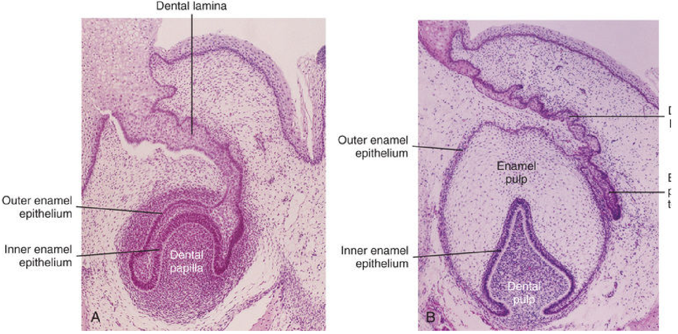
The enamel organ has two distinct cell layers:
- Outer enamel epithelium - the outer cell layer of the cap
- Inner enamel epithelium - the inner cell layer lining the dental papilla
Between them lies the stellate reticulum (enamel reticulum) - a loosely arranged central core of cells.
Here is a histological photomicrograph showing the early (A, 12-week) and late (B, 15-week) bell stage, with the dental lamina, outer and inner enamel epithelia, and dental papilla clearly visible:
4. Bell Stage (Week 10+)
As the enamel organ differentiates further, the tooth assumes a bell shape. This is the most critical stage - it is when the hard tissues (enamel and dentin) are actually formed.
Dentin Formation (Dentinogenesis)
- Mesenchymal cells of the dental papilla, directly adjacent to the inner enamel epithelium, differentiate into odontoblasts (dentin-forming cells).
- Odontoblasts produce predentin, which then calcifies to become dentin - the second hardest tissue in the body.
- As dentin thickens, odontoblasts migrate inward toward the center of the dental papilla, but leave behind long cytoplasmic projections called odontoblastic processes, which remain embedded in the dentin (in microscopic channels called dentinal tubules).
Enamel Formation (Amelogenesis)
- The inner enamel epithelium cells, under the inductive influence of odontoblasts, differentiate into ameloblasts (enamel-forming cells).
- Ameloblasts produce enamel in the form of prisms (rods) deposited over the dentin surface.
- As enamel thickens, ameloblasts migrate outward toward the outer enamel epithelium.
- Enamel is the hardest tissue in the body. Its translucent color is influenced by the underlying dentin.
- Both enamel and dentin formation begin at the cusp (tip) of the tooth and progress toward the future root.
5. Root Formation
Root development begins after dentin and enamel formation of the crown are well advanced:
- The inner and outer enamel epithelia meet at the neck of the tooth (cementoenamel junction) and fold downward, forming the epithelial root sheath (Hertwig's sheath).
- This sheath grows into the mesenchyme and initiates root dentin formation by inducing adjacent odontoblasts.
- As dentin increases, it narrows the dental pulp cavity to a thin root canal carrying vessels and nerves.
- Inner cells of the dental sac differentiate into cementoblasts, which deposit cement (cementum) over the root dentin.
- Outer cells of the dental sac participate in bone formation, surrounding the tooth in an alveolus (tooth socket).
- The tooth is anchored by the periodontal ligament - fibers of which embed into both the cement of the root and the bony wall of the alveolus.
6. Tooth Eruption
- As deciduous teeth develop, they begin a slow, continuous movement toward the oral cavity.
- The mandibular teeth erupt before maxillary teeth; females erupt teeth slightly earlier than males.
- As the permanent tooth grows beneath it, the root of the corresponding deciduous tooth is resorbed by osteoclasts (odontoclasts), so when the tooth is shed, only the crown and upper root remain.
Eruption Timeline
| Tooth | Deciduous (eruption / shedding) | Permanent (eruption) |
|---|---|---|
| Medial incisor | 6-8 months / 6-7 years | 7-8 years |
| Lateral incisor | 8-10 months / 7-8 years | 8-9 years |
| Canine | 16-20 months / 10-12 years | 10-12 years |
| First premolar | - | 10-11 years |
| Second premolar | - | 11-12 years |
| First molar | 12-16 months / 9-11 years | 6-7 years |
| Second molar | 20-24 months / 10-12 years | ~12 years |
| Third molar (wisdom) | - | 13-25 years |
Note: Delayed eruption of all teeth may indicate a systemic or nutritional disturbance such as hypopituitarism or hypothyroidism.
Summary of Tissue Origins
| Tooth Structure | Embryonic Origin |
|---|---|
| Enamel | Oral ectoderm (ameloblasts from inner enamel epithelium) |
| Dentin + Pulp | Neural crest mesenchyme (odontoblasts from dental papilla) |
| Cement | Neural crest mesenchyme (cementoblasts from dental sac) |
| Periodontal ligament | Neural crest mesenchyme (dental sac) |
| Alveolar bone | Neural crest mesenchyme (dental sac) |
Source: The Developing Human: Clinically Oriented Embryology, Chapter 19
This is a shared conversation. Sign in to Orris to start your own chat.