
How to make PRF for the implant placement
PRF platelet rich fibrin preparation protocol implant placement centrifugation
https://pmc.ncbi.nlm.nih.gov/articles/PMC7985567
PRF platelet rich fibrin preparation centrifugation dental implant
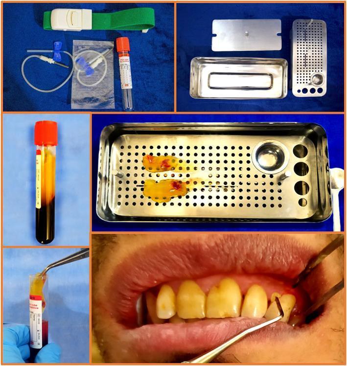
This composite of clinical photographs and diagnostic images details the workflow for Platelet-Rich Fibrin (PRF) preparation and its application in periodontology. The process begins with blood collection equipment, including a green tourniquet, butterfly needle with tubing, and a vacuum collection tube with an orange cap. Post-centrifugation, the blood tube demonstrates distinct layering: an upper clear yellow plasma layer, a middle PRF clot, and a bottom red blood cell mass. A technician is shown using sterile tweezers to extract the yellowish fibrin clot. The central panel illustrates processed PRF membranes resting on a perforated stainless steel PRF box used for compression. The final clinical photograph depicts the intraoral application, where a dental probe is used to place the PRF clot into a 4-6 mm periodontal pocket in a patient with periodontitis. The image demonstrates the transition from biological sample collection to a regenerative surgical adjunct for periodontal ligament and bone healing.
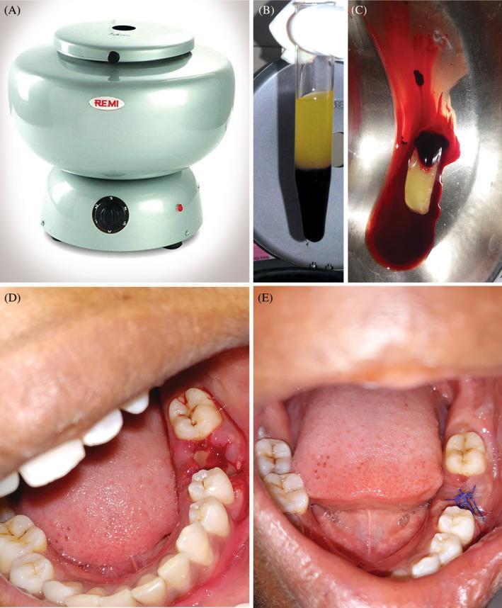
A multi-panel series illustrating the preparation and clinical application of Platelet-Rich Fibrin (PRF) in oral surgery. (A) Shows a clinical centrifuge used for blood processing. (B) Demonstrates a post-centrifugation glass test tube showing the separation of blood into three layers: a bottom layer of red blood cells, a middle 'buffy coat' PRF clot, and an upper layer of acellular plasma. (C) Displays the harvested PRF clot, appearing as a dense, yellowish-white fibrous gel. (D) An intraoral clinical photograph showing the PRF clot being placed into a fresh mandibular molar extraction socket to promote hemostasis and bone regeneration. (E) An intraoral follow-up photograph at the 7th postoperative day, showing the surgical site with blue monofilament sutures in place. The wound demonstrates primary intention healing with healthy pink gingival margins and reduced inflammation compared to the immediate postoperative state. This sequence illustrates the autologous regenerative process used to enhance soft and hard tissue healing in dental procedures.
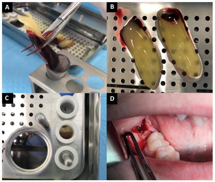
This composite intraoperative clinical photograph illustrates the preparation and application of Advanced Platelet-Rich Fibrin (A-PRF) in oral surgery. (A) Extraction of an A-PRF clot from a centrifugation tube using forceps, showing the distinctive separation between the translucent yellowish fibrin layer and the dark reddish-brown erythrocyte-rich base. (B) Two harvested A-PRF clots placed on a sterile metal tray; the clots exhibit a gelatinous, yellowish texture with a concentrated red corpuscle portion at one pole. (C) Preparation of A-PRF 'corks' within a dedicated PRF box, where the material is compressed into cylindrical shapes for easier socket insertion. (D) Intraoperative view of the A-PRF material being applied into a mandibular extraction socket. The reddish-brown fibrin material is seen packed into the surgical site adjacent to existing molars to promote wound healing and bone regeneration. The sequence demonstrates the autologous blood processing technique used to enhance postoperative recovery and reduce complications like dry socket or excessive pain in dental surgery.
PRF (Platelet-Rich Fibrin) Preparation for Implant Placement
Equipment Needed
- Butterfly needle or venipuncture needle
- 10 mL glass-coated vacuum tubes (no anticoagulant - plain/red top)
- Centrifuge (calibrated for RPM/RCF)
- Sterile forceps/tweezers
- PRF box (stainless steel tray with perforations for compression into membrane)
- Timer
Step-by-Step Protocol
Step 1: Blood Collection
- Draw 10 mL of venous blood per tube directly from the patient (antecubital vein) using a butterfly needle or vacutainer system
- Collect blood immediately before surgery - do NOT let the tubes sit; blood must be centrifuged within 60 seconds to prevent premature clotting
- Use glass-coated plastic tubes without anticoagulant - contact with glass activates coagulation naturally
Step 2: Centrifugation (Protocol varies by PRF type)
| PRF Type | Speed | Time | RCF | Best Use |
|---|---|---|---|---|
| L-PRF (Choukroun original) | 2,700 rpm | 12 min | ~708g | Standard membrane, good fibrin scaffold |
| A-PRF (Advanced) | 1,500 rpm | 14 min | ~230g | More platelets + leukocytes |
| A-PRF+ (Advanced Plus) | 1,300 rpm | 8 min | ~200g | Highest growth factor release |
| i-PRF (Injectable) | 700 rpm | 3 min | ~60g | Injectable form for mixing with bone graft |
The low-speed centrifugation concept (LSCC) is now preferred - lower speeds trap more platelets and leukocytes in the fibrin matrix and produce higher growth factor concentrations. Reducing from 2,400 rpm to 1,200 rpm and further to 600 rpm progressively increases platelet, leukocyte, and growth factor content.
Step 3: Identify the Layers
- Top - Acellular plasma (straw-colored, clear)
- Middle - PRF clot / buffy coat (yellowish fibrin gel - THIS IS WHAT YOU WANT)
- Bottom - Red blood cell mass (dark red)
Step 4: Harvest the PRF Clot
- Open the tube and use sterile forceps to grasp and lift out the yellow fibrin clot
- Separate it from the red blood cell base with scissors or by gentle tearing
Step 5: Processing the PRF
- PRF Membrane: Place the clot in the PRF box and press with the metal lid to compress it into a flat, dense membrane (takes ~1-2 min)
- PRF Plug/Corks: Use the cylindrical molds in the PRF box to form plugs for socket filling
- i-PRF (injectable): Aspirate the yellow liquid layer with a syringe immediately after centrifugation and mix with bone graft material before it solidifies
Application During Implant Placement
| Application | How |
|---|---|
| Socket/osteotomy site | Pack PRF plug directly into the implant osteotomy or extraction socket before/after implant insertion |
| Around implant neck | Wrap PRF membrane around the implant collar at crestal level to enhance soft tissue sealing |
| Bone graft mixing | Mix i-PRF with particulate bone graft to create a "sticky bone" that holds shape |
| Covering the flap | Place PRF membrane under the sutured flap to promote primary healing |
| Peri-implant defects | Fill bony defects around implant with PRF + graft material |
Visual Reference
Key Points to Remember
- No anticoagulants - PRF uses natural clotting activation by glass contact
- Timing is everything - centrifuge within 60 seconds of blood draw; delays reduce quality
- Lower speed = better quality (LSCC principle) - current evidence favors A-PRF/A-PRF+
- Horizontal rotors produce better results than swing-out rotors for PRF
- Growth factors release lasts up to 7-10 days, supporting the critical early healing phase after implant placement
- Benefits for implants include improved osseointegration, enhanced crestal bone preservation, reduced post-operative swelling/pain, and better soft tissue closure