
Acute onset SOB? Pulmonary edema with bilateral pleural effusion evaluation , diagnosis and management and managem
"acute heart failure" AND "pleural effusion"
2025 acute heart failure emergency management pulmonary edema oxygen NIV IV loop diuretic guideline
chest radiograph cardiogenic pulmonary edema bilateral pleural effusions
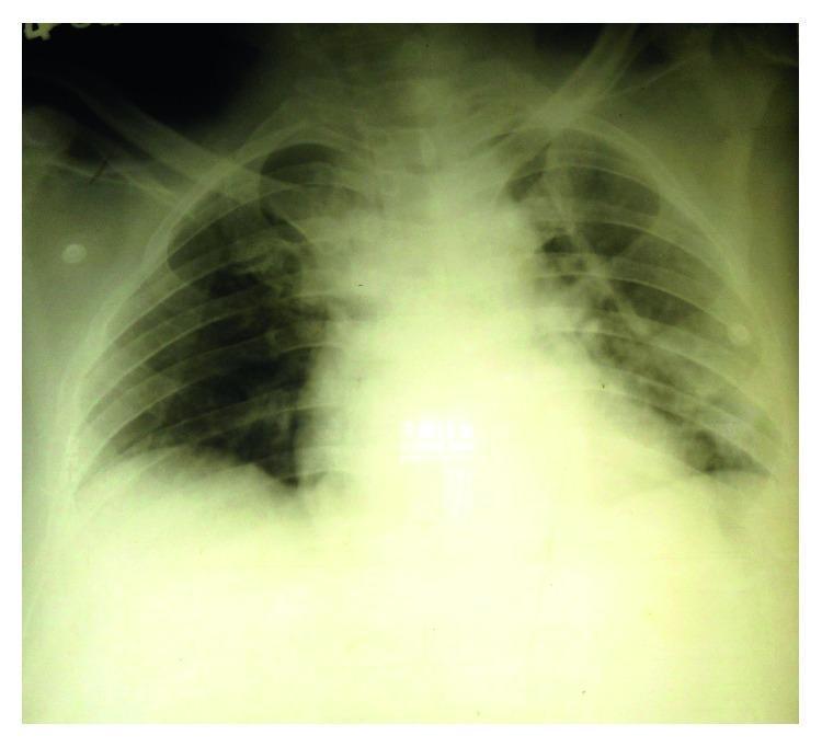
This diagnostic image is a posterior-anterior (PA) chest X-ray demonstrating significant pulmonary pathology. The radiograph shows bilateral, patchy alveolar opacities distributed throughout the upper, middle, and lower zones of both lungs. These opacities exhibit a predominantly peripheral distribution, which is a classic radiographic feature often associated with non-cardiogenic pulmonary edema or acute respiratory distress syndrome (ARDS). Additionally, there is evidence of bilateral blunting of the costophrenic angles, consistent with small bilateral pleural effusions. The cardiac silhouette appears within normal limits for size, further supporting a non-cardiac etiology for the observed pulmonary edema. This clinical presentation is often seen in severe systemic infections like leptospirosis (Weil's disease) or other inflammatory conditions leading to increased capillary permeability. Educational concepts include differentiating cardiogenic from non-cardiogenic edema based on heart size and opacity distribution.
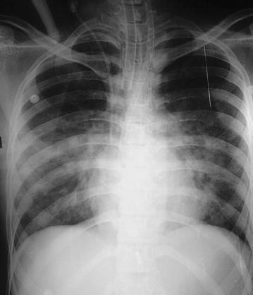
Anteroposterior (AP) chest radiograph demonstrating severe, bilateral, diffuse pulmonary edema. The image shows extensive, fluffy, alveolar opacities with a predominant perihilar (bat-wing) distribution, extending towards the peripheral lung fields and obscuring normal vascular markings. Cardiomegaly is evident, with an increased cardiothoracic ratio. Multiple medical support devices are visible: an endotracheal tube is positioned within the trachea, a central venous catheter is noted via the right jugular approach, and external monitoring electrodes are present on the chest wall. The costophrenic angles appear blunted, consistent with associated pleural effusions. In the context of the clinical history, these findings represent acute cardiogenic or non-cardiogenic pulmonary edema following tricyclic antidepressant (TCA) toxicity and subsequent cardiac arrest. The skeletal structures, including the ribs and clavicles, appear intact. This diagnostic image serves as an educational example of severe pulmonary congestion and the visual manifestations of heart failure or acute respiratory distress in a critical care setting.
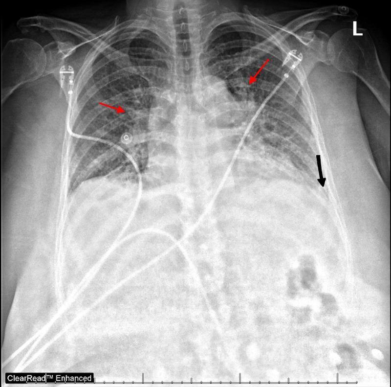
This diagnostic image is an anteroposterior (AP) chest radiograph illustrating acute pulmonary edema and associated pleural findings. The lung fields demonstrate diffuse, bilateral hazy opacities with a predominant perihilar (bat-wing) distribution (highlighted by red arrows), characteristic of alveolar and interstitial fluid accumulation. The pulmonary vasculature appears prominent, and the cardiac silhouette is mildly enlarged, suggesting cardiogenic origin. Small bilateral pleural effusions are evidenced by the blunting of the costophrenic angles (indicated by the black arrow on the left). Several medical support devices are visible, including external electrocardiogram (ECG) leads and a central venous catheter or similar indwelling line coursing through the superior vena cava toward the right atrium. The image serves as a clinical example of congestive heart failure manifestations in an acute setting. Key educational concepts include recognizing patterns of pulmonary vascular congestion, distinguishing between interstitial and alveolar edema, and identifying secondary signs such as pleural effusion and cardiomegaly on a portable x-ray.
https://www.nice.org.uk/guidance/cg187/chapter/1-recommendat…
Acute dyspnea with pulmonary edema and bilateral pleural effusions
Likely diagnosis
- Acute coronary syndrome or acute severe mitral regurgitation
- Hypertensive emergency
- Arrhythmia, especially atrial fibrillation with rapid ventricular response
- Renal failure or missed dialysis with fluid overload
- Iatrogenic fluid overload
- Pulmonary embolism
- Pneumonia/sepsis with ARDS or noncardiogenic pulmonary edema
- Hypoalbuminemia, cirrhosis, nephrotic syndrome
- Malignancy, tuberculosis, or parapneumonic effusion, particularly if effusions are unilateral, markedly asymmetric, febrile, or persistent
Immediate evaluation
1. Stabilize first: ABCDE
- Sit upright.
- Continuous ECG, SpO₂, BP, respiratory-rate, and urine-output monitoring.
- Two IV lines.
- Assess mental status, work of breathing, shock, and perfusion.
- Give oxygen only for hypoxemia and titrate to a safe target, generally SpO₂ 94%-98% unless at risk of hypercapnic respiratory failure.
- Obtain an ABG or VBG if severe distress, suspected hypercapnia/acidemia, or noninvasive ventilation is being considered.
2. Focused history and examination
- Timing: minutes to hours suggests flash edema, acute ischemia, arrhythmia, hypertensive crisis, acute valvular lesion.
- Orthopnea, paroxysmal nocturnal dyspnea, weight gain, reduced urine output, edema.
- Chest pain, palpitations, fever, cough/sputum, pleuritic pain, hemoptysis.
- Drug and fluid history: NSAIDs, missed diuretics, recent IV fluids, alcohol/cocaine, medication nonadherence.
- JVP elevation, peripheral edema, S3, crackles, cool extremities, murmurs.
- Fever, focal chest signs, unilateral leg swelling, ascites, signs of chronic liver or renal disease.
3. Essential investigations
| Test | Purpose |
|---|---|
| ECG and serial troponin | Acute coronary syndrome, ischemia, arrhythmia |
| Portable chest X-ray | Edema pattern, cardiomegaly, pleural effusions, pneumonia, pneumothorax |
| Lung and cardiac POCUS | B-lines, effusion size, LV/RV function, IVC, tamponade, gross valve pathology |
| BNP or NT-proBNP | Supports/rules out HF in the appropriate clinical context. BNP <100 ng/L or NT-proBNP <300 ng/L makes acute HF less likely per NICE guidance |
| CBC, electrolytes, urea/creatinine, glucose, LFTs, albumin | Renal function, anemia, infection, electrolyte status and alternative causes |
| ABG/VBG, lactate | Respiratory failure, acid-base status, hypoperfusion |
| Echocardiography | LV/RV function, acute valvular disease, regional wall-motion abnormalities, pericardial effusion |
| Blood cultures and inflammatory markers | If fever or possible sepsis |
| CT pulmonary angiography | Only if PE is clinically suspected and the patient is stable enough |
Confirming the cause of the pleural effusions
When bilateral effusions are likely due to heart failure
- Bilateral, often right-predominant
- Cardiomegaly, vascular congestion, septal lines, B-lines, elevated JVP, peripheral edema
- Elevated BNP/NT-proBNP
- Improvement after diuresis
Do diagnostic thoracentesis, preferably ultrasound-guided, if:
- The cause is uncertain
- Fever, pleuritic pain, high inflammatory markers, or concern for empyema
- Unilateral or substantially asymmetric effusion
- No cardiomegaly or other convincing evidence of heart failure
- Effusion persists or worsens despite adequate diuresis
- Suspicion of malignancy, tuberculosis, pulmonary embolism, or hemothorax
- Protein and LDH with paired serum samples: Light criteria
- Cell count and differential
- Gram stain and culture
- pH and glucose if infection suspected
- Cytology if malignancy possible
- Triglycerides if chylothorax considered
- ADA/AFB studies where TB is plausible
- Pleural NT-proBNP when the diagnosis remains uncertain. A pleural NT-proBNP >1500 pg/mL supports a heart-failure cause, though interpretation should remain clinical.
Acute management
A. Cardiogenic pulmonary edema with congestion and adequate blood pressure
- IV loop diuretic
- Give IV furosemide promptly.
- A common initial approach is 20-40 mg IV in a diuretic-naive patient, or an IV dose at least equivalent to the patient’s usual outpatient loop-diuretic dose in those already receiving one.
- Reassess urine output, symptoms, BP, creatinine, potassium, magnesium, and sodium. Increase dose or use infusion/sequential nephron blockade only under experienced supervision if response is inadequate.
-
Vasodilator only if hypertensive or normotensive
- For pulmonary edema with marked hypertension, ischemia, or acute mitral/aortic regurgitation, use titrated nitroglycerin according to local protocol.
- Avoid nitrates in hypotension, suspected right-ventricular infarction, severe aortic stenosis, recent PDE-5 inhibitor use, or possible preload-dependent states.
-
Ventilatory support
- CPAP or BiPAP is appropriate for severe dyspnea with hypoxemia or acidaemia, especially when the patient remains in distress despite initial treatment.
- Do not delay intubation if deteriorating consciousness, exhaustion, inability to protect the airway, refractory hypoxemia, or shock. NICE advises NIV in cardiogenic pulmonary edema with severe dyspnea and acidaemia, and invasive ventilation for respiratory failure or exhaustion despite therapy (NICE respiratory support advice).
-
Avoid routine morphine
- It can worsen ventilation and hypotension. It is not routine therapy for acute heart failure.
B. If hypotensive, cold, or in cardiogenic shock
- This is not routine diuretic-and-nitrate management.
- Urgent ICU/cardiology input, bedside echocardiography, lactate and perfusion assessment.
- Identify and treat a cause such as MI, mechanical complication, acute valvular catastrophe, tamponade, or arrhythmia.
- Vasopressor/inotrope and urgent revascularization or mechanical circulatory support may be required in a monitored critical-care setting.
- Avoid vasodilators in hypotension.
C. If renal failure or dialysis-dependent fluid overload
- Give diuretics only if residual renal function is expected to respond.
- Arrange urgent ultrafiltration/hemodialysis when pulmonary edema is refractory, severe, or associated with anuria/advanced renal failure. In severe CKD without useful residual function, dialysis is the fastest effective way to remove volume.
D. Pleural-fluid drainage
- Therapeutic thoracentesis is indicated when a large effusion is itself causing major dyspnea or impaired ventilation, or when diagnostic sampling is needed.
- It is not a substitute for treating the underlying heart failure.
- Use ultrasound guidance. Drain in a controlled manner and stop if chest pain, persistent cough, vasovagal symptoms, or concern for re-expansion pulmonary edema.
Treat the precipitating cause
- C: Acute coronary syndrome
- H: Hypertensive emergency
- A: Arrhythmia
- M: Mechanical cause, such as acute MR, VSD, tamponade
- P: Pulmonary embolism
- I: Infection
- T: Tamponade or treatment/nonadherence/toxins
- STEMI/NSTEMI: urgent ACS pathway and cardiology.
- AF with rapid rate: rate/rhythm strategy based on hemodynamic status.
- Hypertensive pulmonary edema: carefully titrated vasodilator plus diuresis.
- Pneumonia/empyema: antibiotics plus drainage when indicated.
- PE: anticoagulation or reperfusion strategy where indicated.
- Severe valve disease: urgent echocardiography and structural/cardiac-surgical review.
Monitoring and disposition
- Respiratory status and oxygen requirement
- BP, ECG, mental status
- Urine output and daily weight
- Renal function, potassium, magnesium, sodium
- Net fluid balance
- Repeated lung ultrasound or chest imaging when needed
Most probable cause of death in TB pericardial effusion with heart failure
"tuberculous pericarditis" AND mortality
tuberculous pericarditis main cause death cardiac tamponade heart failure mortality guideline
- Acute decompensated heart failure with pulmonary edema and hypoxic respiratory failure
- Effusive-constrictive or chronic constrictive pericarditis, leading to progressive right-sided heart failure
- Sepsis/disseminated tuberculosis, particularly in advanced or immunocompromised patients
- Arrhythmia or acute coronary syndrome, if present
Immediate cause: Cardiac tamponade
Due to: Tuberculous pericardial effusion / tuberculous pericarditis
Contributory condition: Congestive cardiac failure