
Cervicitis
cervicitis cervix inflammation clinical photo or histology
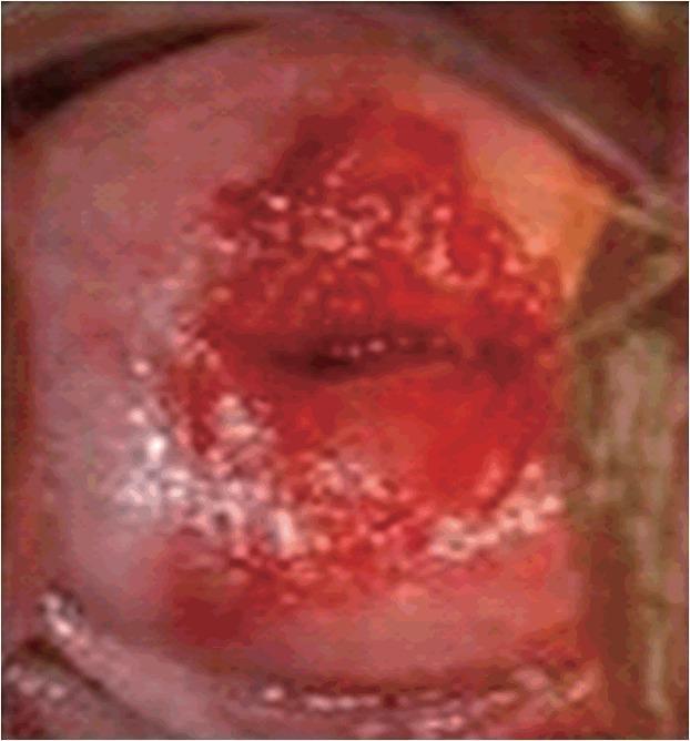
A clinical photograph of a cervix, specifically demonstrating signs of acute cervicitis. The image captures the cervical os and the surrounding ectocervix. The central pathological feature is a prominent, intensely erythematous and friable-looking area concentrated around the cervical opening, characteristic of cervical ectopy or inflammation. The texture in the central region appears raw, irregular, and possibly ulcerated, with deep red saturation. At the periphery of this erythematous lesion, there are multiple small, whitish-yellow spots or raised bumps, which may represent Nabothian cysts or follicular cervicitis findings. The surrounding healthy ectocervical tissue displays a normal pale pink hue and smoother texture. This visual serves as an educational tool for identifying inflammatory gynecological conditions during a pelvic examination, illustrating the distinction between normal squamous epithelium and inflamed columnar epithelium or eroded lesions.
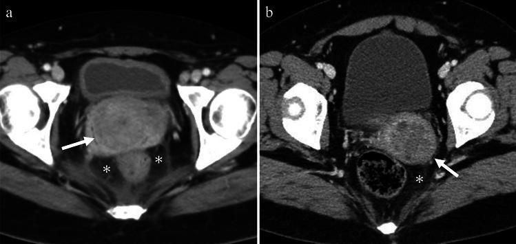
This figure presents two axial contrast-enhanced CT scans of the female pelvis demonstrating imaging features of cervicitis. Image A illustrates acute cervicitis in a patient with lower abdominal pain. Key findings include an enlarged and edematous uterine cervix (white arrow) exhibiting prominent enhancement. Associated inflammatory changes are visible in the surrounding tissue, specifically fat stranding within the parametrium and perirectal spaces (indicated by asterisks). Image B demonstrates cystic cervicitis. The uterine cervix contains multiple small, round cystic lesions (white arrow) characterized by heterogeneous attenuation, which can be seen in the context of hemorrhage or infected mucoid material. There is also a slight haziness of the adjacent pelvic fat (asterisk), reflecting localized inflammation. These images serve as educational examples of how pelvic inflammatory disease (PID) can manifest in the cervix on cross-sectional imaging, highlighting cervical edema, cystic transformation, and secondary infiltration of the surrounding pelvic fat planes.
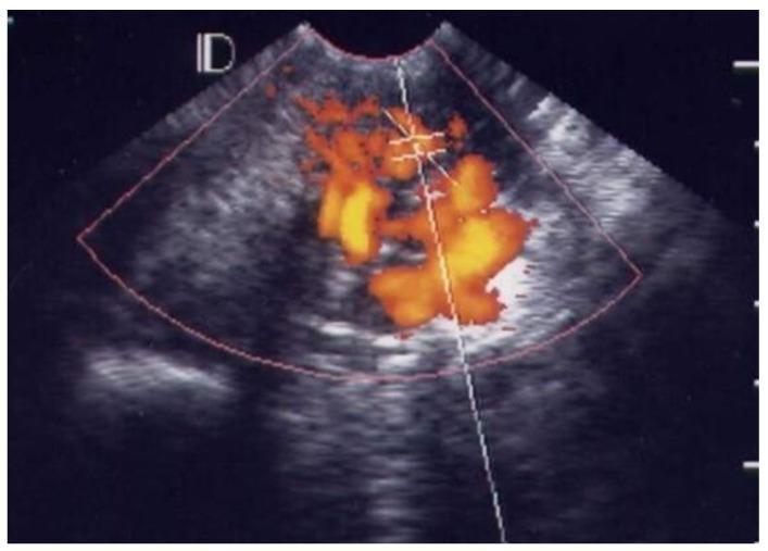
This diagnostic ultrasound image utilizes Color Doppler imaging to visualize the cervix and endocervical canal in a patient diagnosed with Pelvic Inflammatory Disease (PID). The visual demonstrates 'Grade 4' vascularity, which represents the highest clinical category of cervical hypervascularization. The image shows an intense and dense distribution of color Doppler signals (ranging from red to bright yellow) that are spread throughout the entire endocervix and within the endocervical canal. This reticular and confluent pattern of vascular spots signifies severe inflammation and associated angiogenesis within the cervical stroma. Anatomically, the surrounding hypoechoic region represents the cervical tissue, while the central linear echo marks the endocervical canal. This imaging finding is a critical sonographic marker for severe cervicitis, often correlating with decreased Pulsatility Index (PI) and Resistance Index (RI) values during spectral Doppler analysis.
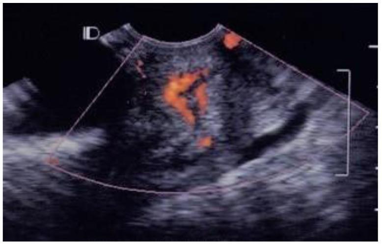
This diagnostic image is a transvaginal color Doppler ultrasound of the cervix in a 22-year-old patient with Pelvic Inflammatory Disease (PID). The image demonstrates Grade 3 cervical vascularity. The grayscale ultrasound shows a sagittal view of the cervix with heterogeneous echogenicity and a clearly visible elongated endocervical canal. The color Doppler overlay reveals multiple prominent vascular spots concentrated within the endocervical canal. These red signals indicate significant blood flow, appearing in a branching and clustered distribution rather than a diffuse pattern throughout the entire stroma. The increased vascularity within the canal is a characteristic finding of acute cervicitis and cervical hyperemia associated with inflammatory conditions. This visualization is used to assess the severity of inflammation and differentiates between simple and complex cervicitis based on the extent of angiogenesis and vessel distribution patterns.
cervicitis diagnosis treatment
Cervicitis
Definition
Anatomy of Relevance
- Ectocervix - stratified squamous epithelium (continuous with vaginal epithelium)
- Endocervix - columnar glandular epithelium
- N. gonorrhoeae and C. trachomatis - infect glandular (columnar) epithelium only
- Trichomonas vaginalis, Candida, HSV - infect squamous ectocervical epithelium
Etiology / Causative Organisms
| Organism | Notes |
|---|---|
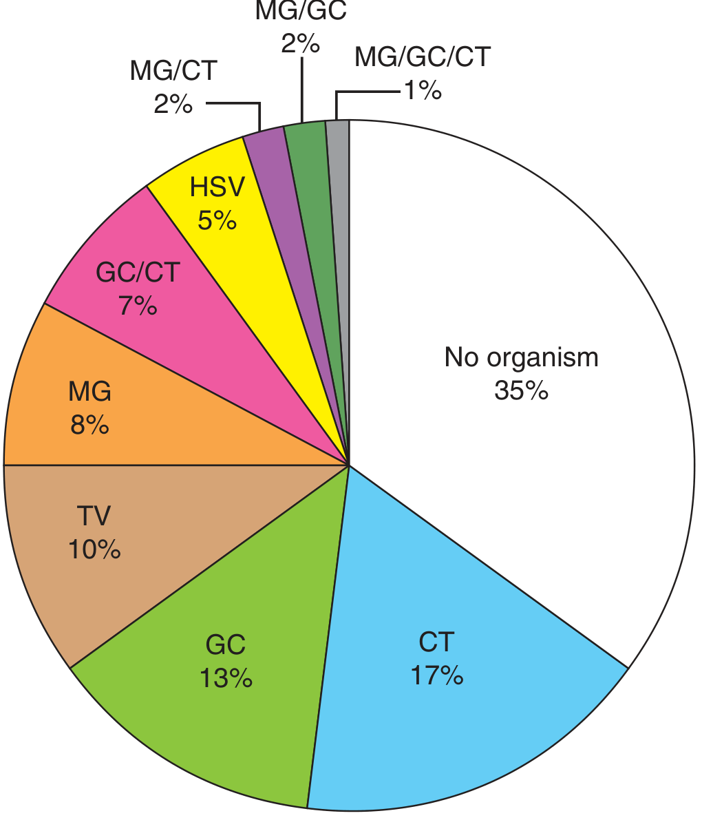
| Chlamydia trachomatis | Most common STI cause; up to 40-50% of cervicitis cases; majority asymptomatic |
| Neisseria gonorrhoeae | Infects columnar epithelium; concurrent treatment always considered |
| Mycoplasma genitalium | Detected in 10-30% of clinical cervicitis; emerging STI |
| HSV-2 | Produces ulcerative lesions on ectocervix AND endocervix; unique among STI causes |
| Trichomonas vaginalis | Ectocervical involvement |
| Ureaplasma urealyticum | Associated but less clearly causative |
| Bacterial vaginosis flora | BV organisms (anaerobes, G. vaginalis) can elicit cervical inflammation |
| No organism identified | ~50% of MPC cases have no identifiable microbiologic etiology even with NAATs |
Clinical Features
- Abnormal vaginal discharge (mucopurulent, yellow/green)
- Intermenstrual vaginal bleeding
- Postcoital bleeding
- Dysuria
- Dyspareunia (pain during intercourse)
- Many cases (especially chlamydial) are entirely asymptomatic
- Yellow mucopurulent discharge from the cervical os (visualized on cotton swab against white/black background) - the hallmark sign
- Endocervical bleeding easily induced by gentle swabbing
- Edematous cervical ectopy - darker red columnar epithelium visible on the ectocervix
- Cervical tenderness on bimanual examination
Diagnosis
- NAAT (Nucleic Acid Amplification Test) - gold standard for N. gonorrhoeae and C. trachomatis; always indicated in MPC evaluation
- Gram stain of endocervical mucus:
- ≥20 PMNs per 1000x field within cervical mucus strands = endocervicitis
- Intracellular gram-negative diplococci = presumptive gonorrhea (specific but only ≤50% sensitive)
- Negative for diplococci = presumptive chlamydial cervicitis
- Wet prep / vaginal pH - evaluate for concurrent BV and Trichomonas
- NAAT for M. genitalium in cases with persistent symptoms after standard therapy
- Consider HSV PCR if ulcerative lesions are seen
Treatment
Empiric Treatment Approach
- Age <25 years
- New or multiple sexual partners
- Unprotected sex
- Poor follow-up expected
Regimens (per Goldman-Cecil and Harrison's 22e):
- Doxycycline 100 mg orally twice daily x 7 days (preferred)
- OR Azithromycin 1 g orally single dose
- Ceftriaxone 500 mg IM single dose (note: current dosing per CDC 2021 guidelines - increased from 250 mg)
- Fluoroquinolones are NO longer recommended due to widespread resistance
- Cefixime no longer a first-line regimen
- Azithromycin (first-line) - but resistance is rising
- Moxifloxacin 400 mg once daily x 7 days (if azithromycin resistance suspected or persistent infection)
- Doxycycline alone frequently fails
Complications
| Complication | Mechanism |
|---|---|
| Pelvic Inflammatory Disease (PID) | Ascent of organisms from endocervix → endometritis → salpingitis → peritonitis |
| Tubo-ovarian abscess | Severe PID extension |
| Ectopic pregnancy | Tubal scarring from salpingitis |
| Infertility | Tubal damage |
| Obstetric complications | Preterm labor, low birthweight, miscarriage in pregnancy |
| Neonatal chlamydia | Neonatal conjunctivitis (~20-40%), neonatal pneumonia (~10-20%) |
| Increased HIV susceptibility | Cervical ectopy and inflammation facilitate HIV entry |
Cervical Ectopy vs. Cervicitis
- In ectopy: cervical os contains clear or slightly cloudy mucus (not yellow mucopus)
- Colposcopy shows intact epithelium
- Ectopy is physiological, common in adolescence/early adulthood and OCP users
- Cauterization of ectopy is not warranted
- Ectopy may increase susceptibility to gonorrhea, chlamydia, and HIV
Key Pearls
- ~50% of MPC cases have no identifiable organism even with NAATs
- C. trachomatis is the most common identified cause (~40-50% of STI clinic cases)
- HSV is unique in causing ulcerative ectocervical lesions unlike other STI agents that cause mucopurulent endocervicitis
- MPC is a risk factor for ascending infection (PID) and obstetric complications in pregnancy
- BV is commonly co-present and must be treated to prevent symptom persistence
- Fluoroquinolones are no longer appropriate for gonococcal cervicitis