
Vitamin d metabolism in medicine
"vitamin D" AND metabolism
vitamin D metabolism synthesis calcitriol pathway diagram
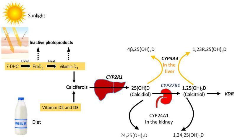
This pathophysiology diagram illustrates the metabolic pathway of Vitamin D homeostasis and its associated genetic enzymes. The process begins with two sources: endogenous synthesis in the skin via Sunlight (UV-B) converting 7-DHC to PreD3 and then to Vitamin D3 (Cholecalciferol) through heat, and dietary intake (Vitamin D2 and D3). These converge into 'Calciferols'. The primary activation pathway shows the liver enzyme CYP2R1 hydroxylating calciferols into 25(OH)D (Calcidiol). This metabolite then moves to the kidney, where CYP27B1 converts it to the active form, 1,25(OH)2D (Calcitriol), which binds to the Vitamin D Receptor (VDR). The diagram also highlights catabolic inactivation pathways: in the kidney, CYP24A1 converts 25(OH)D and 1,25(OH)2D into inactive products (24,25(OH)2D and 1,24,25(OH)3D respectively). In the liver, CYP3A4 facilitates an alternative inactivation route, producing 4β,25(OH)2D and 1,23R,25(OH)3D. Key genes involved in Vitamin D-Dependent Rickets (VDDR) are highlighted in bold, emphasizing the clinical significance of these enzymatic steps in mineral metabolism.
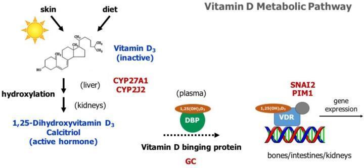
A pathophysiology diagram illustrating the human Vitamin D metabolic pathway from synthesis to gene expression. The flow begins with inactive Vitamin D3 sourced from the skin (via UV photoconversion) and diet. It undergoes two hydroxylation steps: first in the liver, associated with enzymes CYP27A1 and CYP2J2, and then in the kidneys, resulting in the active hormone 1,25-Dihydroxyvitamin D3 (Calcitriol). In the plasma, Calcitriol is shown bound to Vitamin D binding protein (DBP, encoded by the GC gene). The final stage depicts the Vitamin D Receptor (VDR) complex binding to DNA in target tissues such as bones, intestines, and kidneys. This molecular interaction triggers gene expression, specifically highlighting SNAI2 and PIM1. The diagram serves as an educational tool for understanding endocrinology, calcium homeostasis, and the genomic effects of steroid hormones.
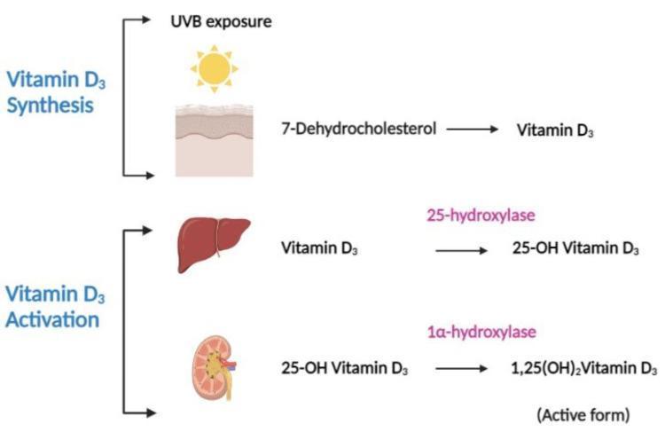
A pathophysiology diagram illustrating the multi-organ process of Vitamin D3 synthesis and activation. The diagram is divided into two primary stages: Synthesis and Activation. In the Synthesis stage, ultraviolet B (UVB) exposure acts on the skin, facilitating the conversion of the precursor 7-dehydrocholesterol into Vitamin D3 (cholecalciferol). The Activation stage depicts a sequential two-step hydroxylation process. First, within the liver, the enzyme 25-hydroxylase converts Vitamin D3 into 25-hydroxyvitamin D3 (calcifediol). Second, within the kidney, the enzyme 1̠-hydroxylase converts 25-OH Vitamin D3 into 1,25-dihydroxyvitamin D3 (calcitriol), which is labeled as the physiologically active form. The visual utilizes anatomical icons for the skin, liver, and kidney alongside biochemical flowcharts to demonstrate the endocrine pathway of Vitamin D metabolism, making it a key educational resource for understanding calcium homeostasis and metabolic bone disease foundations.
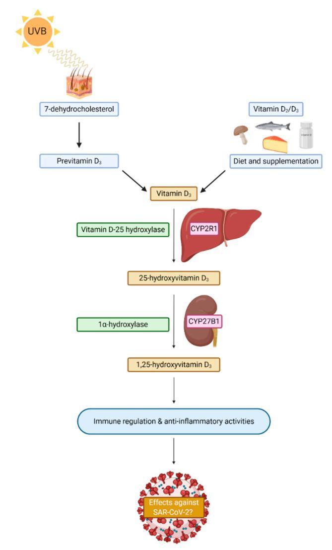
This pathophysiology diagram illustrates the metabolic pathway of Vitamin D synthesis and activation in humans, emphasizing its role in immune modulation. The flow begins with two primary sources: endogenous synthesis in the skin (UVB radiation converting 7-dehydrocholesterol to Previtamin D3 and subsequently Vitamin D3) and exogenous intake (dietary sources like fish, eggs, and mushrooms or supplementation). The pathway then details the two-step hydroxylation process: first in the liver, where Vitamin D-25 hydroxylase (CYP2R1) converts Vitamin D3 into 25-hydroxyvitamin D3; and second in the kidney, where 1α-hydroxylase (CYP27B1) converts it into the bioactive form, 1,25-dihydroxyvitamin D3. The diagram concludes by highlighting the clinical significance of active Vitamin D in promoting immune regulation and anti-inflammatory activities, specifically questioning its potential protective effects against SARS-CoV-2. This schematic serves as an educational tool for understanding endocrine metabolism and its systemic physiological impacts.
Vitamin D Metabolism in Medicine
1. Sources of Vitamin D
- Endogenous synthesis: UV-B radiation (290-315 nm) acts on 7-dehydrocholesterol in the skin, converting it to previtamin D3, which is then thermally isomerized to cholecalciferol (vitamin D3). This process is reduced by dark skin (high melanin), sunscreen (SPF ≥8), aging, and winter latitude.
- Dietary intake: Vitamin D2 (ergocalciferol) from plant sources (fungi, fortified foods) and vitamin D3 (cholecalciferol) from animal sources (fish liver oil, egg yolk, dairy).
2. The Metabolic Activation Pathway (Sequential Hydroxylation)
Step 1 - 25-Hydroxylation in the Liver
- This step is essentially unregulated - calcidiol levels reflect vitamin D status accurately
- Calcidiol is the major circulating and storage form (t½ ~ 2-3 weeks)
- DBP has greater affinity for 25(OH)D than for calcitriol
Step 2 - 1α-Hydroxylation in the Kidney (Activation)
- The plasma half-life of calcitriol is 3-5 days
- CYP27B1 is also expressed extrarenally (macrophages, monocytes, skin, breast, prostate, colon, placenta), producing calcitriol for local/paracrine action
- This second hydroxylation is the tightly regulated, rate-limiting step
Step 3 - 24-Hydroxylation (Catabolism/Inactivation)
- 25(OH)D → 24,25(OH)₂D (likely inactive)
- 1,25(OH)₂D → 1,24,25(OH)₃D → further oxidized and excreted as calcitroic acid
3. Regulation of 1α-Hydroxylase (CYP27B1)
| Stimulus | Effect on CYP27B1 | Mechanism |
|---|---|---|
| Low serum Ca²⁺ | ↑ (stimulates) | Via PTH |
| Low serum phosphate | ↑ (stimulates) | Direct + via reduced FGF23 |
| PTH | ↑ (stimulates) | cAMP-mediated gene transcription |
| FGF23 (bone-derived) | ↓ (inhibits) | Direct suppression + CYP24A1 induction |
| Calcitriol itself | ↓ (negative feedback) | Suppresses CYP27B1, induces CYP24A1, suppresses PTH |
| Hypercalcemia | ↓ (inhibits) | Suppresses PTH; 24-hydroxylation shunts |
| Prolactin / estrogen / GH | ↑ (stimulates) | Relevant during pregnancy/lactation |
4. Transport and Cellular Uptake
- Calcitriol circulates bound to DBP (main carrier) and albumin
- DBP-null mice remain normocalcemic, suggesting DBP primarily maintains stable serum stores rather than being essential for tissue delivery
- At the cell surface, both 25(OH)D and calcitriol are internalized by megalin/cubilin-mediated endocytosis in renal tubular cells
5. Mechanism of Action
Genomic (Nuclear) Actions (slow, hours)
- Calcitriol binds cytosolic VDR (Vitamin D Receptor) - a member of the nuclear receptor superfamily
- The VDR-calcitriol complex translocates to the nucleus
- It heterodimerizes with RXR (retinoid X receptor)
- The complex binds VDREs (Vitamin D Response Elements) in target gene promoters
- Coactivators/corepressors are recruited, modulating transcription
Rapid Non-Genomic Actions (seconds to minutes)
- Rapid mobilization of calcium transporters in intestinal mucosa
- Signal transduction via membrane-associated VDR (alternative binding site, planar configuration)
- Activates PKC, MAPK, phospholipase C pathways
6. Physiological Functions of Calcitriol
Calcium and Phosphate Homeostasis (Primary Role)
- Without calcitriol: Ca²⁺ absorption is inefficient, via passive paracellular diffusion
- Calcitriol induces TRPV6 and Ca1.3 channels (luminal entry), calbindin-D9K/D28K (intracellular diffusion), and PMCA (Ca²⁺-ATPase) (basolateral extrusion)
- Also enhances phosphate absorption via NaPi transporters
- Works synergistically with PTH to mobilize Ca²⁺ and phosphate from bone
- In physiological doses: supports osteoblast differentiation and bone mineralization
- In pharmacological excess: can stimulate bone resorption
- Stimulates Ca²⁺ and phosphate reabsorption in distal tubules
- Decreases urinary calcium losses
Extra-Skeletal / Pleiotropic Actions
| System | Effect |
|---|---|
| Immune | Inhibits IL-2 production by T-lymphocytes; inhibits immunoglobulin by B-lymphocytes; promotes monocyte differentiation |
| Endocrine | Stimulates insulin secretion; modulates PTH and thyroid hormone synthesis |
| Cell growth | Inhibits proliferation; promotes differentiation; anti-tumor effects in prostate, colorectal, breast tissue |
| Cardiovascular | Renin suppression; cardiomyocyte function |
| Neuromuscular | Muscle fiber function; large proximal muscle weakness in deficiency |
7. Clinical Correlates of Disordered Vitamin D Metabolism
Vitamin D Deficiency
- Low 25(OH)D → ↓ calcitriol → ↓ intestinal Ca²⁺ absorption → hypocalcemia → ↑ PTH secretion → PTH restores Ca²⁺ at the expense of bone, causes phosphaturia → hypophosphatemia → ↑ FGF23
- Children: Rickets - failure to mineralize growth plates; bowing of limbs, rachitic rosary, craniotabes, widened metaphyses
- Adults: Osteomalacia - undermineralized bone matrix; bone pain, proximal myopathy, stress fractures. Serum 25(OH)D <8 ng/mL highly predictive.
- Lab finding: ↑ ALP, ↑ PTH, low/normal calcium, low phosphate
Vitamin D Toxicity (Hypervitaminosis D)
- Daily doses ≥50,000 IU sustained intake can cause toxicity
- Mechanism: excess 25(OH)D saturates DBP, allowing free calcitriol to act unregulated
- Presents as hypercalcemia: nausea, polyuria, nephrocalcinosis, calcinosis of soft tissues, hypertension
- Infants may be sensitive at doses as low as 50 µg/day (2000 IU)
Metabolic Rickets and Osteomalacia (Genetic Disorders)
| Disorder | Defect | Key Feature |
|---|---|---|
| VDDR type 1 (PDDR) | CYP27B1 mutation (1α-hydroxylase deficiency) | No calcitriol produced; AR; responds to calcitriol supplementation |
| VDDR type 2 (HVDDR) | VDR mutation | End-organ resistance; hypocalcemia + total alopecia; does not respond to calcitriol |
| X-linked hypophosphatemic rickets (XLH) | PHEX gene loss → excess FGF23 → phosphaturia | Most common hereditary rickets; treated with phosphate + calcitriol or burosumab (anti-FGF23 antibody) |
| CKD-MBD (renal rickets) | ↓ CYP27B1 activity + ↑ FGF23 | Secondary hyperparathyroidism, adynamic bone disease |
Vitamin D in Chronic Kidney Disease (CKD)
- Reduced 25(OH)D substrate delivery to proximal tubule
- Loss of CYP27B1-expressing tubular mass
- FGF23 rise (early in CKD): directly inhibits CYP27B1 and upregulates CYP24A1
- Phosphate retention directly suppresses CYP27B1
8. Assessment of Vitamin D Status
| Metabolite | Significance |
|---|---|
| 25(OH)D (calcidiol) | Best marker of overall vitamin D status; long half-life reflects intake + synthesis |
| 1,25(OH)₂D (calcitriol) | Active hormone; NOT a reliable marker of vitamin D stores (tightly regulated by PTH/FGF23; can be normal even in frank deficiency) |
- Deficiency: <20 ng/mL (50 nmol/L)
- Insufficiency: 20-29 ng/mL
- Sufficiency: ≥30 ng/mL
- Toxicity concern: >150 ng/mL
9. Pharmacological Vitamin D Preparations
| Preparation | Form | Key Use |
|---|---|---|
| Cholecalciferol (D3) | Native | Deficiency prevention and treatment |
| Ergocalciferol (D2) | Native | Deficiency (plant-based); equivalent to D3 once hydroxylated |
| Calcidiol (25-OHD) | Partially activated | Liver disease (bypasses first hydroxylation) |
| Calcitriol (1,25(OH)₂D) | Fully activated | CKD, VDDR-1, hypoparathyroidism; requires careful Ca²⁺ monitoring |
| Alfacalcidol (1α-OHD3) | Prodrug (requires 25-hydroxylation only) | CKD; converted in liver |
| Paricalcitol, Doxercalciferol | Synthetic analogs | CKD-MBD; selective VDR agonists with less hypercalcemia |
- Goodman & Gilman's Pharmacological Basis of Therapeutics (Chapter 52)
- Brenner and Rector's The Kidney (Chapter 53)
- Harper's Illustrated Biochemistry, 32nd Ed. (Chapter 44)
- Comprehensive Clinical Nephrology, 7th Ed. (Chapter 88)