
Which artery passes through the two roots of auriculotemporal nerve
Reading File
Finding Sources
The middle meningeal artery passes through (between) the two roots of the auriculotemporal nerve.
The auriculotemporal nerve arises from the posterior division of the mandibular nerve (V3) as two roots, which encircle the middle meningeal artery just below the foramen spinosum before reuniting into a single trunk. This close anatomical relationship is why the artery is described as passing "between" or "through" the two roots at their point of origin.
- Gray's Anatomy for Students, p. 2313/2447
- Scott-Brown's Otorhinolaryngology Head & Neck Surgery, p. 1830
Histology of parotid with diagram
Finding Sources
Searching Images
histology of parotid gland serous acini diagram
Finding Sources
query_images_db
Finding Sources
query_images_db
Finding Sources
Histology of the Parotid Gland
General Architecture
The parotid is the largest major salivary gland, enclosed in a dense connective tissue capsule that sends septa inward, dividing the gland into lobules. It is a classic compound tubuloacinar (branched acinar) gland.
Key Histological Features
1. Acini - purely serous
The parotid is composed entirely of serous acini (unlike the submandibular and sublingual glands, which are mixed). Each acinus is a rounded cluster of pyramidal serous cells with:
- Basally located, round nuclei
- Basophilic cytoplasm (due to abundant rough endoplasmic reticulum) at the base
- Apical zymogen (secretory) granules containing alpha-amylase and proline-rich proteins
2. Myoepithelial cells
Star-shaped contractile cells sit between the acinar cell base and the basal lamina, helping expel secretions into the duct system.
3. Duct system (in order of drainage)
- Intercalated ducts - short, lined by low cuboidal epithelium; drain acini
- Striated ducts - lined by columnar cells with basal striations (infoldings of the basolateral membrane packed with mitochondria) that modify saliva's ionic composition
- Interlobular (excretory) ducts - within connective tissue septa, lined by stratified columnar/cuboidal epithelium, eventually becoming pseudostratified/stratified as they approach the main parotid (Stensen's) duct
4. Stroma
A characteristic feature of the parotid is abundant adipose tissue interspersed between lobules, which increases with age and is a normal finding, not pathology.
Diagram
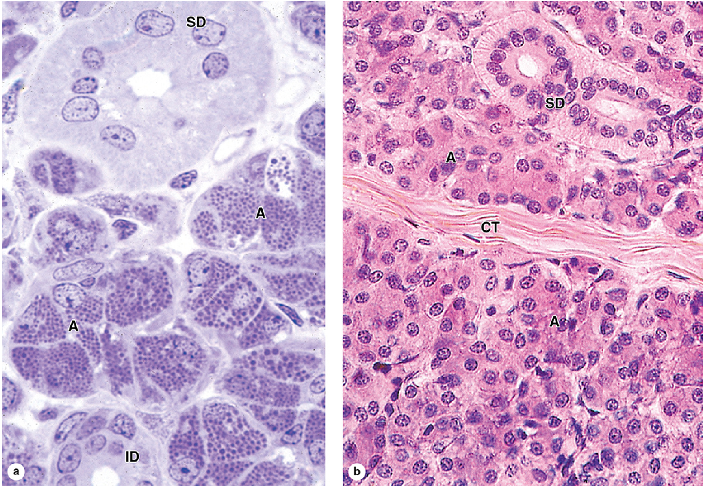
Figure: Parotid gland showing densely packed serous acini with labeled duct structures.
- Junqueira's Basic Histology and Atlas, 17e, Fig. 16-3
Normal H&E Section
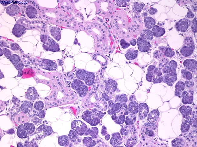
Normal parotid parenchyma: tubuloacinar architecture with serous acini (pyramidal cells, basophilic cytoplasm, basal nuclei), small ductal profiles, and interlobular adipose tissue - a typical age-related feature.
- Source: webpathology
Summary Table
| Feature | Parotid Gland |
|---|---|
| Acinar type | Purely serous |
| Secretion | Watery, amylase-rich |
| Myoepithelial cells | Present, surround acini |
| Intercalated ducts | Long and well-developed |
| Striated ducts | Prominent, ion-modifying |
| Stroma | Abundant adipose tissue (increases with age) |
Sources:
- Junqueira's Basic Histology and Atlas, 17e - Salivary Glands, p. 906-925, 1399
- Histology: A Text and Atlas (Gartner & Hiatt) - Major Salivary Glands, p. 4327-4353
- Cummings Otolaryngology Head and Neck Surgery - Anatomy and Physiology, p. 1771
This is a shared conversation. Sign in to Orris to start your own chat.