
Highlight the contrast in parameters of pulmonary function test between obstructive and restrictive conditions answer this according to 10 marker question in cvts for physiotherapy students
Reading File
Now let me search the medical library for PFT content on obstructive and restrictive conditions.
Finding Sources
Finding Sources
Excellent results. Let me now read the most relevant sections in parallel.
Reading File
Reading File
Reading File
Excellent! I have very rich content. Let me also fetch the PFT diagram and get the Murray & Nadel restrictive section for completeness.
Loading Image
Finding Sources
Reading File
I now have all the content needed for a comprehensive 10-marker answer. Let me also quickly get the restrictive ventilatory defect section and the DLCO data.
Reading File
Finding Sources
I now have all the content I need for a thorough 10-marker answer. Let me compile it.
Pulmonary Function Tests: Obstructive vs. Restrictive Conditions
(CVTS Physiotherapy - 10 Marker Answer)
Introduction (1 mark)
Pulmonary Function Tests (PFTs) are a battery of non-invasive tests used to assess lung mechanics, volumes, capacities, and gas exchange. They are the cornerstone of evaluating cardiopulmonary disorders in physiotherapy practice. PFTs allow differentiation of two fundamental patterns of ventilatory dysfunction: obstructive and restrictive defects - each with distinct pathophysiology and characteristic parameter changes.
Definition of Patterns
Obstructive defect: Characterized by small airway obstruction to expiratory flow, resulting in difficulty emptying the lungs. Seen in conditions such as COPD, asthma, bronchiectasis, and chronic bronchitis.
Restrictive defect: Characterized by a proportional decrease in all lung volumes due to reduced lung compliance or chest wall expansion. Seen in interstitial lung disease (IPF, sarcoidosis), neuromuscular diseases, kyphoscoliosis, and obesity.
Spirometric Parameters Compared (4 marks)
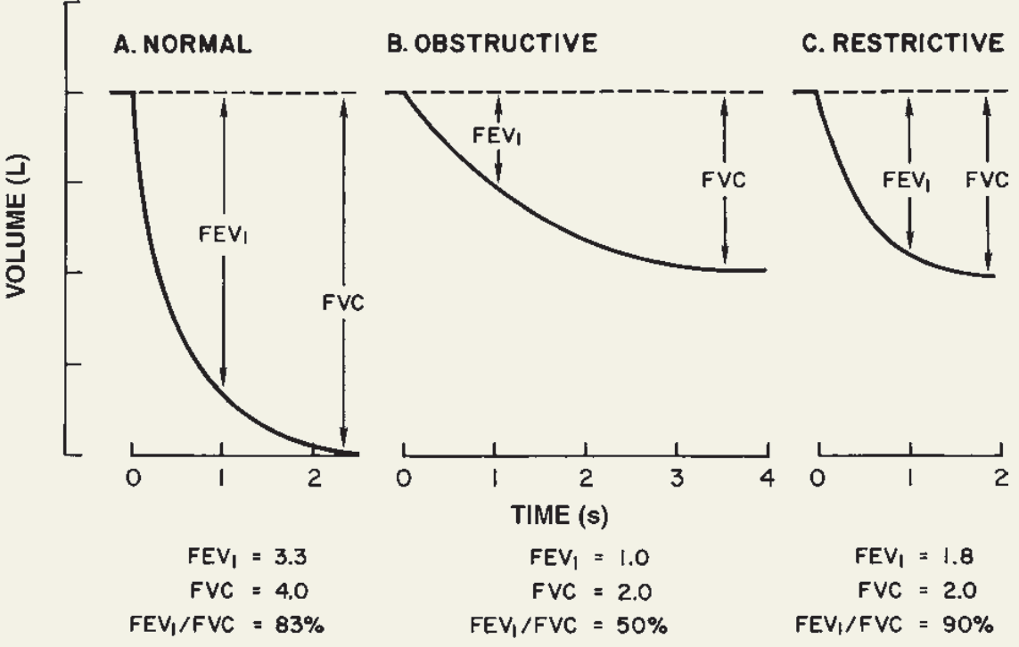
The PFT spirogram below illustrates the key differences visually:
1. FVC (Forced Vital Capacity)
| Obstructive | Restrictive | |
|---|---|---|
| Change | Normal or slightly decreased (air trapping may reduce it) | Markedly decreased (↓↓↓) |
| Reason | Air trapping increases RV, compressing FVC; but early obstruction may preserve FVC | Loss of lung tissue, stiff lungs, or chest wall limitation reduces total lung volume |
2. FEV₁ (Forced Expiratory Volume in 1 second)
| Obstructive | Restrictive | |
|---|---|---|
| Change | Markedly decreased (↓↓↓) | Decreased proportionally with FVC |
| Reason | Airway narrowing and collapse during forced expiration severely limits flow in the first second | All volumes are reduced equally; FEV₁ decreases because FVC is smaller, not because flow is impaired |
3. FEV₁/FVC Ratio (THE KEY DIFFERENTIATOR)
| Obstructive | Restrictive | |
|---|---|---|
| Change | Decreased (<70% or <LLN) | Normal or increased (>70-80%, often ~90%) |
| Why it matters | The hallmark diagnostic criterion; FEV₁ falls disproportionately more than FVC | Both FEV₁ and FVC fall proportionally, preserving or even raising the ratio |
| Example values | FEV₁=1.0L, FVC=2.0L → ratio=50% | FEV₁=1.8L, FVC=2.0L → ratio=90% |
Normal FEV₁/FVC = ~80-83% (Ganong's Review of Medical Physiology, 26th ed., p. 629)
This single parameter is the primary step in all PFT interpretation algorithms - as outlined in Murray & Nadel's Textbook of Respiratory Medicine.
4. FEF₂₅₋₇₅% (Forced Expiratory Flow at mid-expiration)
| Obstructive | Restrictive | |
|---|---|---|
| Change | Markedly decreased (↓↓↓) - earliest indicator of small airway disease | Normal |
| Significance | Reflects small airway patency; sensitive early marker even before FEV₁ falls | Mid-expiratory flow is preserved because airway calibre is normal |
Lung Volume Parameters (3 marks)
5. TLC (Total Lung Capacity)
| Obstructive | Restrictive | |
|---|---|---|
| Change | Normal or increased (hyperinflation / gas trapping) | Decreased (↓↓↓) - GOLD STANDARD for confirming restriction |
| Reason | Air trapping leads to barrel chest and elevated resting lung volume | Stiff lungs (high elastic recoil), chest wall restriction, or neuromuscular weakness limits maximal inspiration |
"A TLC less than the lower limit of normal (LLN) defines a restrictive ventilatory defect." - Murray & Nadel's, p. 763
6. FRC (Functional Residual Capacity)
| Obstructive | Restrictive | |
|---|---|---|
| Change | Normal or increased (↑ if gas trapping) | Decreased (↓↓↓) |
| Reason | Dynamic hyperinflation pushes end-expiratory lung volume upward | Increased elastic recoil collapses lungs toward a lower equilibrium volume |
7. RV (Residual Volume)
| Obstructive | Restrictive | |
|---|---|---|
| Change | Increased (↑↑) | Decreased or normal |
| Reason | Air trapping - small airways collapse before full exhalation | Stiff lungs are smaller but can still empty effectively; RV/TLC ratio may be elevated in extraparenchymal restriction |
8. RV/TLC Ratio
| Obstructive | Restrictive | |
|---|---|---|
| Change | Markedly increased | Normal or may be elevated in extraparenchymal diseases (e.g., neuromuscular) |
| Significance | Quantifies degree of gas trapping | Parenchymal restriction: ratio normal; extraparenchymal: ratio elevated due to early airway closure |
Gas Exchange Parameter (1 mark)
9. DLCO (Diffusing Capacity of Lung for Carbon Monoxide)
| Obstructive (COPD/Emphysema) | Restrictive | |
|---|---|---|
| Change | Decreased - due to loss of alveolar surface area, capillary bed destruction, V/Q mismatch | Decreased - due to loss of functioning alveoli, thickened alveolar-capillary membrane |
| Note | In asthma (obstructive), DLCO may be normal or even elevated | In fibrotic restrictive disease, DLCO is often the earliest and most sensitive abnormality |
In COPD: DLCO decreases due to "decreased alveolar surface area, loss of capillary bed, and increased distance from the terminal bronchiole to the alveolar-capillary membrane." - Barash Clinical Anesthesia, 9th ed., p. 1139
Flow-Volume Loop Pattern (1 mark)
10. Flow-Volume Loop Morphology
| Obstructive | Restrictive | |
|---|---|---|
| Expiratory limb shape | Concave upward (scooped out) - hallmark sign. Flow is relatively preserved at high volumes but falls steeply at low volumes (effort-independent zone) | Convex upward (steep, vertical) - increased elastic recoil maintains high flows; curve is narrow and tall |
| Overall loop size | Wider (increased TLC) but scooped | Smaller (reduced TLC and FVC) but shape preserved |
| Inspiratory limb | Usually preserved (variable intrathoracic) | Reduced proportionally |
Summary Comparison Table (Key for exam)
| PFT Parameter | Normal | Obstructive | Restrictive |
|---|---|---|---|
| FVC | 100% | Normal/↓ | ↓↓↓ |
| FEV₁ | 100% | ↓↓↓ | ↓ (proportional) |
| FEV₁/FVC | ~80% | ↓↓↓ (<70%) | Normal/↑ (>80%) |
| FEF₂₅₋₇₅% | 100% | ↓↓↓ | Normal |
| TLC | 100% | Normal/↑ | ↓↓↓ |
| FRC | 100% | Normal/↑ | ↓↓↓ |
| RV | 100% | ↑↑ | ↓/Normal |
| RV/TLC ratio | ~30% | ↑↑ | Normal/↑ (extrapulm) |
| DLCO | 100% | ↓ (emphysema) | ↓ (fibrosis) |
| Flow-volume loop | Normal | Concave (scooped) | Convex (narrow, tall) |
Clinical Significance for Physiotherapy
- In obstructive conditions, PFTs guide bronchodilator therapy decisions, exercise prescription (6MWT capacity), and airway clearance techniques (ACBT, autogenic drainage).
- In restrictive conditions, PFTs inform breathing retraining (diaphragmatic breathing, incentive spirometry), postural correction (kyphoscoliosis), and pulmonary rehabilitation targets.
- Severity grading (mild/moderate/severe/very severe) of both patterns relies on percent-predicted FEV₁ values, which guide physiotherapy intensity and goals.
- Post-bronchodilator reversibility testing (≥12% and ≥200 mL improvement in FEV₁) distinguishes asthma (reversible) from COPD (irreversible obstruction).
Sources:
- Barash, Cullen & Stoelting's Clinical Anesthesia, 9th ed., Table 15-2 (p. 1139-1140)
- Ganong's Review of Medical Physiology, 26th ed., Clinical Box 34-2 (p. 629)
- Murray & Nadel's Textbook of Respiratory Medicine, 2-Volume Set, Chapters 32 & 33
This is a shared conversation. Sign in to Orris to start your own chat.