
Hypoxic pulmonary vasoconstriction for 5 marks anesthesia residency
"hypoxic pulmonary vasoconstriction" mechanism
hypoxic pulmonary vasoconstriction mechanism diagram biphasic response
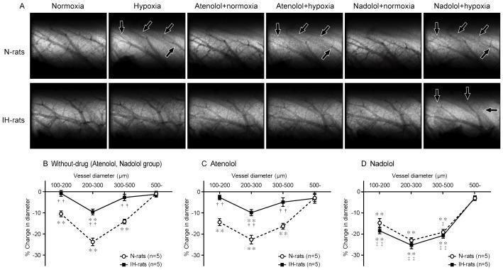
This composite educational graphic presents microangiographic imaging and quantitative data regarding Hypoxic Pulmonary Vasoconstriction (HPV). Panel A displays a matrix of selective pulmonary microangiograms comparing N-rats (normoxic-raised) and IH-rats (intermittent hypoxia-raised) under various conditions: normoxia, acute hypoxia, and hypoxia following pretreatment with beta-blockers Atenolol (beta-1 selective) or Nadolol (non-selective beta-1/beta-2). In N-rats, acute hypoxia induces visible narrowing of small pulmonary arteries (indicated by black arrows). This response is blunted in IH-rats but significantly restored after Nadolol administration, suggesting beta-2 adrenergic receptor involvement in IH-induced HPV attenuation. Panels B, C, and D provide quantitative line graphs plotting '% change in diameter' against 'vessel diameter (micrometers)' for these cohorts. The data highlights that HPV is most pronounced in vessels measuring 200–300 µm. The graphs statistically confirm that while Atenolol does not correct the blunted HPV in IH-rats, Nadolol returns the vasoconstrictive response to levels comparable to N-rats. This visual illustrates the pathophysiology of pulmonary vascular reactivity and the role of peripheral beta-adrenergic receptors in chronic hypoxia adaptations.
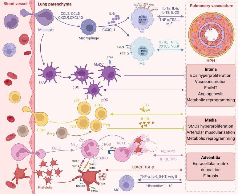
This pathophysiology diagram illustrates the immune-inflammatory landscape of Hypoxic Pulmonary Hypertension (HPH), divided into three compartments: blood vessel, lung parenchyma, and pulmonary vasculature. The left section shows the transendothelial migration of immune cells, including monocytes, T cells, and neutrophils, from the circulation into the lung tissue. The central 'Lung parenchyma' section details the differentiation and signaling pathways of resident and recruited cells, such as M1/M2 macrophages (modulated by HIF-1α, NF-κB, and PPARγ), dendritic cells (cDC, pDC), γδT cells, Th17 cells, and regulatory T/B cells (Treg, Breg). Key cytokines and chemokines (CCL2, CCL5, CXCL9, CXCL10, IL-6, IL-17, IL-10) are mapped to specific cellular interactions. The right section focuses on the 'Pulmonary vasculature,' detailing structural remodeling in HPH across three vessel layers: the intima (endothelial cell hyperproliferation, EndMT, vasoconstriction), the media (smooth muscle cell hyperproliferation, arteriolar muscularization), and the adventitia (extracellular matrix deposition, fibrosis). The diagram emphasizes the cross-talk between inflammatory mediators (like TNF-α, IL-1β, ROS, and NETs) and metabolic reprogramming in the progression of pulmonary vascular lesions.
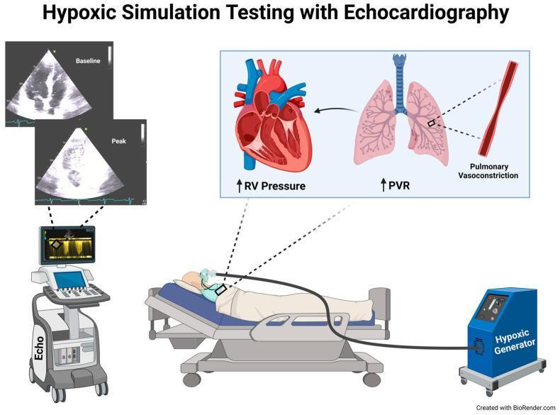
Educational medical infographic illustrating the setup and pathophysiology of Hypoxic Simulation Testing using Transthoracic Echocardiography (TTE). The clinical setup shows a patient in a semi-recumbent position on a hospital bed, breathing a low-oxygen gas mixture delivered via a face mask from a hypoxic generator. A bedside echocardiography machine is actively monitoring cardiac function. The visual includes two comparative four-chamber view ultrasound frames: 'Baseline' (normal cardiac morphology) and 'Peak' (showing changes during maximum hypoxia). A physiological inset diagram explains the underlying mechanism: inhalation of the hypoxic mixture triggers pulmonary vasoconstriction and increased Pulmonary Vascular Resistance (PVR), which subsequently leads to elevated Right Ventricular (RV) pressure. This illustration is used to teach diagnostic approaches for evaluating pulmonary hypertension susceptibility and right-to-left shunting (e.g., patent foramen ovale) under physiological stress.
Hypoxic Pulmonary Vasoconstriction (HPV)
Definition
- Miller's Anesthesia, 10e, p. 2105
Stimulus
- Primary stimulus: Alveolar O2 tension (PAO2). HPV is triggered when PAO2 falls below ~60 mmHg and is maximal at PAO2 ~30 mmHg.
- Secondary (weaker) stimulus: Mixed venous PO2 (PvO2).
- Augmented by: Hypercapnia, acidosis (local CO2 and H+ potentiate vasoconstriction, especially during hypoxia), and low pH.
- HPV is more potent when affecting a smaller lung region and is less effective when a large portion of the lung is hypoxic (as in global hypoxemia).
Mechanism
| Step | Mechanism |
|---|---|
| O2 sensing | Pulmonary artery smooth muscle cells act as intrinsic O2 sensors |
| K+ channels | Hypoxia inhibits voltage-gated K+ channels (Kv), leading to membrane depolarization |
| Ca2+ influx | Depolarization activates L-type voltage-gated Ca2+ channels → Ca2+ influx |
| SR Ca2+ release | Hypoxia releases Ca2+ from smooth muscle sarcoplasmic reticulum via ryanodine receptors |
| Ca2+ sensitization | Hypoxia enhances myofilament Ca2+ sensitivity (Rho kinase pathway) |
| Connexin-40 | Retrograde endothelial signal conduction via connexin-40 is required for HPV |
| TRP channels | Ca2+ influx at TRPV4 channels on pulmonary arterioles contributes |
Biphasic Temporal Response
- Phase 1 (rapid): Begins immediately, peaks/plateaus by 20-30 minutes
- Phase 2 (sustained/delayed): Begins after ~40 minutes, plateaus at ~2 hours
Physiological Significance
- HPV can reduce blood flow to the non-ventilated lung by up to 50% during one-lung ventilation (OLV)
- Maintains arterial oxygenation in conditions of: atelectasis, pneumonia, ARDS, reactive airway disease, OLV
- Under normal conditions in supine healthy humans, HPV contributes minimally to blood flow heterogeneity
Factors That Inhibit HPV (Clinically Important)
| Factor | Mechanism |
|---|---|
| Volatile anesthetics (halothane > isoflurane > sevoflurane/desflurane) | Reduce Ca2+ sensitivity, inhibit K+ channels, reduce free cytosolic Ca2+ |
| Vasodilators (nitroglycerin, nitroprusside, dobutamine) | Direct pulmonary vasodilation |
| High FiO2 to hypoxic lung | Removes hypoxic stimulus |
| Low cardiac output / high PVR | Alters perfusion pressure, may reverse HPV |
| Alkalosis | Reduced H+ reduces vasoconstriction |
| Hypothermia | Depresses metabolic response |
| Lung surgery/trauma | Release of local vasoactive metabolites |
Factors That Augment HPV
- Hypercapnia / acidosis (local)
- Alveolar hypoxia (progressive)
- Vasoconstrictors (phenylephrine, norepinephrine - modest effect)
- Almitrine bismesylate (augments HPV, used in thoracic surgery)
Chronic Effects
Anesthetic Relevance - Summary Box
| Scenario | HPV Relevance |
|---|---|
| One-lung ventilation (OLV) | HPV reduces shunt by diverting blood from collapsed lung |
| ARDS | HPV partially limits shunting through consolidated lung |
| Volatile vs. TIVA | TIVA preserves HPV; volatile agents blunt it |
| Vasodilators intraoperatively | May worsen hypoxemia by inhibiting HPV |
| Thoracic epidural | No direct effect on HPV; indirect via hypotension/CO reduction |
Recent evidence note: A 2024 review (Archer et al., PMID 39589299, Physiol Res) reinforces HPV as part of the homeostatic oxygen-sensing system, with updated discussion of mitochondrial ROS and KATP channel interplay - consistent with the textbook framework above.