
Ovarian torsion
ovarian torsion ultrasound diagnosis
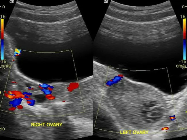
Pelvic ultrasound with color and power Doppler was performed to evaluate acute left lower quadrant pain in a young female. Gray-scale (B-mode) imaging demonstrates a normal right ovary with preserved parenchymal echotexture and a small left ovarian hemorrhagic cyst, with several tiny peripheral follicles. A pivotal radiologic finding is a twisted vascular pedicle just cranial to the left ovary, compatible with ovarian torsion. Color Doppler evaluation shows absence of detectable arterial and venous flow within the left ovary, while the right ovary maintains normal perfusion. The left ovary is tender to transducer pressure, and pedicle twist explains the acute presentation with vomiting. The absence of Doppler flow is highly specific for torsion, although a normal flow pattern does not completely exclude it. The exam emphasizes searching for an underlying ovarian mass or hemorrhagic cyst as a possible precipitating lesion. The radiologic diagnosis supports urgent surgical exploration to preserve ovarian viability. Clinical correlation includes abrupt onset abdominal pain in a reproductive-age patient, nausea, vomiting, and localized adnexal tenderness. Differential considerations include early torsion with intermittent flow, hemorrhagic corpus luteum cyst, adnexal mass, or adnexitis. This case underscores the diagnostic value of color Doppler in acute pelvic pain and the need for prompt gynecologic consultation.
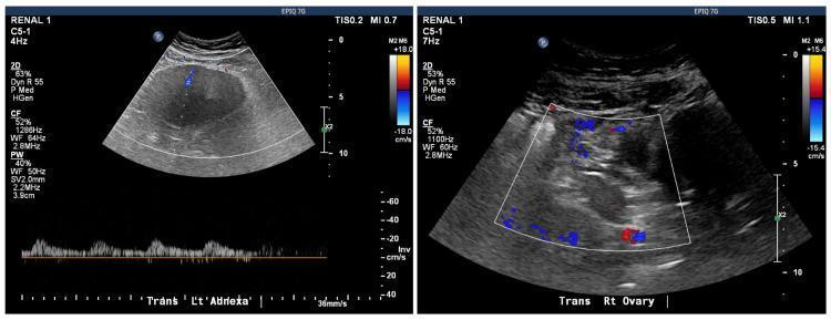
This diagnostic image consists of two side-by-side transabdominal pelvic ultrasound panels with color Doppler flow assessment, comparing the bilateral adnexa. The left panel, labeled 'Trans L’t Adnexa,' displays a large, hypoechoic, and relatively homogeneous mass. The superimposed color Doppler reveals a significant absence of internal vascularity, with only a negligible blue signal visible, and the corresponding pulsed-wave Doppler at the bottom shows minimal spectral activity, a finding highly suggestive of ovarian torsion. In contrast, the right panel, labeled 'Trans Rt Ovary,' shows a more heterogeneous ovarian structure with robust color Doppler signals (red and blue), indicating preserved arterial and venous blood flow. This side-by-side comparison serves as a classic educational example of how Doppler sonography is used to evaluate suspected adnexal torsion by demonstrating the loss of blood flow to the affected organ compared to the normal contralateral ovary. The image is relevant for gynecology and emergency medicine training regarding the diagnosis of acute pelvic pain.
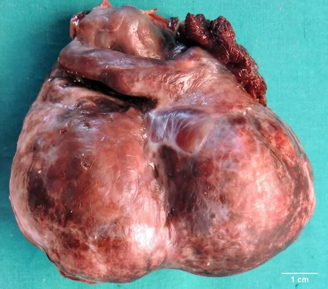
Gross pathology photograph of a salpingo-oophorectomy specimen from an adult female with acute pelvic pain. The excised adnexa measures approximately 10 x 8 x 6 cm and comprises an ovary adherent to the fallopian tube. The external surface is bosselated and variably dark bluish black in color. The parenchyma contains multiple cysts filled with altered blood and clots, separated by conspicuous fibrous septae, consistent with hemorrhagic degeneration. On gross inspection, ischemic necrosis predominates in most areas, with focal viable zones displaying serous cystadenoma-like features. The specimen is multiloculated with a thick capsule and torsion-related vascular compromise. Ovarian torsion, or adnexal torsion, involves rotation of the ovary and part of the tube around its vascular pedicle and is a surgical emergency requiring prompt intervention to prevent necrosis. Hemorrhagic infarction reflects obstruction of arterial inflow with venous outflow stasis. The narrative notes torsion's bimodal age distribution, affecting reproductive-age and postmenopausal women, with risk factors including benign and malignant neoplasms and corpus luteum cysts. Clinically, these findings align with severe abdominal pain, acute pelvic condition, and ultrasound-detected complex adnexal mass. This specimen illustrates torsion with hemorrhagic cysts and a benign serous neoplasm component, valuable for teaching, differential diagnosis, and surgical pathology education.
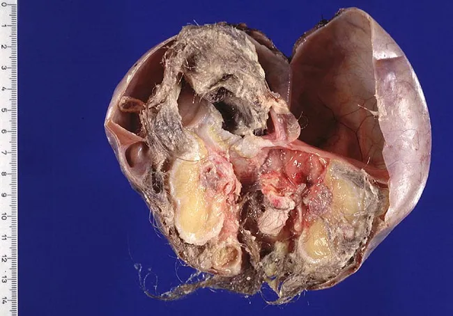
Gross pathology photograph of an ovarian mass from a 16-year-old female, consistent with a mature cystic teratoma (dermoid cyst). The specimen is opened to reveal classic dermoid contents: hair, sebaceous material, and adipose tissue arranged within a fluctuant multilocular cyst. The cyst wall is lined by stratified squamous epithelium with keratinous debris; surrounding ovarian stroma shows fibrous tissue without invasion. Internal components demonstrate ectodermal derivatives including hair shafts and sebaceous glands; adipose tissue and occasionally calcified fragments may be present. The mass is largely benign in behavior, typically unilateral, and often presents with abdominal pain or mass effect in adolescence; torsion is a known complication. Histologically, mature tissues from ectodermal, mesodermal, and endodermal origins can be seen, but in this specimen no immature neural elements or malignant transformation are evident. The gross appearance and cut-open configuration strongly support diagnosis of a mature cystic teratoma, which is the most common ovarian neoplasm in young girls. Clinically, this lesion requires surgical excision with preservation of ovarian tissue when possible; prognosis is excellent after complete resection. Radiologic imaging (ultrasound, CT, MRI) typically demonstrates a fat-containing cyst with echogenic sebaceous material, aiding differential diagnosis prior to pathology.
"ovarian torsion" diagnosis management
Ovarian Torsion
Definition and Pathophysiology
- Venous and lymphatic obstruction first - causes ovarian congestion, edema, and enlargement
- Decreased arterial inflow - from rising interstitial pressure; arteries are more resistant due to thicker walls
- Arterial thrombosis and ischemia - with prolonged torsion
- Necrosis and potential sepsis - if diagnosis and treatment are delayed
- Tintinalli's Emergency Medicine, p. 654
- Gray's Anatomy for Students, p. (Case 3 - OVARIAN TORSION)
Risk Factors
| Risk Factor | Notes |
|---|---|
| Ovarian cysts or tumors | Most common precipitant; 83% of torsion occurs in ovaries ≥5 cm |
| Benign cystic teratoma (dermoid) | Most common neoplasm to undergo torsion |
| Polycystic ovary | Can undergo torsion even without a discrete mass |
| Ovarian hyperstimulation syndrome | Chemical induction of ovulation |
| Pregnancy | Enlarged corpus luteum; increased risk in first trimester |
| Previous torsion | Ligament laxity after prior episode |
| Tubal ligation | Altered anatomy |
| Para-ovarian or paratubal cyst | Acts as fulcrum for rotation |
| Hydrosalpinx | Adds mass to adnexa |
- Tintinalli's Emergency Medicine, p. 654
- Bailey and Love's Short Practice of Surgery 28th Edition, p. 1602
- Berek & Novak's Gynecology, p. 561
Clinical Features
Symptoms
- Acute-onset, severe, unilateral lower abdominal/pelvic pain - the classic presentation
- Pain may begin after exertion (lifting, exercise, intercourse) as the ovary twists
- Atypical presentations are common: ~50% of patients have gradual or intermittent pain (intermittent torsion and detorsion)
- Nausea and vomiting in ~70% of cases
- Diarrhoea occasionally
Signs
-
Unilateral lower abdominal tenderness with guarding
-
Unilateral adnexal tenderness on bimanual examination
-
Palpable latero-uterine or adnexal mass
-
Mild temperature elevation, tachycardia, and leukocytosis may accompany tissue necrosis
-
Important: ~30% of patients have bilateral adnexal tenderness; a minority have no tenderness at all
-
50% of patients are initially misdiagnosed
-
Tintinalli's Emergency Medicine, p. 655
-
Berek & Novak's Gynecology, p. 562
Diagnosis
Imaging
- Ovarian enlargement (>4 cm) - most common finding; most constant finding is ≥7 cm per Grainger & Allison
- Echogenic stroma with peripherally displaced follicles ("follicular ring sign")
- Whirlpool/swirl sign - twisted vascular pedicle visible on grey-scale and color Doppler
- Absent or reduced ovarian vascularity on Doppler
- Free fluid in the pelvis (pouch of Douglas)
- Deviation of the uterus to the affected side
- A positive Doppler study has 100% positive predictive value for adnexal torsion
- However, up to 60% of torsions can be missed on arterial Doppler alone - arterial disruption is a late finding
- Venous Doppler may be the only abnormality in early torsion
- Normal blood flow does NOT exclude torsion
- Up to 26% of ultrasound studies show normal adnexa in confirmed torsion cases
Laboratory Tests
- Beta-hCG: must be checked to exclude ectopic pregnancy
- CBC: leukocytosis may be present but is non-specific (can also be normal)
- Inflammatory markers (CRP, ESR): may be raised or normal
- Type and screen if hemodynamic compromise is suspected
Ovarian Torsion Composite Index (OT-CI)
Definitive Diagnosis
Differential Diagnosis
- Ruptured ectopic pregnancy
- Appendicitis
- Ruptured ovarian cyst (hemorrhagic corpus luteum)
- Pelvic inflammatory disease / tubo-ovarian abscess
- Endometriosis / ruptured endometrioma
- Ureteral colic
- Acute intestinal obstruction
- Mesenteric ischemia
Management
Surgical Steps
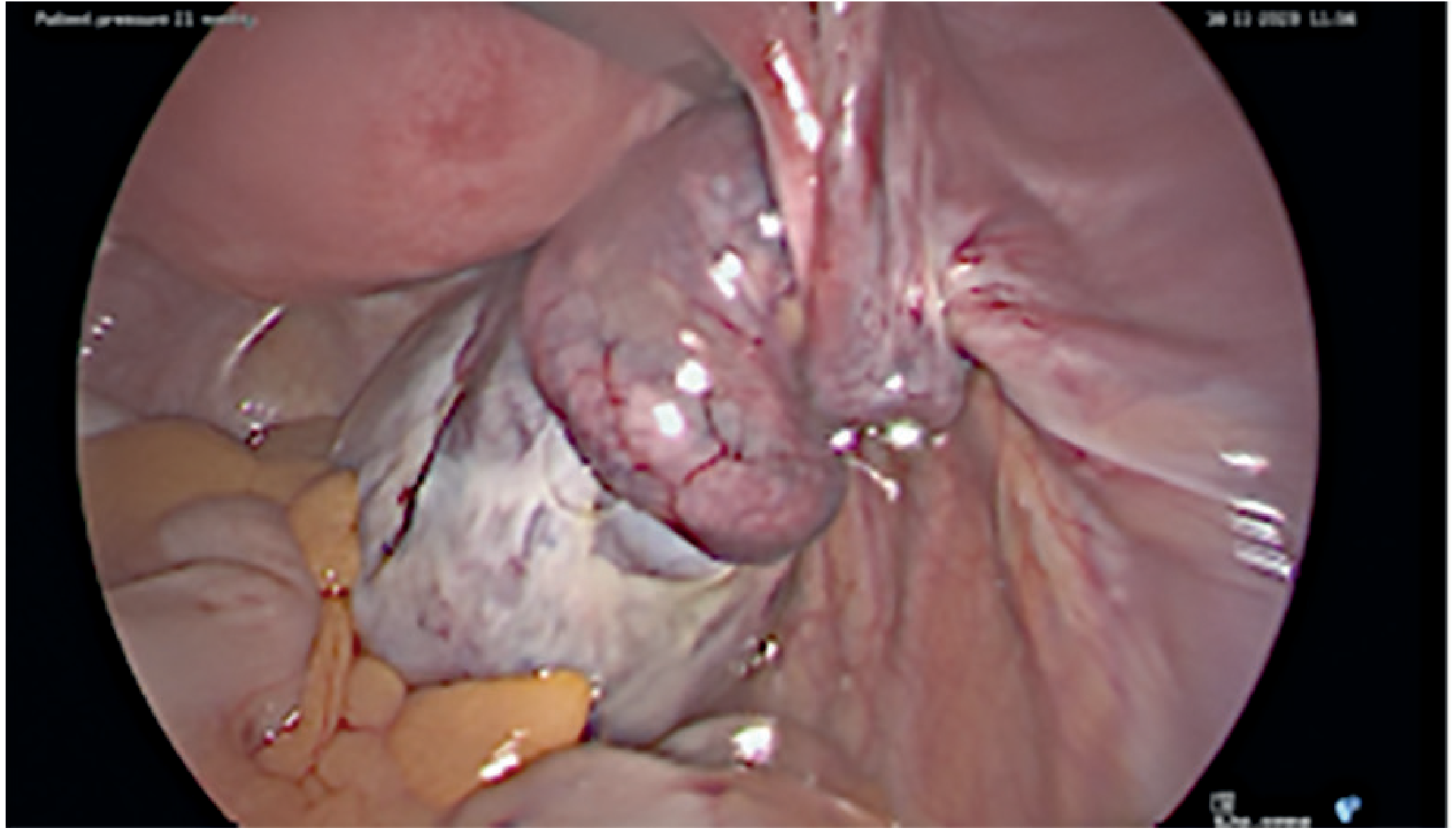
- Detorsion (untwisting) - the ovary and adnexa are unwound
- Ovarian cystectomy - to address the underlying mass; may be done at the same time or at a later date once edema has resolved (reducing the likelihood of needing oophorectomy)
- Salpingo-oophorectomy - reserved for clearly necrotic, non-viable tissue
Special Populations
Adolescents and Premenarchal Girls
- Ovarian torsion can occur at any age, including infants, neonates, and postmenopausal women
- Peak pediatric risk periods: first year of life, at menarche, and during pregnancy
- Torsion of a normal ovary (without an underlying cyst) is more common in adolescents due to elongated pelvic ligaments and more mobile adnexa
- Premenarchal girls more often present with fever, restlessness, and palpable pelvic mass
- Longer delay between symptom onset and diagnosis in premenarchal girls
- Pediatric torsion is rare but frequently missed, leading to delayed treatment
Pregnancy
- Corpus luteum enlargement in the first trimester is a significant risk factor
- High clinical suspicion must be maintained as normal pregnancy complaints can mask symptoms
Postmenopausal Women
- Lower incidence but usually associated with a true ovarian neoplasm rather than a functional cyst
Prognosis and Complications
- Delayed diagnosis leads to irreversible ovarian necrosis and loss of the ovary
- Risk of thromboembolic events from venous stasis in the twisted pedicle (though rare, pulmonary embolism from detorsion has been described)
- Recurrence risk - particularly in patients with predisposing anatomy or polycystic ovaries; oophoropexy (fixation of the ovary) can be considered in high-risk cases
Summary
| Feature | Key Point |
|---|---|
| Mechanism | Torsion of vascular pedicle → venous obstruction → arterial ischemia → necrosis |
| Most common side | Right (70%) |
| Most common precipitant | Ovarian cyst or benign cystic teratoma |
| Classic symptom | Acute unilateral lower abdominal pain + nausea/vomiting |
| Primary imaging | TVUS with Doppler (limitations: normal flow does not exclude torsion) |
| Most constant US finding | Ovarian enlargement (≥4-7 cm) |
| Gold standard | Surgical diagnosis (laparoscopy) |
| Treatment principle | Detorsion first; preserve ovary even if it looks necrotic |
| Misdiagnosis rate | ~50% initially misdiagnosed |
- Tintinalli's Emergency Medicine: A Comprehensive Study Guide, Chapter 97
- Berek & Novak's Gynecology, Chapter on Acute Pelvic Pain
- Sabiston Textbook of Surgery, Chapter on Gynecologic Emergencies
- Bailey and Love's Short Practice of Surgery 28th Edition, Table 87.3
- Grainger & Allison's Diagnostic Radiology, Chapter 35
- Gray's Anatomy for Students, Clinical Case 3
Recent evidence note: A 2024 systematic review and meta-analysis (PMID 39519217) evaluated non-invasive biomarkers for ovarian torsion detection. No single reliable serum biomarker has been identified to date, reinforcing that clinical and sonographic assessment with surgical confirmation remains the standard of care.