
Mucosal folds of ear, refer from scott brown, hazarika, shamberg, zakir hussain.
Reading File
Now let me search all the relevant books simultaneously for mucosal folds of the ear.
Finding Sources
Finding Sources
Finding Sources
Let me check what ENT/otology books are available and search more specifically for ear mucosal folds.
Finding Sources
Finding Sources
Excellent! I found the key sections. Let me now read them in parallel - Scott-Brown Vol 2, Shambaugh, and also search for more content.
Reading File
Reading File
Reading File
I have Scott-Brown Vol 2 content on mucosal folds. Now let me read the full Cummings section and also search for Shambaugh's dedicated mucosal fold content and look for Hazarika and Zakir Hussain content.
Reading File
Finding Sources
Finding Sources
Excellent content found from Cummings and KJ Lee. Now let me get the detailed sections - particularly from Cummings block32 (Epitympanum section) and KJ Lee's "Middle Ear Folds of Significance".
Reading File
Reading File
Let me now get the specific "Middle Ear Folds of Significance" from KJ Lee and more from Cummings block32, then also check the KJ Lee block3 for the epitympanum section.
Reading File
Reading File
Reading File
Now let me get more from Cummings block32 (Prussak's space details and key points) and also fetch the relevant diagrams.
Reading File
Reading File
Loading Image
Loading Image
Now let me also fetch the Cummings diagram of the epitympanic diaphragm and check for Shambaugh's mucosal fold content and Scott-Brown Vol 1 for any additional content.
Loading Image
Finding Sources
I now have comprehensive content from all available sources. Note: Hazarika and Zakir Hussain are not in the library database - only Scott-Brown (Vol 1 & 2), Shambaugh, Cummings, and KJ Lee are available. Let me compile a complete, well-organized answer.
Mucosal Folds of the Ear (Middle Ear)
The mucosal folds of the middle ear are thin membranous structures - some thickening to form ligaments - that partially divide the tympanic cavity into distinct compartments, determining ventilation pathways and cholesteatoma spread patterns.
From Scott-Brown's Otorhinolaryngology Head & Neck Surgery (Vol 2)
Scott-Brown's Otorhinolaryngology Head & Neck Surgery - Vol 2, The middle ear mucosal folds section
The mucosal folds in the middle ear, which thicken in some instances to form ligaments, divide the middle ear cavity into a superior epitympanum and an inferior mesotympanum and hypotympanum.
The ligaments forming the epitympanic diaphragm arise from malleal folds:
- The lateral malleoincudal fold - posteriorly
- The tensor fold - anteriorly
The connection between the two is the tympanic isthmus - the only conduit for a ventilation stream in the epitympanum from the Eustachian tube (unlike the mesotympanum, which is flooded by air at all times).
Clinical significance: Congenital defects in the folds, or folds affected by inflammatory processes (e.g. mucosal disease or cholesteatoma), can block the narrow isthmus, leading to loss of air from the epitympanum and provoking further inflammation.
From Shambaugh Surgery of the Ear
Shambaugh Surgery of the Ear, Revision stapes surgery section
Shambaugh describes the plica mallearis (also called the mucosal fold of the malleus):
"Along most of its length the manubrium is attached to the drum with a mucosal fold: the plica mallearis."
In the context of revision stapes surgery, the plica mallearis is elevated from the malleus to create a space for prosthesis placement. A malleus attachment prosthesis is an option in patients with a normal tympanic membrane and intact plica mallearis. The periosteum and plica mallearis are separated from the manubrium, creating a space through which the hook of a prosthesis is introduced. The laser is particularly helpful in revision surgery for dividing adhesions, mucosal folds, and soft tissue surrounding prostheses in the oval window.
From Cummings Otolaryngology Head and Neck Surgery
Cummings Otolaryngology, Chapter 126 (Middle ear anatomy) and Chapter 144 (Endoscopic ear surgery)
The Epitympanic Diaphragm and its Folds
The epitympanum contains the head of the malleus, from which three ligamental folds arise:
- Anterior malleal fold (amlf)
- Posterior malleal fold (plm) / posterior malleal ligament and fold
- Lateral malleal fold (mlf)
The body and short process of the incus contribute:
4. Lateral incudomalleal fold (imlf) - arises from the posterior end of the short process of the incus, extends anteriorly between the incus body and proximal portion of the long process, joins the head of the malleus, and then projects inferiorly.
Together with the tensor fold (tf), these ligaments and folds form the epitympanic diaphragm that determines ventilation pathways in the middle ear and mastoid.
The Tensor Fold (Fig. 144.3 & 144.5)
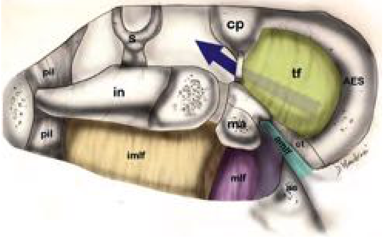
The tensor fold is a membranous fold that arises from the tensor tympani canal and cochleariform process, separating the anterior epitympanum from the supratubal recess (a superior/anterior extension of the mesotympanum near the Eustachian tube).
Key facts about the tensor fold:
- Absent or incomplete in 27% of cadaveric temporal bones (Palva et al.) - this provides an alternate ventilation pathway for the anterior epitympanum
- Present and complete in 73% - in this case, the tympanic isthmus becomes the sole pathway for attic ventilation
- When the tensor fold is vertical, the supratubal recess is large and wide
- When the tensor fold is horizontal, the supratubal recess is inferiorly displaced and narrow
- Surgical implication: Opening a complete tensor fold during chronic ear surgery provides an alternate route for epitympanic ventilation
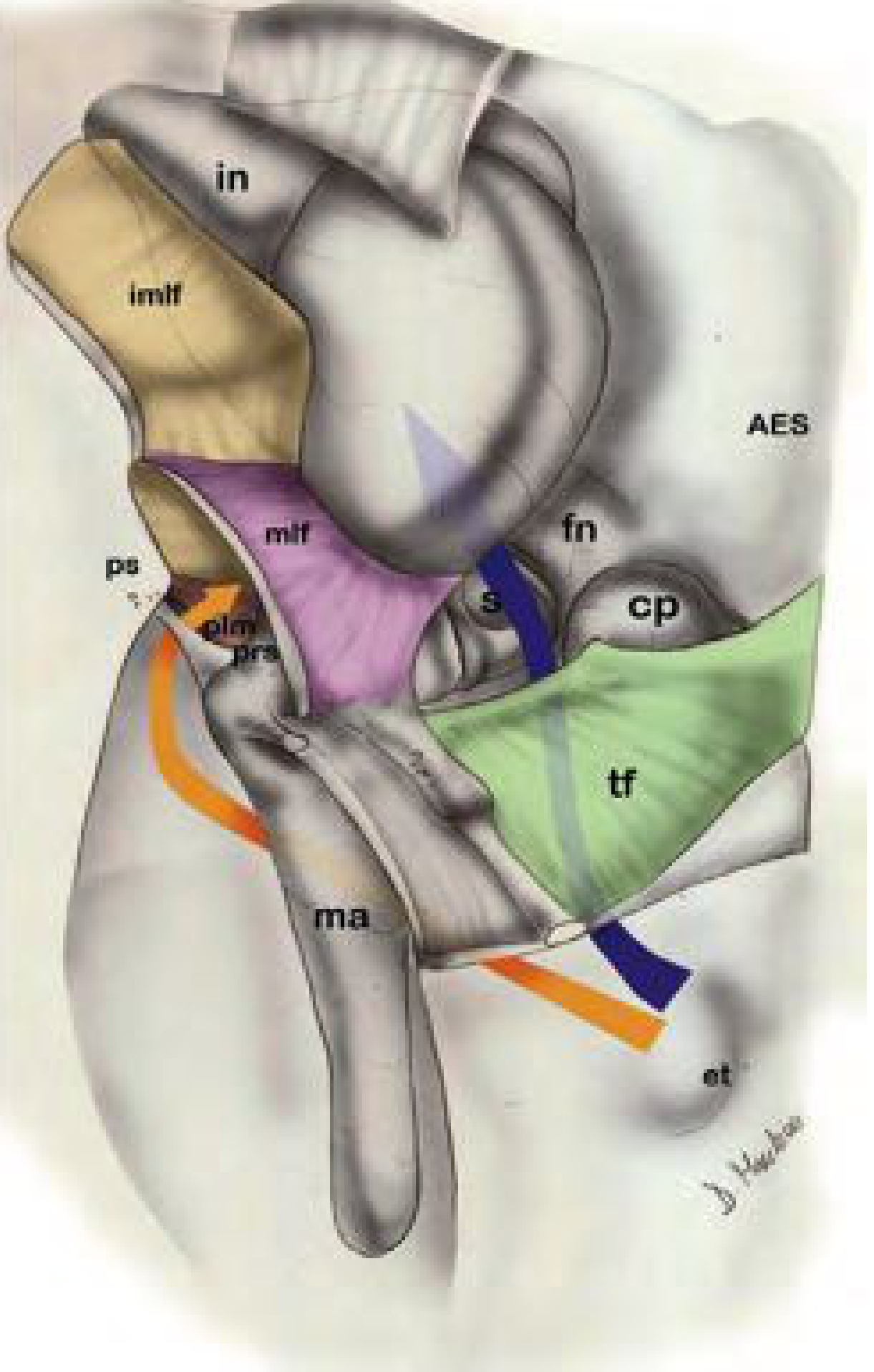
Prussak's Space and the Lateral Malleal Fold
The lateral malleal fold (mlf) is the key fold defining Prussak's space (the most lateral compartment in the posterior epitympanum):
- Superior: lateral malleal fold (separates Prussak's space from the rest of the posterior epitympanum)
- Inferior: posterior malleal fold
- Anterior: anterior malleal fold
- Posterior: lateral malleal fold (continuous arch)
- Medial: neck of the malleus
- Lateral: pars flaccida / Shrapnell's membrane
Ossicular chain mucosal folds listed in Cummings (Fig. 126.10):
| Fold | Number | Location/Course |
|---|---|---|
| Obturatoria stapedi | 5 | Stapes region |
| Plica stapedi | 8 | Around stapes |
| Medial incudal fold | 9 | Medial incus |
| Superior incudal fold | 11 | Superior incus |
| Tensor fold | 13 | Tensor tympani canal to cochleariform process |
| Anterior malleolar fold | 14 | Anterior malleus neck |
| Interosseous fold | 17 | Between ossicles |
| Superior malleolar fold | 18 | Superior malleus |
| Lateral incudomalleal fold | (imlf) | Incus to malleus junction |
| Lateral malleal fold | (mlf) | Neck of malleus, arch form |
The ossicular chain, ligaments, and mucosal folds almost completely partition the epitympanic space from the mesotympanum, leaving only the isthmus tympani anticus (anterior tympanic isthmus, #20) and isthmus tympani oticus (posterior tympanic isthmus, #21) as the remaining openings.
Tympanic Isthmus
- The isthmus is the ventilation pathway starting at the Eustachian tube, proceeding through the mesotympanum medial to the malleus, incus, and lateral incudomalleal fold
- When the tensor fold is complete and the isthmus is obstructed by inflammatory tissue, the anterior epitympanum is left without ventilation - leading to negative pressure, TM retraction, and cholesteatoma
From KJ Lee's Essential Otolaryngology
KJ Lee's Essential Otolaryngology, "Middle Ear Folds of Significance" and "Epitympanum (Attic)" sections
Middle Ear Folds of Significance
There are five malleal folds and four incudal folds:
Malleal folds:
| Fold | Attachment |
|---|---|
| Anterior malleal fold | Neck of malleus → anterosuperior margin of tympanic sulcus |
| Posterior malleal fold | Neck → posterosuperior margin of tympanic sulcus |
| Lateral malleal fold | Neck to neck in an arch form, attaching to Shrapnell membrane |
| Anterior pouch of von Troeltsch | Between anterior malleal fold and TM anterior to handle |
| Posterior pouch of von Troeltsch | Between posterior malleal fold and TM posterior to handle |
(Note: The anterior and posterior pouches of von Troeltsch are recesses bounded by malleal folds.)
Incudal folds (from KJ Lee block3, Epitympanum section):
The diaphragm components include:
- (a) Lateral incudomalleal fold
- (b) Medial incudal fold
- (c) Lateral malleal fold
- (d) Lateral malleal ligament
- (e) Anterior malleal fold
- (f) Anterior malleal ligament
- (g) Tensor tympani fold (= posterior malleal fold)
- (h) Tensor tympani muscle tendon
Prussak's Space (KJ Lee)
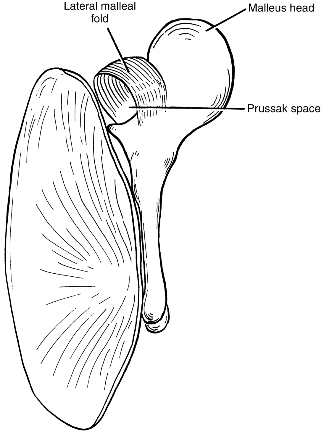
- Superior: lateral malleal fold (= tympanic diaphragm)
- Inferior: malleus neck
- Medial: malleus head
- Lateral: pars flaccida (Shrapnell membrane)
- Anterior: anterior malleal fold
- Posterior: posterior malleal fold
- Ventilation: independent, rough, narrow pathway through the posterior pocket of von Troeltsch; thick, viscous secretions may close this pocket, leading to sectorial dysventilation of Prussak space, pars flaccida retraction, and adhesion to malleus neck - without involving the anterior/posterior epitympanum, aditus, or mastoid cells
Tympanic Isthmus (KJ Lee)
A 2.5-mm opening in the tympanic diaphragm that ventilates the entire attic:
- (a) Anterior tympanic isthmus: between the incudostapedial joint and tensor tympanic muscle tendon - the largest and most important ventilation route to the attic. Granulation tissues and web blockages here lead to attic selective dysventilation, chronic edema, exudate, inflammation, retraction pockets, and cholesteatoma.
- (b) Posterior tympanic isthmus: posterior to the incudostapedial joint, between the stapedial muscle tendon, pyramidal eminence, and incus short process.
Summary Table of Key Mucosal Folds
| Fold | Origin | Insertion | Clinical Significance |
|---|---|---|---|
| Lateral malleal fold | Neck of malleus | Tympanic notch / Shrapnell membrane (arch form) | Roof of Prussak's space; part of epitympanic diaphragm |
| Anterior malleal fold | Neck of malleus | Anterosuperior tympanic sulcus | Anterior wall of Prussak's space |
| Posterior malleal fold | Neck of malleus | Posterosuperior tympanic sulcus | Posterior wall of Prussak's space; = tensor tympani fold |
| Tensor fold | Tensor tympani canal + cochleariform process | Anterior epitympanum | Separates AES from supratubal recess; absent in 27%; sole attic ventilation route when complete |
| Lateral incudomalleal fold | Short process of incus → malleus head | Inferiorly aligned with posterior malleal fold | Separates superior & inferior lateral attic; part of tympanic isthmus boundary |
| Plica mallearis | Manubrium (along most of length) | Tympanic membrane | Attaches TM to malleus; elevated in malleus-grip prosthesis placement (Shambaugh) |
Notes on Hazarika and Zakir Hussain
Hazarika (P. Hazarika's Textbook of ENT & Head and Neck Surgery) and Zakir Hussain (Clinical ENT by Zakir Hussain) are not present in the digital textbook library available here, so direct citations from those sources cannot be provided. The content above comprehensively covers the topic as described in Scott-Brown's, Shambaugh, Cummings, and KJ Lee (the four major ENT references available). For Hazarika and Zakir Hussain, you would need to cross-reference those physical texts - they generally cover the same anatomical framework (malleal folds, tensor fold, tympanic isthmus, Prussak's space) with minor terminological variations.
This is a shared conversation. Sign in to Orris to start your own chat.