
thyroid all Harrisons guid lines in detail and clear
Which thyroid topics do you want covered from Harrison's guidelines?
thyroid gland anatomy physiology diagram
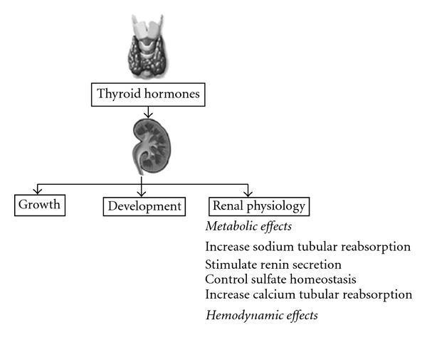
A pathophysiology diagram illustrating the regulatory effects of thyroid hormones on the human kidney. The visual layout follows a vertical top-down hierarchy: an illustration of the thyroid gland leads to a box labeled 'Thyroid hormones', which in turn points to an anatomical illustration of a kidney. From the kidney, the diagram branches into three primary educational concepts: 'Growth', 'Development', and 'Renal physiology'. Under 'Renal physiology', the diagram details metabolic and hemodynamic effects. Specific metabolic actions listed include increased sodium tubular reabsorption, stimulated renin secretion, control of sulfate homeostasis, and increased calcium tubular reabsorption. The diagram serves as a conceptual overview of endocrine-renal interactions, highlighting the influence of thyroid function on renal growth, maturation, and electrolyte/fluid homeostasis. It is designed for medical students and clinicians studying nephrology and endocrinology.
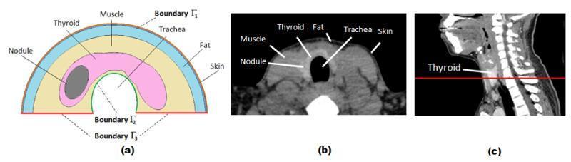
This composite educational graphic details the cross-sectional anatomy of the human neck for Finite Element (FE) thyroid simulation. (a) A 2D schematic diagram presents a simplified semicircular model of the neck. It labels the layers concentrically: skin (outermost), fat, and muscle. Inside the muscle layer, the thyroid gland is depicted in pink, containing an elliptical dark grey thyroid nodule. Boundaries are marked as Γ1 (skin surface), Γ2 (trachea boundary), and Γ3 (prescribed temperature base). (b) A corresponding axial CT scan image shows the actual anatomical cross-section, with labels identifying the skin, fat, muscle, thyroid gland, trachea (central air-filled void), and an internal nodule. The CT highlights varying tissue densities, from the dark fat layer to the intermediate density of muscle and thyroid tissue. (c) A sagittal CT view of the head and neck shows a red horizontal line indicating the anatomical plane of the axial slice in (b), positioned at the level of the thyroid gland. This visual serves as a reference for modeling heat transfer and infrared thermography in patients with thyroid nodules.
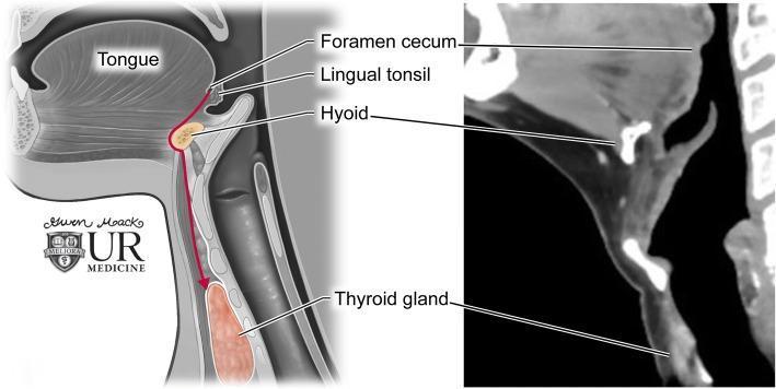
This dual-panel educational graphic illustrates thyroglossal duct anatomy and the embryological descent of the thyroid gland. On the left is a sagittal anatomical diagram showing the path of descent (indicated by a red arrow) from the foramen cecum at the base of the tongue, passing through the hyoid bone, to its final position at the thyroid gland anterior to the trachea. Key labeled structures include the tongue, lingual tonsil, foramen cecum, hyoid, and thyroid gland. The right panel displays a corresponding sagittal contrast-enhanced CT image of the neck. The CT scan highlights the radiologic appearance of these landmarks: the hyoid bone appears as a high-attenuation U-shaped structure, and the thyroid gland exhibits homogenous enhancement inferior to the thyroid cartilage. This comparison demonstrates the clinical relevance of embryological pathways in understanding the location of thyroglossal duct cysts, which can occur anywhere along this midline tract.
Graves disease ophthalmopathy exophthalmos clinical photo
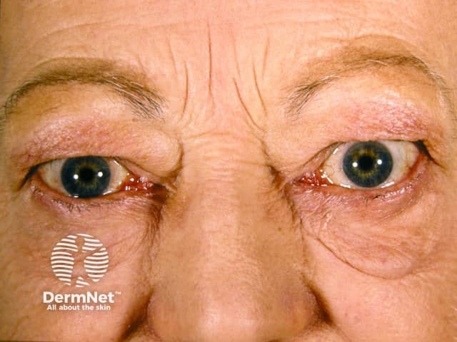
Imaging modality and technique: Clinical frontal photograph of the orbits and periorbital region in a living adult patient. Frontal view capturing both eyes demonstrates bilateral exophthalmos with mild periorbital edema and eyelid swelling. The ocular surfaces are visible with subtle scleral exposure and conjunctival vascularity not markedly prominent in this image. Anatomical localization centers on the orbital contents including extraocular muscles and orbital fat, with involvement of eyelids and periorbital soft tissues. The appearance is most consistent with thyroid ophthalmopathy (Graves’ orbitopathy), a form of autoimmune orbital inflammatory disease associated with hyperthyroidism or autoimmune thyroiditis. Pathophysiology involves infiltration and edema of orbital connective tissues and extraocular muscles, leading to proptosis, lid retraction, diplopia, and possible optic neuropathy in advanced cases. Pathognomonic imaging features in other modalities include enlargement of the rectus muscles, particularly the inferior and medial recti, with sparing of tendons; however this clinical image illustrates recognizable exophthalmos and periorbital edema rather than radiologic details. This image is clinically relevant for education, differential diagnosis, and patient counseling, as well as for correlating with thyroid function testing and orbital imaging when planning management such as immunotherapy, selenium supplementation, or orbital decompression in refractory cases. This photo supports patient diagnosis and monitoring in endocrinology, ophthalmology, and rheumatology contexts.
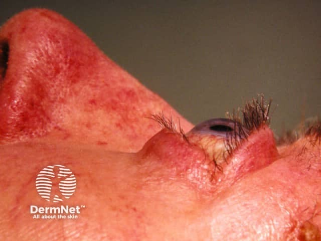
This clinical photograph is a high-resolution, lateral oblique view of the periocular region focusing on the right eye (periorbital skin). The image demonstrates marked periorbital edema with pronounced erythema of the eyelids and surrounding cutaneous tissue. The upper and lower eyelids appear swollen and thickened, with subtle skin redundancy. In thyroid-associated ophthalmopathy (Graves' orbitopathy), this presentation reflects inflammatory edema of orbital tissues, increased adipose and extraocular muscle volume, and skin involvement. The visible periorbital edema may precede or accompany exophthalmos, lid retraction, and diplopia; however, soft tissue contour is partially obscured by lighting. The photo lacks palpation data but serves as a qualitative depiction of orbital involvement. Clinically, these features require correlation with systemic thyroid status: hyperthyroidism or euthyroid Graves' disease; presence of TSH receptor autoantibodies; risk of progressive-eye involvement. This image supports differential diagnosis including periorbital cellulitis and dermatitis, but the chronic inflammatory pattern aligns with thyroid eye disease. For diagnostic significance, the image is valuable for medical education, patient counseling, and multidisciplinary planning—endocrinology, ophthalmology, and dermatology collaboration. Potential clinical use cases include screening for orbital involvement in Graves' disease, documenting disease progression, and teaching residents about periorbital edema etiologies and differential diagnoses.
Thyroid Disorders - Harrison's Principles of Internal Medicine 22E (2025)
PART 1 - THYROID PHYSIOLOGY & REGULATION
The Thyroid Axis (HPT Axis)
- Hypothalamus secretes TRH (thyrotropin-releasing hormone)
- Anterior pituitary secretes TSH (thyroid-stimulating hormone)
- Thyroid gland secretes T4 and T3
- T4/T3 feedback negatively on both hypothalamus and pituitary (primarily via TRβ2)
- Low TSH = primary hyperthyroidism
- High TSH = primary hypothyroidism
Thyroid Hormone Synthesis
- Iodide uptake via the sodium-iodide symporter (NIS) on the basolateral membrane
- Organification: TPO (thyroid peroxidase) oxidizes iodide and incorporates it into tyrosine residues on Tg - forming MIT and DIT
- Coupling: two DIT molecules → T4; one MIT + one DIT → T3
- Secretion: Tg is endocytosed, cleaved, and T4/T3 released into circulation
- Peripheral conversion: Most T3 is generated by peripheral deiodination of T4 (T4 is a prohormone)
Thyroid Function in Pregnancy
- Estrogen increases TBG → total T4/T3 rise, but free hormone remains normal
- hCG is a weak TSH agonist → mildly stimulates thyroid in first trimester → TSH transiently falls
- Iodine requirements increase; fetal thyroid begins functioning at ~12 weeks
- Maternal T4 is critical for fetal neurological development before the fetal thyroid functions
PART 2 - HYPOTHYROIDISM (Chapter 395)
Epidemiology & Causes
| Category | Examples |
|---|---|
| Primary (most common) | Hashimoto's thyroiditis, iatrogenic (radioiodine, surgery, XRT), drugs, iodine excess/deficiency, infiltrative disease |
| Transient | Silent/postpartum thyroiditis, subacute thyroiditis |
| Secondary (central) | Pituitary adenoma, hypothalamic disease, hypophysitis |
| Congenital | Thyroid dysgenesis (65%), dyshormonogenesis (30%), TSH-R antibody (5%) |
Autoimmune Hypothyroidism (Hashimoto's Thyroiditis)
- Goitrous (Hashimoto's thyroiditis) - goiter present
- Atrophic thyroiditis - minimal residual thyroid tissue
- Anti-TPO antibodies (most sensitive): present in ~95% of cases
- Anti-Tg antibodies: less specific
- TSH-R blocking antibodies: cause atrophic thyroiditis, especially in Asian patients; can cross the placenta causing transient neonatal hypothyroidism
Clinical Features of Hypothyroidism
| System | Features |
|---|---|
| General | Fatigue, cold intolerance, weight gain, slow movements/speech |
| Skin | Dry coarse skin, hair loss, periorbital puffiness, myxedema |
| Cardiovascular | Bradycardia, hypertension (diastolic), pericardial effusion |
| Neurological | Cognitive slowing, depression, carpal tunnel syndrome, cerebellar ataxia |
| Reproductive | Menorrhagia, infertility, hyperprolactinemia (from elevated TRH) |
| Metabolic | Hyponatremia, hyperlipidemia, elevated CPK |
| Severe | Myxedema coma (hypothermia, stupor, respiratory failure) |
Congenital Hypothyroidism
- Prevalence: ~1 in 2,000-4,000 newborns
- Causes: dysgenesis (65%), dyshormonogenesis (30%), TSH-R antibody-mediated (5%)
- Neonatal screening is performed in most industrialized countries
- Untreated leads to irreversible neurodevelopmental delay (cretinism)
- Treatment must begin within weeks of birth
Laboratory Diagnosis of Hypothyroidism
- Elevated TSH is the most sensitive test for primary hypothyroidism
- Low free T4 confirms overt hypothyroidism
- Normal TSH excludes primary hypothyroidism
- Anti-TPO antibodies confirm autoimmune etiology
Treatment of Hypothyroidism
| Patient | Starting Dose |
|---|---|
| Young adults without heart disease | 50-100 μg/day |
| Full replacement (no residual function) | ~1.6 μg/kg body weight (typically 100-150 μg/day) |
| After Graves' disease treatment | Often lower (75-125 μg/day) - underlying autonomous function |
| Elderly / cardiac disease | Start low: 25-50 μg/day, titrate slowly |
- LT4 ideally taken ≥30 min before breakfast on an empty stomach
- TSH goal: normal range, ideally lower half
- Recheck TSH 6-8 weeks after any dose change
- Adjust in 12.5-25 μg increments
- Annual TSH monitoring once stable
- Suppressed TSH (overtreatment) increases risk of atrial fibrillation and osteoporosis
Myxedema Coma
- Hypothermia, stupor/coma, bradycardia, hypoventilation, hyponatremia
- Precipitants: cold exposure, infection, sedatives, surgery
- Treatment: IV LT4 (200-400 μg loading then 50-100 μg/day) or T3, glucocorticoids (until adrenal insufficiency excluded), supportive care (ventilation, rewarming, fluids)
PART 3 - HYPERTHYROIDISM & THYROTOXICOSIS (Chapter 396)
Key Distinction
- Thyrotoxicosis: state of thyroid hormone excess (not synonymous with hyperthyroidism)
- Hyperthyroidism: excessive thyroid hormone production/secretion
- Graves' disease (60-80%)
- Toxic multinodular goiter (MNG)
- Toxic adenoma
- Subacute thyroiditis
- Silent thyroiditis / postpartum thyroiditis
- Drug-induced thyroid destruction (amiodarone, cytokines, tyrosine kinase inhibitors, immune checkpoint inhibitors)
- Thyrotoxicosis factitia (exogenous T4/T3 ingestion)
- TSH-secreting pituitary adenoma
- Thyroid hormone resistance syndrome
- hCG-secreting tumors
- Gestational thyrotoxicosis
GRAVES' DISEASE
| Feature | Details |
|---|---|
| Goiter | Diffuse, smooth, bruit (from increased vascularity) |
| Ophthalmopathy | Lid retraction, periorbital edema, conjunctival injection, proptosis; occurs in up to 50% |
| Dermopathy | Pretibial myxedema (non-pitting) over lateral shins; occurs 1-2 years after thyrotoxicosis |
| Acropachy | Clubbing + periostitis; rare |
| General thyrotoxicosis | Heat intolerance, sweating, palpitations, tremor, weight loss despite increased appetite, diarrhea, anxiety |
| Cardiovascular | Tachycardia, atrial fibrillation, high-output heart failure |
| Reproductive | Menstrual irregularity, gynecomastia in men |
| Eye | Thyroid acropachy - clubbing + periostitis, occasionally |
- TSH suppressed (often undetectable)
- Elevated free T4, free T3
- In 2-5% (more in iodine-deficient areas): T3 toxicosis - only T3 elevated
- TRAb (TSH receptor antibody) elevated - most specific test
General Clinical Features of Thyrotoxicosis
| System | Features |
|---|---|
| General | Weight loss, heat intolerance, excessive sweating, fatigue |
| Cardiovascular | Palpitations, sinus tachycardia, AF, high-output failure |
| GI | Increased appetite, diarrhea, nausea |
| Neurological | Anxiety, tremor, emotional lability, proximal myopathy |
| Musculoskeletal | Osteoporosis, proximal muscle weakness, periodic hypokalemic paralysis (in Asian men) |
| Skin | Fine hair, onycholysis, palmar erythema |
| Reproductive | Menstrual irregularity, fertility problems |
Treatment of Graves' Hyperthyroidism
1. Antithyroid Drugs (ATDs)
- Typical MMI dose: 10-30 mg/day (single dose)
- PTU: 100-200 mg three times daily
- Euthyroid in 4-8 weeks; continue 12-18 months for remission attempt
- Remission rate: 40-50% after 12-18 months ATD
- Factors predicting remission: small goiter, mild disease, normalized TRAb at end of treatment
- Minor: rash, urticaria, arthralgia, fever (5%)
- Major: agranulocytosis (~0.3%), hepatotoxicity (PTU >> MMI), vasculitis (PTU), aplastic anemia
- Check WBC if fever/sore throat; stop drug if agranulocytosis confirmed
2. Radioactive Iodine (RAI / ¹³¹I)
- Most common treatment in North America
- Contraindicated in pregnancy and breastfeeding
- Most patients become hypothyroid within 6-12 months (this is expected and treated with LT4)
- Can worsen ophthalmopathy - use selenium + glucocorticoid cover
- Pre-treat with ATDs if severe thyrotoxicosis or elderly; stop ATDs 3-5 days before RAI
3. Surgery (Total or Near-Total Thyroidectomy)
- Pre-operative preparation: euthyroid with ATDs; Lugol's iodine 10 days before surgery (to reduce vascularity)
- Complications: hypoparathyroidism, recurrent laryngeal nerve injury
- Outcome: high cure rate; hypothyroidism requires LT4 replacement
Graves' Ophthalmopathy Treatment
Thyroid Storm (Thyrotoxic Crisis)
- PTU 500-1000 mg loading dose, then 250 mg every 4 hours (oral/NG/PR) - drug of choice (also inhibits T4→T3 conversion). If PTU unavailable: methimazole 20 mg q6h
- Wait 1 hour, then give stable iodide (SSKI 5 drops every 6 hours) - Wolff-Chaikoff effect blocks new hormone synthesis; the delay prevents iodine incorporation into new hormone
- Propranolol 60-80 mg PO q4h or 2 mg IV q4h (reduces tachycardia, adrenergic symptoms; high doses also decrease T4→T3 conversion). Or IV esmolol if monitoring for heart failure
- Glucocorticoids: hydrocortisone 300 mg IV bolus, then 100 mg every 8 hours (inhibit T4→T3 conversion, treat possible adrenal insufficiency)
- Cholestyramine - sequesters thyroid hormones
- Supportive: cooling blankets, oxygen, IV fluids, antibiotics if infection present, antipyretics (avoid aspirin - displaces T4 from binding proteins)
Other Causes of Thyrotoxicosis
- Typically older patients with long-standing goiter
- Multiple autonomously functioning nodules
- RAI or surgery preferred (less likely to remit with ATDs alone)
- Single autonomously functioning nodule
- TSH suppressed; hot nodule on scan
- Treat with RAI or surgery
- Type 1 AIT: underlying thyroid abnormality (Graves', nodular goiter) + Jod-Basedow iodine excess. Color-flow Doppler: increased vascularity. Treat with ATDs (high dose MMI) ± potassium perchlorate
- Type 2 AIT: destructive thyroiditis from direct toxicity; no underlying thyroid disease. Color-flow Doppler: decreased vascularity. Treat with glucocorticoids (prednisolone 30-40 mg/day)
- RAI and thyroid scan difficult to interpret (high endogenous iodine suppresses tracer uptake)
- Amiodarone is 39% iodine by weight; 200 mg/d = very high iodine load; persists >6 months after stopping (stored in adipose)
PART 4 - THYROIDITIS
Subacute (De Quervain's) Thyroiditis
- Most common cause of painful thyroid
- Usually follows viral upper respiratory infection (viral etiology: mumps, Coxsackie, adenovirus)
- Pathology: granulomatous inflammation with giant cells
- Thyrotoxic phase (2-8 weeks): painful/tender goiter, fever, elevated T4/T3, suppressed TSH, elevated ESR; thyroidal uptake very low (destructive release, not new synthesis)
- Euthyroid phase: brief transition
- Hypothyroid phase (weeks to months): transient hypothyroidism as hormone stores depleted
- Recovery: most return to euthyroid; 5-15% develop permanent hypothyroidism
- Mild: NSAIDs/aspirin for pain
- Moderate-severe: prednisone 40 mg/day, taper over 6-8 weeks
- Beta-blockers for thyrotoxic symptoms
- LT4 if hypothyroid phase symptomatic
Silent Thyroiditis (Lymphocytic Thyroiditis)
- Painless goiter + transient thyrotoxicosis
- Histology: lymphocytic infiltration (no giant cells)
- Low radioiodine uptake (destructive release)
- More common in women; often postpartum (postpartum thyroiditis)
- Postpartum thyroiditis: occurs in ~5-10% of women, typically 1-6 months postpartum; positive TPO antibodies in most
- Course: thyrotoxic phase → hypothyroid phase → usually recovery (but 20-30% develop permanent hypothyroidism)
- Treatment: beta-blockers for thyrotoxic phase; LT4 if hypothyroid phase symptomatic or trying to conceive
Hashimoto's Thyroiditis
- Lymphocytic infiltration with germinal centers, Hürthle cell change
- Anti-TPO and anti-Tg antibodies elevated
- May rarely present with transient thyrotoxicosis (Hashitoxicosis) early in the disease
- Treatment: LT4 when hypothyroid; no role for glucocorticoids routinely
Riedel's Thyroiditis (Fibrous Thyroiditis)
- Rare; dense fibrosis obliterating thyroid and adjacent structures
- Associated with IgG4-related disease
- Presents as rock-hard, painless goiter; may cause compression symptoms (dysphagia, hoarseness, stridor)
- Surgery may be needed to relieve compression; glucocorticoids, tamoxifen used medically
PART 5 - THYROID NODULES & CANCER (Chapter 397)
Thyroid Nodules - Approach
- History and physical exam: risk factors for malignancy (childhood irradiation, family history MTC/MEN2, rapid growth, hard consistency, fixed nodule, cervical lymphadenopathy, dysphonia)
- TSH: if low → hot nodule → radionuclide scan to assess function
- Ultrasound: mainstay of evaluation; assesses size, echogenicity, borders, calcifications, vascularity, lymph nodes. Risk stratification by sonographic features (ATA/ACR TIRADS guidelines)
- FNA (Fine Needle Aspiration): gold standard for cytological diagnosis. FNA indicated based on size + sonographic risk. Size thresholds have been raised under recent guidelines to avoid overdiagnosis.
| Category | Description | Malignancy Risk | Action |
|---|---|---|---|
| I | Non-diagnostic | ~5-10% | Repeat FNA |
| II | Benign | ~0-3% | Clinical follow-up |
| III | Atypia of undetermined significance (AUS/FLUS) | ~10-30% | Repeat FNA or molecular testing |
| IV | Follicular neoplasm | ~25-40% | Surgical lobectomy |
| V | Suspicious for malignancy | ~50-75% | Surgery |
| VI | Malignant | ~97-99% | Surgery |
Thyroid Cancer
Well-Differentiated Thyroid Cancer (DTC)
Papillary Thyroid Cancer (PTC) - Most Common (~80-85%)
- Derived from follicular epithelium
- Spreads via lymphatics → cervical lymph nodes
- Nuclear features: Orphan Annie eye nuclei, nuclear grooves, intranuclear inclusions, psammoma bodies
- Often multifocal
- May harbor BRAF V600E mutation (associated with worse prognosis) or RET/PTC rearrangements
- Generally excellent prognosis (>98% 10-year survival for low-risk)
- Microcarcinoma (PTC <1 cm): very low malignant potential; active surveillance is an option
Follicular Thyroid Cancer (FTC) - ~10%
- Encapsulated tumor; spreads hematogenously (lung, bone, brain)
- Cannot be distinguished from follicular adenoma by FNA alone - requires assessment of capsular/vascular invasion on histology (hence lobectomy → pathology review)
- RAS mutations common; PAX8-PPARγ translocation
- Hürthle cell carcinoma is a variant (oxyphilic cells)
Staging of Differentiated Thyroid Cancer
- Patients <55 years: only Stage I (no distant mets) or Stage II (distant mets) - no T or N classification affects stage
- Patients ≥55 years: full TNM staging; local extension and nodal involvement matter more
- Low risk: intrathyroidal PTC, no distant mets, no vascular invasion, no aggressive histology
- Intermediate risk: microscopic extrathyroidal extension, vascular invasion, N1 disease, BRAF V600E mutation with other risk factors
- High risk: gross extrathyroidal extension (T4), incomplete resection, distant metastases, Tg disproportionately elevated
Treatment of Differentiated Thyroid Cancer
- Lobectomy: acceptable for low-risk unifocal PTC <4 cm, no extrathyroidal extension, no lymphadenopathy
- Total thyroidectomy: for tumors >4 cm, bilateral disease, extrathyroidal extension, known metastases, high-risk histology
- Used postoperatively for intermediate/high-risk patients
- Not routinely for low-risk disease (limited benefit; increases risk of second malignancies)
- Requires TSH stimulation (stop LT4, allow TSH to rise to >30 mIU/L, or use recombinant human TSH - rhTSH)
- Low iodine diet prior to RAI
- LT4 in doses to suppress TSH below normal in high-risk patients
- Low-risk patients: TSH maintained at low-normal (0.5-2 mIU/L)
- Intermediate risk: TSH 0.1-0.5 mIU/L
- High risk: TSH <0.1 mIU/L
- Thyroglobulin (Tg) + anti-Tg antibodies + neck ultrasound
- Rising Tg suggests recurrence; imaging with I-131 whole-body scan or ¹⁸F-FDG PET if Tg rising but no structural disease detected
- RAI-avid disease: RAI treatment
- RAI-refractory: Tyrosine kinase inhibitors (sorafenib, lenvatinib for DTC; vandetanib, cabozantinib for MTC)
Anaplastic Thyroid Cancer (ATC)
- <2% of thyroid cancers but highly aggressive (among the most lethal of all cancers)
- Median survival: 3-5 months; <20% 1-year survival
- Presents as rapidly enlarging neck mass, often with compression symptoms and distant metastases
- All ATC is considered Stage IV by definition
- BRAF V600E mutation in ~45%; targeted therapy with dabrafenib + trametinib (BRAF/MEK inhibition) has shown responses in BRAF-mutated ATC and is now an important treatment option
- Multimodal: surgery if resectable + external beam radiation + chemotherapy ± targeted therapy
- Immune checkpoint inhibitors being investigated
Medullary Thyroid Carcinoma (MTC)
- Derived from parafollicular C cells (calcitonin-secreting)
- Not follicular-derived - does NOT concentrate iodine; RAI has no role
- Accounts for ~5% of thyroid cancers
- 25% are hereditary (associated with MEN2A, MEN2B, or familial MTC) via RET proto-oncogene mutations
- MEN2A: MTC + pheochromocytoma + primary hyperparathyroidism
- MEN2B: MTC + pheochromocytoma + mucosal neuromas + marfanoid habitus; most aggressive MTC
- Sporadic MTC (~75%): usually presents 5th-6th decade as solitary nodule
- Calcitonin is the tumor marker (elevated; very specific)
- CEA also elevated (useful for monitoring)
- All patients with MTC should undergo RET mutation testing
- Screen for pheochromocytoma before surgery (24-hour urine metanephrines)
- FNA may show amyloid (from calcitonin deposits, stains with Congo red)
- Total thyroidectomy + bilateral central neck dissection
- No role for RAI
- Vandetanib or cabozantinib (RET/VEGFR inhibitors) for advanced/metastatic MTC
- Selpercatinib or pralsetinib (highly selective RET inhibitors): more recently approved, better tolerability
Thyroid Lymphoma
- Rare; usually diffuse large B-cell lymphoma (DLBCL) arising in the setting of Hashimoto's thyroiditis
- Presents as rapidly enlarging goiter
- Diagnosis: large-needle core biopsy (FNA often inadequate for lymphoma typing)
- Treatment: chemotherapy (CHOP) ± radiation; excellent response compared to ATC
PART 6 - MEASUREMENT OF THYROID FUNCTION
Thyroid Function Tests Summary
| Test | Clinical Use |
|---|---|
| TSH | Primary screening; most sensitive test; low in hyperthyroid, high in hypothyroid |
| Free T4 | Confirms overt hypo/hyperthyroid; used when TSH abnormal |
| Free T3 | T3 toxicosis; monitor severe hyperthyroidism; rarely needed |
| Total T4/T3 | Affected by binding protein changes; less useful clinically |
| Anti-TPO antibodies | Autoimmune thyroiditis (Hashimoto's, Graves'); best screening antibody |
| Anti-Tg antibodies | Less specific; interfere with Tg measurement |
| TRAb (TBI assay) | Graves' disease (elevated TSI); TSH-R blocking Abs in atrophic thyroiditis |
| Thyroglobulin (Tg) | Thyroid cancer surveillance post-thyroidectomy |
| Calcitonin | MTC diagnosis and surveillance |
| Radioiodine uptake | Distinguishes causes of thyrotoxicosis (high uptake = hyperthyroidism; low uptake = destructive thyroiditis) |
PART 7 - AMIODARONE & THYROID
| Effect | Mechanism |
|---|---|
| Transient suppression of thyroid function | Iodine excess - initial Wolff-Chaikoff effect |
| Euthyroid hyperthyroxinemia | Inhibits deiodinase (↓T4→T3, ↑T4, ↑rT3) |
| Hypothyroidism (up to 13% in iodine-replete areas) | Escape failure from Wolff-Chaikoff in autoimmune thyroiditis |
| AIT Type 1: Thyrotoxicosis | Jod-Basedow; underlying nodular/Graves' thyroid; ↑Doppler vascularity |
| AIT Type 2: Thyrotoxicosis | Destructive thyroiditis; histiocyte accumulation; ↓Doppler vascularity |
- Type 1: high-dose MMI ± potassium perchlorate
- Type 2: prednisolone 30-40 mg/day, taper
- Mixed: combination therapy; consider thyroidectomy if refractory and amiodarone cannot be stopped (often amiodarone is lifesaving and cannot be discontinued)
PART 8 - SUBCLINICAL THYROID DISEASE
Subclinical Hypothyroidism
- TSH elevated, free T4 normal
- No universally accepted treatment threshold
- Generally treat if: TSH >10, symptoms present, antibodies positive, pregnancy
- Risk of progression to overt hypothyroidism: ~2-5%/year (higher with positive TPO antibodies)
Subclinical Hyperthyroidism
- TSH suppressed, free T4/T3 normal
- Increased risks: atrial fibrillation (especially in elderly), osteoporosis, cardiac mortality
- Generally treat if: TSH persistently <0.1 mIU/L, elderly, cardiac disease, osteoporosis
- TSH 0.1-0.4 mIU/L: observe vs treat based on clinical context