
python3 -c " import pdfplumber pdf_path = '/tmp/workspace/attachments/b69f2255-c4ed-4994-b7c8-935a47354a87/DOC-20260803-WA0002..pdf' with pdfplumber.open(pdf_path) as pdf: print(f'Total pages: {len(pdf.pages)}') for i, page in enumerate(pdf.pages): text = page.extract_text() print(f'--- Page {i+1} ---') print(text[:2000] if text else '[No text extracted]') print() "
prolonged QT interval ECG 12 lead
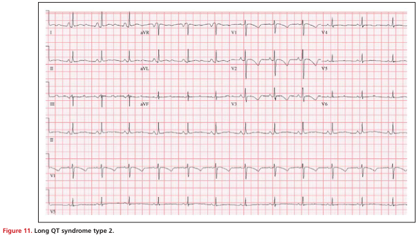
Summary : This figure presents a 12-lead electrocardiogram (ECG) tracing from a patient with Long QT syndrome type 2, illustrating the characteristic prolonged QT interval. ecg tracing: # Leads & Layout : • Standard 12-lead ECG format: I, II, III, aVR, aVL, aVF, V1–V6. • Each lead is displayed in a horizontal row, with time progressing left to right. • Vertical grid lines represent time intervals (small squares = 0.04 s, large squares = 0.2 s). # Key Features : • Regular sinus rhythm is present. • QT interval is visibly prolonged in multiple leads (notably V2–V5). • T waves are broad and notched in some leads, consistent with Long QT syndrome type 2. • No evidence of acute ischemia, bundle branch block, or significant arrhythmia. # Technical Details : • Paper speed: standard 25 mm/s. • Amplitude: standard 10 mm/mV. • Calibration marker visible at the start of each lead row. # Analysis : • The ECG demonstrates a prolonged QT interval, a hallmark of Long QT syndrome type 2. • T wave morphology (broad, notched) is typical for this subtype. • No other acute abnormalities are apparent; the tracing is otherwise unremarkable except for the QT prolongation.
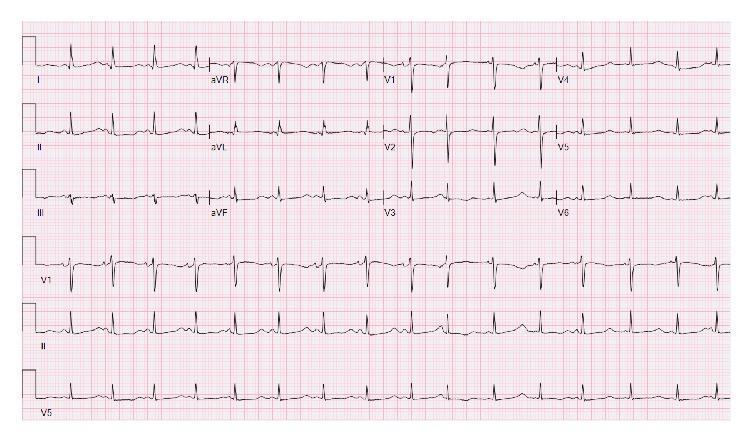
A standard 12-lead electrocardiogram (ECG) demonstrating a sinus rhythm with a significantly prolonged QT interval. The QTc is calculated at 632 ms, which represents a high risk for the development of Torsades de Pointes. The T waves are notably broad and prominent across most leads, appearing upright in leads I, II, aVF, and V2-V6, while inverted in lead aVR. The QRS complexes maintain a normal duration and morphology with appropriate R-wave progression in the precordial leads (V1 to V6), showing no signs of bundle branch blocks or acute ST-segment elevation. This diagnostic image serves as a clinical example of severe drug-induced or electrolyte-related QT prolongation, commonly associated with conditions like hypokalemia or hypomagnesemia in a clinical setting. The visual illustrates the critical importance of interval measurement in ECG analysis for identifying life-threatening arrhythmia risks.
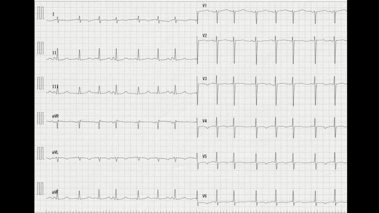
A standard 12-lead electrocardiogram (ECG) showing a regular sinus rhythm at approximately 75 beats per minute. The primary diagnostic finding is a prolonged QT interval, with a corrected QT (QTc) calculated at 458 ms. The limb leads (I, II, III, aVR, aVL, aVF) demonstrate relatively normal axis and morphology, though the T waves appear notably tall and peaked in multiple leads. In the precordial leads (V1-V6), the QRS complexes remain narrow with a normal R-wave progression and a transition zone occurring between V3 and V4. The ST segments are isoelectric without significant elevation or depression. The tall, peaked T waves and prolonged QTc interval are the most prominent educational features, which in this clinical context may be associated with metabolic disturbances such as hypocalcemia or primary hypoparathyroidism. This ECG serves as a visual reference for diagnosing interval abnormalities and T-wave morphology variations in an acute clinical setting.
ECG Report Interpretation - Mrs. Maheshwari, 62F
Recorded Parameters
| Parameter | Value | Normal Range |
|---|---|---|
| Atrial Rate (AR) | 74 bpm | 60-100 bpm |
| Ventricular Rate (VR) | 74 bpm | 60-100 bpm |
| QRS Duration (QRSD) | 106 ms | <120 ms |
| QT Interval | 498 ms | <440 ms |
| QTcB (Bazett's corrected QT) | 553 ms | <440 ms (F), <450 ms (F) |
| PR Interval (PRI) | 148 ms | 120-200 ms |
| P-R-T Axis | 56° / 69° / 0° | P: 0-90°, QRS: 0-90° |
Key Findings
- AR = VR = 74 bpm, confirming 1:1 AV conduction with normal sinus rhythm.
- PR interval of 148 ms is within normal limits (120-200 ms), so no AV block or pre-excitation.
- At 106 ms, the QRS is slightly widened but below the 120 ms threshold for bundle branch block. This could represent an incomplete bundle branch block or intraventricular conduction delay - borderline finding requiring clinical correlation.
- Normal QTc for women: <450 ms
- Borderline prolonged: 450-480 ms
- Prolonged (moderate risk): 480-500 ms
- Severely prolonged (high risk): >500 ms ← This patient is at 553 ms
Reference ECG - Prolonged QT Interval
Differential Diagnosis for Prolonged QTc
- Drug-induced (most common cause): antiarrhythmics (sotalol, amiodarone, dofetilide), antibiotics (erythromycin, clarithromycin, fluoroquinolones), antipsychotics, antidepressants (escitalopram), antifungals (fluconazole), antimalarials
- Electrolyte disturbances: hypokalemia, hypomagnesemia, hypocalcemia
- Cardiac conditions: acute MI, myocarditis, cardiomyopathy
- Hypothyroidism
- Intracranial pathology (subarachnoid hemorrhage)
- Long QT Syndrome (LQTS) - less likely in a 62-year-old without prior history, but possible
Clinical Recommendations
-
Urgent medication review - Identify and stop any QT-prolonging drugs immediately. Cross-reference against the patient's full medication list (Maudsley Guidelines lists erythromycin, clarithromycin, fluoroquinolones, chloroquine, antipsychotics, antidepressants as common offenders).
-
Urgent electrolyte panel - Check serum K⁺, Mg²⁺, Ca²⁺ and correct any deficiencies. Hypokalemia and hypomagnesemia are common reversible causes.
-
Full 12-lead ECG review - This report shows calculated values; the actual 12-lead tracing should be reviewed for T-wave morphology (notching, bifid T-waves), ST-segment changes, and QRS morphology.
-
Thyroid function tests (TSH) and basic metabolic panel.
-
Consider cardiology referral - A QTcB of 553 ms warrants specialist input. Continuous cardiac monitoring may be appropriate depending on symptoms (syncope, palpitations, near-syncope).
-
Symptom assessment - Ask specifically about syncope, presyncope, palpitations, and family history of sudden cardiac death.
-
Avoid triggers - Until the cause is identified, avoid QT-prolonging drugs, correct electrolytes, and advise against intense physical exertion.
Important disclaimer: This interpretation is based on the computed ECG parameters only - the actual waveform tracing was not available for visual analysis. A qualified cardiologist must review the full 12-lead ECG tracing in conjunction with the patient's clinical history, medications, and symptoms before any clinical decisions are made. The machine-calculated QTcB using Bazett's formula can overcorrect at faster heart rates - though at 74 bpm this is less of a concern here.
Clonofit
Clonofit clonazepam tablet uses dose side effects
Clonofit (Clonazepam)
Mechanism of Action
- Plasma protein binding: ~85%
- Half-life: 20-60 hours (mean ~30 hours) - one of the longer-acting benzodiazepines
- Peak plasma levels: 1-4 hours after oral dosing
- Metabolism: Hepatic - reduction of the nitro group to inactive 7-amino derivatives; less than 1% excreted unchanged in urine
Indications
| Indication | Notes |
|---|---|
| Epilepsy / Seizures | Absence seizures, myoclonic seizures, atonic seizures, Lennox-Gastaut syndrome |
| Panic Disorder | FDA-approved; preferred over alprazolam by some due to longer half-life and slower onset |
| Anxiety disorders | Social anxiety, generalized anxiety (adjunct) |
| REM Sleep Behavior Disorder (RBD) | First-line or second-line (with melatonin) |
| Akathisia | Restlessness due to antipsychotic medications |
| Acute catatonia | Short-term management |
Dosing
| Indication | Adult Starting Dose | Usual Range | Max |
|---|---|---|---|
| Seizure disorders | 0.5 mg TID | 0.5-2 mg TID | 20 mg/day |
| Panic disorder | 0.25 mg BD | 0.5-2 mg/day | 4 mg/day |
| Adults (general) | Should not exceed 1.5 mg/day initially | Titrate slowly | Depends on indication |
| Children (seizures) | 0.01-0.05 mg/kg/day | Divided doses | 0.2 mg/kg/day |
Note: Tolerance to antiseizure effects often develops after 1-6 months; some patients no longer respond at any dose after this period. (Goodman & Gilman's)
Available Formulations (Clonofit)
- Tablets: 0.25 mg, 0.5 mg, 1 mg, 2 mg
- Liquid/oral solution also available in some markets
Side Effects
- Drowsiness / sedation (~50% of patients initially - tolerance usually develops)
- Dizziness and unsteadiness
- Ataxia and muscular incoordination
- Memory problems and cognitive slowing
- Fatigue and depression
- Respiratory depression - especially dangerous with opioids or alcohol (can be fatal)
- Paradoxical disinhibition - increased agitation, aggression, impulsivity (especially in children, elderly, or those with cognitive impairment)
- Suicidal ideation (class warning for all antiseizure medications)
- Unusual involuntary eye movements
Important Warnings
1. Physical Dependence and Withdrawal (FDA Boxed Warning)
- Severe anxiety, insomnia, irritability
- Tremor, sweating, confusion
- Seizures - potentially life-threatening
- Withdrawal symptoms can last several weeks to more than 12 months
2. Opioid Combination - Black Box Warning
3. Alcohol
4. Tolerance
Contraindications
- Severe hepatic impairment
- Acute narrow-angle glaucoma
- Myasthenia gravis
- Severe respiratory insufficiency
- Known hypersensitivity to benzodiazepines
- Use in pregnancy (crosses placenta; neonatal withdrawal / "floppy infant" syndrome)
Drug Interactions
| Interacting Drug/Class | Effect |
|---|---|
| Opioids | Severe CNS/respiratory depression, death |
| Alcohol | Enhanced CNS depression |
| Other CNS depressants (antihistamines, antipsychotics, other benzodiazepines) | Additive sedation |
| CYP3A4 inhibitors (ketoconazole, erythromycin, clarithromycin) | Increased clonazepam levels |
| CYP3A4 inducers (rifampicin, phenytoin, carbamazepine) | Decreased clonazepam levels |
| Valproate | May cause absence status epilepticus when combined |
Special Populations
- Elderly: Prolonged sedation; higher fall/fracture risk; use lowest effective dose
- Pregnancy: Avoid if possible - neonatal withdrawal, cleft palate risk (teratogen Category D)
- Renal impairment: Use with caution; monitor
- Children: Efficacy established for seizures; behavioural paradoxical reactions more common
Equivalence
Clinical Note (relevant to the ECG above): Clonazepam itself does not significantly prolong the QT interval and is generally considered safe from a cardiac repolarization standpoint - unlike many other psychotropics. If this patient (Mrs. Maheshwari) is being prescribed Clonofit, it is unlikely to be the cause of her prolonged QTc of 553 ms. Other medications in her regimen should be reviewed first.