
Shock. Etiology and classification. Pathogenesis of different types of shock (decompensation). Morphological features.
Reading File
Finding Sources
Finding Sources
Searching PubMed
"shock"[MeSH Terms] AND "classification"
Reading File
Searching Images
shock pathogenesis morphology acute tubular necrosis or diffuse alveolar damage shock lung
Reading File
Reading File
Reading File
Reading File
Reading File
Shock
Definition. Shock is circulatory failure with impaired tissue perfusion and cellular hypoxia, caused by reduced cardiac output and/or reduced effective circulating volume. Early cell injury is reversible. Prolonged shock causes irreversible injury and death (Robbins, Cotran & Kumar Pathologic Basis of Disease, p. 133).
1. Etiology and classification
Robbins groups the causes into three major types. Neurogenic and anaphylactic shock are listed separately (Robbins Pathologic Basis of Disease, p. 134, Table 4.3). Many courses use the broader hemodynamic scheme below.
| Type | Main mechanism | Causes |
|---|---|---|
| Hypovolemic | Low blood or plasma volume | Hemorrhage, trauma, burns, vomiting, diarrhea, polyuria |
| Cardiogenic | Pump failure from intrinsic myocardial damage, extrinsic compression, or outflow obstruction | Myocardial infarction, ventricular rupture, arrhythmia, cardiac tamponade, pulmonary embolism |
| Septic | Dysregulated host response to infection with cytokine cascades, vasodilation, endothelial injury, DIC | Gram-positive bacteria (most frequent), gram-negative bacteria, fungi, viruses (e.g. SARS-CoV-2) |
| Neurogenic | Loss of vascular tone with acute vasodilation | Spinal cord injury, anesthesia |
| Anaphylactic | IgE-mediated systemic mast cell degranulation with vasodilation and increased permeability | Drugs, venom, food |
- Obstructive shock. Some classifications make this a separate group: tamponade, tension pneumothorax, massive pulmonary embolism. Robbins places these under cardiogenic shock.
- Distributive shock. This group covers septic, neurogenic and anaphylactic shock. The shared problem is vasodilation and maldistribution of flow.
- Related terms.
- Sepsis is life-threatening organ dysfunction caused by a dysregulated host response to infection.
- Septic shock is the subset of sepsis with profound circulatory, cellular and metabolic abnormalities and higher mortality.
- SIRS is a sepsis-like state triggered by non-infectious insults such as burns, trauma and pancreatitis.
2. Pathogenesis: stages and decompensation
Shock evolves through three stages. They are best documented in hypovolemic shock but are common to the other forms (Robbins Pathologic Basis of Disease, p. 136).
Stage I: Nonprogressive (compensated)
- Reflex compensation keeps perfusion of vital organs.
- Mechanisms are baroreceptor reflexes, catecholamines, ADH, activation of the renin-angiotensin-aldosterone axis, and generalized sympathetic stimulation.
- Net effects are tachycardia, peripheral vasoconstriction and renal fluid conservation.
- Cutaneous vasoconstriction gives cool, pale skin. Early septic shock may instead give warm, flushed skin from vasodilation.
- Coronary and cerebral vessels respond less to sympathetic signals, so blood is shunted to the heart and brain.
Stage II: Progressive (decompensation)
- Compensation fails and hypoperfusion becomes widespread.
- Persistent oxygen deficit shifts cells from aerobic respiration to anaerobic glycolysis. Lactic acid accumulates and causes metabolic lactic acidosis.
- Low tissue pH blunts the vasomotor response. Arterioles dilate and blood pools in the microcirculation.
- Pooling lowers venous return and cardiac output. It also exposes endothelium to anoxic injury, which can lead to DIC.
- Vital organs begin to fail.
Stage III: Irreversible
- Cell injury is severe enough that survival is not possible even if the hemodynamics are corrected.
- Lysosomal enzymes leak and worsen the shock state.
- Myocardial contractility falls further, partly from increased nitric oxide synthesis.
- Ischemic bowel lets intestinal flora enter the circulation, so bacteremic shock may be superimposed.
- Progression to renal failure, with multiorgan failure and death, is common. The textbook text for this last point was partly truncated in my retrieval, so I filled it in from general knowledge.
Pathogenesis by type
Hypovolemic shock
- Volume loss lowers preload and cardiac output, and the stages above follow.
- Compensation can maintain blood pressure after about 10 to 15% volume loss. Beyond that, decompensation sets in.
Cardiogenic shock
- Pump failure lowers cardiac output despite adequate volume.
- Venous pressures are raised, with congestion.
- Compensatory vasoconstriction raises afterload, which increases myocardial oxygen demand.
- This creates a vicious circle of ischemia and worsening pump failure.
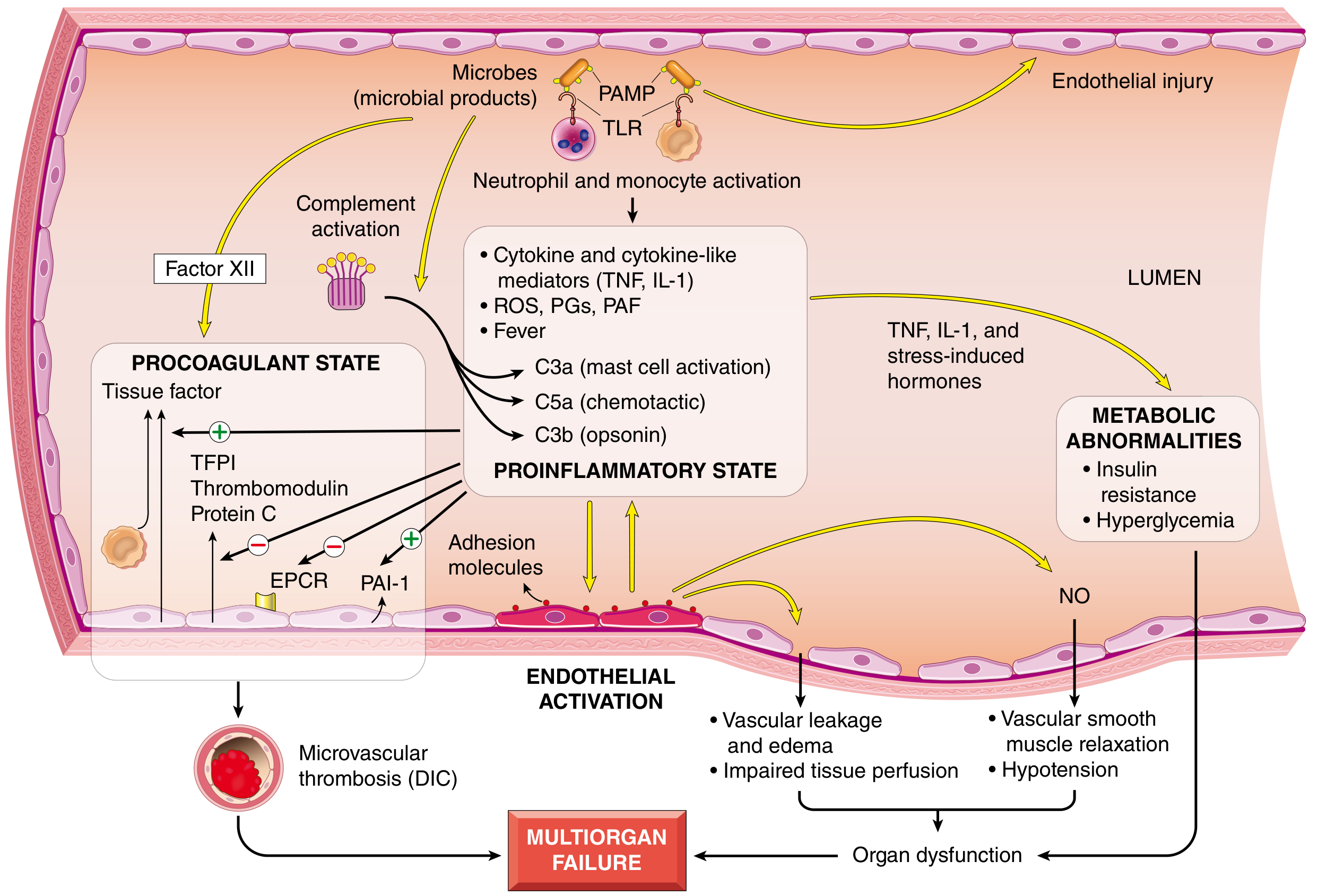
Septic shock (Robbins Pathologic Basis of Disease, pp. 134-136; Fig. 4.20)
- Inflammatory and counter-inflammatory responses.
- PAMPs and DAMPs engage TLRs, G-protein-coupled receptors and C-type lectin receptors. NF-kB is activated.
- TNF, IL-1, IL-12, IL-18, IFN-γ and HMGB1 are released.
- Complement is activated, producing C3a and C5a.
- A counter-regulatory immunosuppression follows, with IL-10, soluble TNF receptor, lymphocyte apoptosis and anergy. Patients may oscillate between hyperinflammatory and immunosuppressed states.
- Endothelial activation and injury.
- Tight junctions loosen, causing vascular leak and protein-rich edema.
- NO, C3a, C5a and PAF cause vasodilation and hypotension.
- Microvascular flow is heterogeneous and autoregulation is lost, so oxygen delivery does not match need.
- Procoagulant state.
- Tissue factor rises. Thrombomodulin, protein C and TFPI fall. PAI-1 rises, which suppresses fibrinolysis.
- Neutrophil extracellular traps may also promote coagulation.
- The result is fibrin thrombi in small vessels and DIC in up to half of septic patients. Consumption of factors and platelets then causes bleeding.
- Metabolic abnormalities.
- Insulin resistance with hyperglycemia, raised triglycerides and lactic acidosis.
- Adrenal insufficiency may follow an initial glucocorticoid surge. It can be functional or from adrenal hemorrhagic necrosis (Waterhouse-Friderichsen syndrome).
- Organ dysfunction.
- Hypotension, edema, microvascular dysfunction and thrombosis reduce oxygen delivery.
- Mitochondrial damage impairs oxygen use.
- Cytokines depress myocardial contractility.
- Increased permeability and endothelial injury can lead to ARDS.
- Superantigens (e.g. toxic shock syndrome) cause polyclonal T-cell activation and a similar picture.
Neurogenic and anaphylactic shock
- Both cause acute vasodilation, hypotension and tissue hypoperfusion.
- In neurogenic shock, sympathetic tone is lost.
- In anaphylaxis, mast cell mediators (histamine, leukotrienes) act.
3. Morphological features
The changes are those of hypoxic injury, caused by hypoperfusion plus microvascular thrombosis (Robbins Pathologic Basis of Disease, p. 136). The organs most involved are the brain, heart, kidneys, adrenals and gastrointestinal tract.
- Kidney. Acute tubular necrosis (acute kidney injury) is the key lesion and leads to oliguria. Fibrin thrombi are most readily seen in the glomeruli.
- Heart. Hypoxic injury with possible subendocardial hemorrhage and focal or coagulative necrosis of myocytes.
- Brain. Ischemic encephalopathy with neuronal loss, especially in watershed zones.
- Adrenals. Lipid depletion of the cortical cells, a stress reaction from use of stored lipids for steroid synthesis. In DIC with sepsis, hemorrhagic necrosis can occur (Waterhouse-Friderichsen syndrome).
- Lungs.
- They resist hypoxic injury in hemorrhagic shock.
- Sepsis or trauma can cause diffuse alveolar damage, called "shock lung": edema, hyaline membranes, then type II pneumocyte hyperplasia.
- Gastrointestinal tract. Mucosal hemorrhagic necrosis ("hemorrhagic enteropathy") and stress ulcers. Barrier loss allows bacterial translocation.
- Liver. Centrilobular hemorrhagic necrosis and fatty change.
- Microvasculature. Fibrin thrombi from DIC may be present in any tissue.
- Recovery. If the patient survives, tissues recover completely except for neuronal and cardiomyocyte loss.
Clinical features and prognosis
- Hypovolemic and cardiogenic shock give hypotension, a weak rapid pulse, tachypnea, and cool, clammy, cyanotic skin.
- Septic shock may give warm, flushed skin.
- More than 90% of young, healthy patients with hypovolemic shock survive with treatment. Septic and cardiogenic shock have much poorer outcomes. Sepsis mortality is about 40%.
Recent evidence check. A PubMed search of systematic reviews and meta-analyses from the last two years found nothing that contradicts these pathophysiology fundamentals. The hits were on shock index in trauma (PMID 42854533) and in acute coronary syndrome (PMID 42183638), and on point-of-care ultrasound in shock (PMID 40603759).
This is a shared conversation. Sign in to Orris to start your own chat.