from Guyton and Hall textbook of medical physiology, 4th South Asia edition, write a 5-mark short note on Intrinsic Pathway of Coagulation

Reading File
Finding Sources
Finding Sources
Finding Sources
Finding Sources
Reading File
Reading File
Reading File
Finding Sources
Searching Images

intrinsic pathway blood coagulation cascade factor XII XI IX VIII diagram
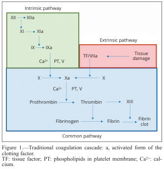
Summary : This diagram illustrates the traditional coagulation cascade, detailing the sequence of events and factors involved in blood clotting via the intrinsic, extrinsic, and common pathways.

flowchart:
# Pathways :
  • Intrinsic pathway (green box): Initiated by factor XII, proceeds through XI, IX, and VIII, with activation steps indicated by "a" (e.g., XIIa, XIa, IXa).
  • Extrinsic pathway (red box): Initiated by tissue damage, which activates tissue factor (TF) and factor VIIa.
  • Common pathway (blue box): Both intrinsic and extrinsic pathways converge at factor X, leading to the activation of prothrombin to thrombin, which then converts fibrinogen to fibrin, forming a fibrin clot.

# Nodes :
  • XII → XIIa (Intrinsic)
  • XI → XIa (Intrinsic)
  • IX → IXa (Intrinsic)
  • Ca²⁺, PT, V (Intrinsic, Common)
  • TF/VIIa (Extrinsic)
  • Tissue damage (Extrinsic)
  • X → Xa (Common)
  • Prothrombin → Thrombin (Common)
  • Fibrinogen → Fibrin (Common)
  • XIII (Common)
  • Fibrin clot (Common)

# Connectors :
  • Arrows indicate the direction of activation or conversion between factors.
  • Intrinsic and extrinsic pathways both feed into the activation of factor X in the common pathway.
  • Ca²⁺ and PT, V are required cofactors at specific steps.

# Layout :
  • Three main colored sections: green (intrinsic, top left), red (extrinsic, top right), blue (common, bottom).
  • Flow proceeds from top (initiation) to bottom (clot formation).
  • Arrows show sequential activation and convergence of pathways.

# Analysis :
  • The diagram clearly separates the intrinsic and extrinsic initiation mechanisms, both converging on the common pathway at factor X.
  • The cascade is highly sequential, with each activated factor catalyzing the next step.
  • Calcium ions (Ca²⁺) and phospholipids (PT) are essential cofactors at multiple steps.
  • The final outcome is the formation of a stable fibrin clot, highlighting the coordinated nature of the coagulation process.
Summary : This diagram illustrates the traditional coagulation cascade, detailing the sequence of events and factors involved in blood clotting via the intrinsic, extrinsic, and common pathways. flowchart: # Pathways : • Intrinsic pathway (green box): Initiated by factor XII, proceeds through XI, IX, and VIII, with activation steps indicated by "a" (e.g., XIIa, XIa, IXa). • Extrinsic pathway (red box): Initiated by tissue damage, which activates tissue factor (TF) and factor VIIa. • Common pathway (blue box): Both intrinsic and extrinsic pathways converge at factor X, leading to the activation of prothrombin to thrombin, which then converts fibrinogen to fibrin, forming a fibrin clot. # Nodes : • XII → XIIa (Intrinsic) • XI → XIa (Intrinsic) • IX → IXa (Intrinsic) • Ca²⁺, PT, V (Intrinsic, Common) • TF/VIIa (Extrinsic) • Tissue damage (Extrinsic) • X → Xa (Common) • Prothrombin → Thrombin (Common) • Fibrinogen → Fibrin (Common) • XIII (Common) • Fibrin clot (Common) # Connectors : • Arrows indicate the direction of activation or conversion between factors. • Intrinsic and extrinsic pathways both feed into the activation of factor X in the common pathway. • Ca²⁺ and PT, V are required cofactors at specific steps. # Layout : • Three main colored sections: green (intrinsic, top left), red (extrinsic, top right), blue (common, bottom). • Flow proceeds from top (initiation) to bottom (clot formation). • Arrows show sequential activation and convergence of pathways. # Analysis : • The diagram clearly separates the intrinsic and extrinsic initiation mechanisms, both converging on the common pathway at factor X. • The cascade is highly sequential, with each activated factor catalyzing the next step. • Calcium ions (Ca²⁺) and phospholipids (PT) are essential cofactors at multiple steps. • The final outcome is the formation of a stable fibrin clot, highlighting the coordinated nature of the coagulation process.

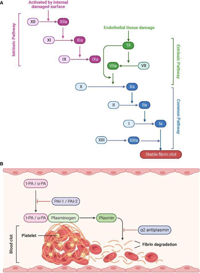
This medical schematic illustrates the physiological processes of hemostasis and fibrinolysis. Part A depicts the Coagulation Cascade, divided into the Intrinsic, Extrinsic, and Common pathways. The Intrinsic pathway (purple) is triggered by internal damaged surfaces, activating Factor XII to XIIa, followed by XI and IX. The Extrinsic pathway (green) begins with endothelial tissue damage and Tissue Factor (TF), activating Factor VII to VIIa. Both pathways converge at the Common pathway (blue) starting with the activation of Factor X to Xa. This leads to the conversion of prothrombin (II) to thrombin (IIa), and fibrinogen (I) to fibrin (Ia), culminating in a stable fibrin clot stabilized by Factor XIIIa. Activated factors are denoted by an 'a' suffix. Part B illustrates Fibrinolysis, where tissue-type (t-PA) and urokinase-type (u-PA) plasminogen activators convert plasminogen to plasmin, which then degrades the fibrin network of the blood clot. Inhibitory regulators including PAI-1, PAI-2, and ̡2-antiplasmin are also shown. The diagram represents the structural breakdown of a clot containing platelets and red blood cells into fibrin degradation products.
This medical schematic illustrates the physiological processes of hemostasis and fibrinolysis. Part A depicts the Coagulation Cascade, divided into the Intrinsic, Extrinsic, and Common pathways. The Intrinsic pathway (purple) is triggered by internal damaged surfaces, activating Factor XII to XIIa, followed by XI and IX. The Extrinsic pathway (green) begins with endothelial tissue damage and Tissue Factor (TF), activating Factor VII to VIIa. Both pathways converge at the Common pathway (blue) starting with the activation of Factor X to Xa. This leads to the conversion of prothrombin (II) to thrombin (IIa), and fibrinogen (I) to fibrin (Ia), culminating in a stable fibrin clot stabilized by Factor XIIIa. Activated factors are denoted by an 'a' suffix. Part B illustrates Fibrinolysis, where tissue-type (t-PA) and urokinase-type (u-PA) plasminogen activators convert plasminogen to plasmin, which then degrades the fibrin network of the blood clot. Inhibitory regulators including PAI-1, PAI-2, and ̡2-antiplasmin are also shown. The diagram represents the structural breakdown of a clot containing platelets and red blood cells into fibrin degradation products.

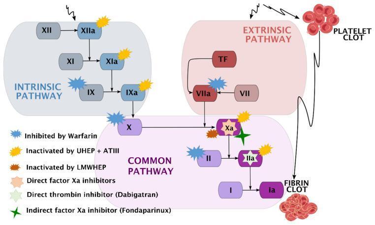
A comprehensive pathophysiology diagram illustrating the human coagulation cascade and the specific pharmacodynamic sites of action for various anticoagulants. The diagram is divided into three functional zones: the Intrinsic Pathway (blue), starting with Factor XII; the Extrinsic Pathway (red), initiated by Tissue Factor (TF) and Factor VII; and the Common Pathway (purple), where the pathways converge at Factor X. The cascade terminates in the conversion of Fibrinogen (I) to Fibrin (Ia), forming a fibrin clot. A secondary visual element depicts a platelet clot associated with the extrinsic pathway. Key clinical pharmacotherapeutic targets are marked with color-coded symbols: Warfarin inhibits Factors IX, VII, X, and II; Unfractionated Heparin (UHEP) + Antithrombin III (ATIII) inactivates Factors XIIa, XIa, IXa, Xa, and IIa; Low-Molecular-Weight Heparin (LMWHEP) and Fondaparinux target Factor Xa; Direct Factor Xa inhibitors act on Factor Xa; and Dabigatran acts as a direct thrombin inhibitor (Factor IIa). This schematic is designed for medical education regarding hemostasis and thromboprophylaxis.

A comprehensive pathophysiology diagram illustrating the human coagulation cascade and the specific pharmacodynamic sites of action for various anticoagulants. The diagram is divided into three functional zones: the Intrinsic Pathway (blue), starting with Factor XII; the Extrinsic Pathway (red), initiated by Tissue Factor (TF) and Factor VII; and the Common Pathway (purple), where the pathways converge at Factor X. The cascade terminates in the conversion of Fibrinogen (I) to Fibrin (Ia), forming a fibrin clot. A secondary visual element depicts a platelet clot associated with the extrinsic pathway. Key clinical pharmacotherapeutic targets are marked with color-coded symbols: Warfarin inhibits Factors IX, VII, X, and II; Unfractionated Heparin (UHEP) + Antithrombin III (ATIII) inactivates Factors XIIa, XIa, IXa, Xa, and IIa; Low-Molecular-Weight Heparin (LMWHEP) and Fondaparinux target Factor Xa; Direct Factor Xa inhibitors act on Factor Xa; and Dabigatran acts as a direct thrombin inhibitor (Factor IIa). This schematic is designed for medical education regarding hemostasis and thromboprophylaxis.

Reading File
Loading Image
I now have the original Guyton figure (Fig. 37.6) and all the textbook content needed. Here is the complete 5-mark short note:

Intrinsic Pathway of Coagulation

(Guyton and Hall Textbook of Medical Physiology, 4th South Asia Edition, Chapter 37)

Definition / Introduction

The intrinsic pathway is one of two mechanisms by which prothrombin activator is formed, ultimately leading to blood coagulation. It is called "intrinsic" because it begins entirely within the blood itself - triggered by trauma to the blood or by contact of blood with collagen exposed in a damaged vascular wall. It is slower than the extrinsic pathway, typically requiring 1 to 6 minutes to produce a clot.

Steps of the Intrinsic Pathway (5-Step Cascade)
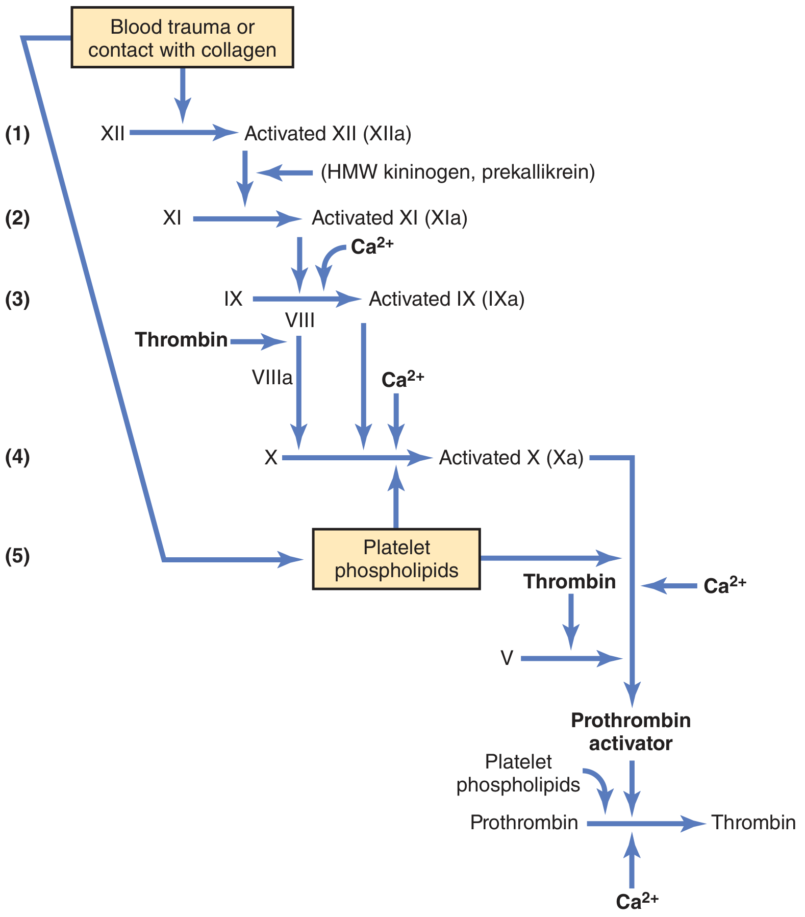
Figure 37.6 - Intrinsic pathway for initiating blood clotting (Guyton and Hall)
Figure 37.6 - Intrinsic pathway for initiating blood clotting. HMW, high-molecular weight. (Guyton and Hall Textbook of Medical Physiology)

Step 1 - Activation of Factor XII and Release of Platelet Phospholipids

When blood is traumatized or comes in contact with vascular wall collagen, two important events occur:
  • Factor XII is disturbed and undergoes a conformational change, converting it into a proteolytic enzyme called activated factor XII (XIIa).
  • The platelets adhere to collagen (or are otherwise damaged), releasing platelet phospholipids containing the lipoprotein platelet factor 3, which plays a role in subsequent reactions.

Step 2 - Activation of Factor XI

Activated factor XII (XIIa) acts enzymatically on factor XI to produce activated factor XI (XIa). This reaction also requires high-molecular-weight (HMW) kininogen and is accelerated by prekallikrein.

Step 3 - Activation of Factor IX

Activated factor XI (XIa) acts enzymatically on factor IX in the presence of Ca2+ to produce activated factor IX (IXa).

Step 4 - Activation of Factor X (Role of Factor VIII)

Activated factor IXa, acting together with:
  • Activated factor VIII (VIIIa)
  • Platelet phospholipids (platelet factor 3)
  • Ca2+
...activates factor X to produce activated factor X (Xa).
Clinical note: Factor VIII is the factor missing in classic hemophilia (Hemophilia A), making this step deficient in that condition. When platelets are deficient, the disease is called thrombocytopenia.

Step 5 - Formation of Prothrombin Activator (Role of Factor V)

Activated factor Xa combines with factor V and platelet/tissue phospholipids to form the complex called prothrombin activator. This step is identical to the last step in the extrinsic pathway (the two pathways converge here). Prothrombin activator then cleaves prothrombin into thrombin (in the presence of Ca2+), which sets off the final clotting process.

Role of Calcium Ions

Except for the first two steps (activation of factor XII and factor XI), calcium ions (Ca2+) are required for promotion or acceleration of all subsequent reactions. In the absence of Ca2+, clotting by the intrinsic pathway does not occur. This is exploited clinically - blood removed from the body can be prevented from clotting by deionizing calcium using citrate ions (calcium chelation) or precipitating calcium with oxalate ions.

Positive Feedback by Thrombin

Once thrombin is formed, it amplifies its own production by activating factors VIII, IX, X, XI, and XII, and by promoting platelet aggregation. This positive feedback ensures the clot grows rapidly until bleeding stops.

Comparison with Extrinsic Pathway

FeatureIntrinsic PathwayExtrinsic Pathway
TriggerBlood trauma / contact with collagenTissue trauma / tissue factor release
SpeedSlow (1-6 min)Rapid (as little as 15 sec)
Initial factorFactor XIITissue factor + Factor VII
ConvergenceBoth converge at Factor X activation

Source: Guyton and Hall Textbook of Medical Physiology, 4th South Asia Edition, Chapter 37 - Hemostasis and Blood Coagulation, pp. 484-486.
This is a shared conversation. Sign in to Orris to start your own chat.