
Polyps
Reading File
Reading File
Reading File
Which type of polyp do you mean?
- Nasal polyps
- Colon/rectal polyps
- Uterine/endometrial polyps
- Gallbladder polyps
- Cervical or stomach polyps
Tell me the location or any symptoms, and I’ll explain causes, risks, tests, and treatment.
Nasal polyps essay
Reading File
Finding Sources
Finding Sources
Finding Sources
Searching PubMed
"nasal polyps" AND treatment
Searching the Web
EPOS 2020 chronic rhinosinusitis with nasal polyps intranasal corticosteroids surgery biologics
Reading File
Reading File
Reading File
Finding Sources
Finding Sources
Reading File
Reading File
Reading File
Searching Images
endoscopic appearance bilateral inflammatory nasal polyps
Reading File
Reading File
Reading File
Nasal Polyps
Nasal polyps are benign, soft, painless, oedematous protrusions of the nasal and paranasal sinus mucosa. They are most often a manifestation of chronic rhinosinusitis with nasal polyps (CRSwNP). They commonly arise around the middle meatus and ethmoidal sinuses, and are usually bilateral, pale, translucent, mobile, and insensitive to touch. - Scott-Brown's Otorhinolaryngology Head & Neck Surgery, Vol. 1, p. 1085
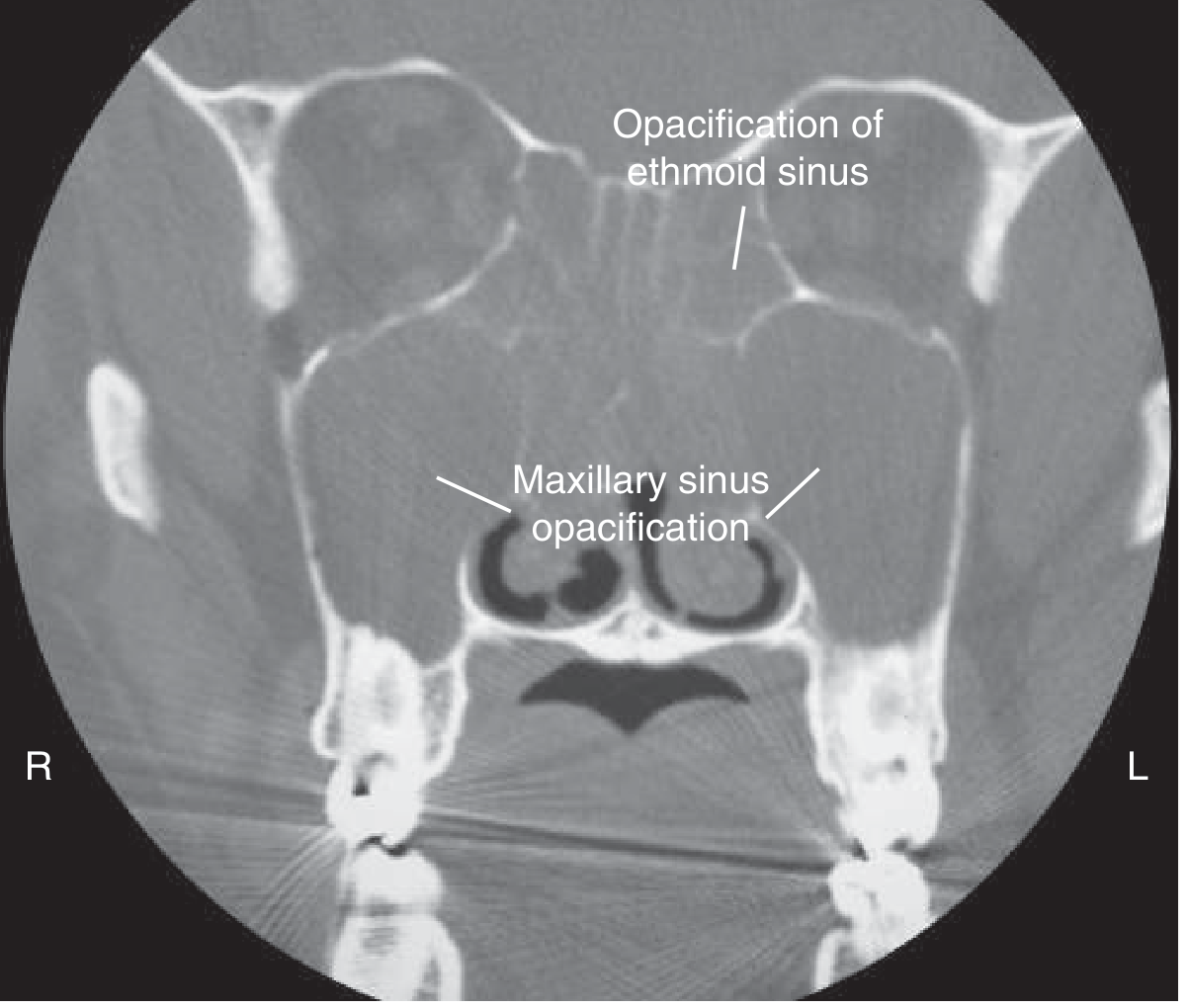
CT appearance of bilateral nasal polyposis. - Goldman-Cecil Medicine, p. 4169
Epidemiology
Nasal polyps are found in about 1% to 4% of adults and are uncommon in children. They occur more often in men, usually develop in adulthood, and tend to follow a chronic relapsing course. In a child, their presence should prompt assessment for disorders such as cystic fibrosis and, where appropriate, exclusion of congenital lesions such as meningocele or encephalocele. - Scott-Brown's Otorhinolaryngology Head & Neck Surgery, Vol. 1, p. 1085; Bailey and Love's Short Practice of Surgery, p. 793
Etiology and associations
The exact cause is not fully understood. Polyps result from persistent inflammation, mucosal oedema, and abnormal interaction between sinonasal epithelium and immune cells. Most CRSwNP has a type 2 inflammatory pattern involving IL-4, IL-5, IL-13, IgE, and eosinophils.
Important associations include:
- Chronic rhinosinusitis
- Asthma
- Aspirin or NSAID-exacerbated respiratory disease, formerly called Samter triad
- Allergic fungal rhinosinusitis
- Cystic fibrosis
- Eosinophilic granulomatosis with polyangiitis
- Occasionally, allergy and infection as contributing factors
A minority have a neutrophil-predominant, IL-17-mediated inflammatory pattern. - Goldman-Cecil Medicine, p. 4169
Pathology
Grossly, nasal polyps resemble pale grey, glistening, grape-like masses. Microscopically, they consist of loose oedematous connective tissue containing inflammatory cells and fluid, usually covered by pseudostratified ciliated columnar respiratory epithelium. Eosinophils are prominent in the common type 2 inflammatory form. - Scott-Brown's Otorhinolaryngology Head & Neck Surgery, Vol. 1, p. 1085
Clinical features
Small polyps can be asymptomatic. Larger polyps commonly cause:
- Progressive bilateral nasal obstruction or congestion
- Persistent watery rhinorrhoea or postnasal drip
- Reduced smell, hyposmia, or complete loss of smell
- Mouth breathing, snoring, and disturbed sleep
- Dull facial pressure or headache
- Visible pale mass at the nostril in advanced cases
- Recurrent or persistent sinus symptoms
Benign polyps do not usually bleed. Epistaxis, marked pain, facial swelling, eye symptoms, or a unilateral mass should raise concern for another diagnosis. - Scott-Brown's Otorhinolaryngology Head & Neck Surgery, Vol. 1, p. 1085
Diagnosis
Diagnosis is based on history and nasal examination, ideally with nasal endoscopy. Endoscopy shows pale, smooth, translucent masses, usually arising from the middle meatus. CT of the nose and paranasal sinuses demonstrates the extent of polyps, sinus disease, and anatomy before surgery.
Chronic rhinosinusitis is diagnosed when symptoms such as nasal blockage or discharge, with or without facial pressure and loss of smell, persist along with endoscopic evidence of polyps or characteristic CT changes. - Scott-Brown's Otorhinolaryngology Head & Neck Surgery, Vol. 1, p. 1085
Differential diagnosis
A unilateral nasal mass is not assumed to be a simple inflammatory polyp until other causes are excluded. Differential diagnoses include:
- Antrochoanal polyp
- Inverted papilloma
- Allergic fungal rhinosinusitis
- Malignancy of the nose or paranasal sinuses
- Meningoencephalocele in children
- Foreign body or granulomatous disease
Early biopsy or histopathology is advised for unilateral, bleeding, atypical, or suspicious lesions. - Goldman-Cecil Medicine, p. 4169; Bailey and Love's Short Practice of Surgery, p. 793
Treatment
Treatment aims to improve nasal airflow and smell, control inflammation, reduce polyp size, treat associated disease, and prevent recurrence.
-
Saline irrigation
Regular saline nasal washes help clear mucus and improve delivery of topical medicines. -
Intranasal corticosteroids
These are first-line long-term therapy. They reduce nasal blockage, secretions, sneezing, polyp size, and may improve smell. Benefit generally requires several weeks of regular use. - Scott-Brown's Otorhinolaryngology Head & Neck Surgery, Vol. 1, pp. 331-332 -
Short course of oral corticosteroids
Oral corticosteroids can give rapid improvement in severe obstruction and loss of smell, but benefit may be temporary and repeated courses carry important adverse effects. They should be prescribed only under clinician supervision. - Scott-Brown's Otorhinolaryngology Head & Neck Surgery, Vol. 1, p. 332 -
Management of associated conditions
Asthma, aspirin sensitivity, allergic fungal disease, and cystic fibrosis should be identified and managed. Aspirin desensitisation may help selected patients with aspirin-exacerbated respiratory disease. -
Endoscopic sinus surgery
Functional endoscopic sinus surgery with polypectomy is indicated when symptoms remain troublesome despite appropriate medical therapy, when sinus drainage needs restoration, or when diagnosis is uncertain. Surgery improves symptoms but does not remove the inflammatory tendency, so recurrence is common. Topical nasal steroids after surgery reduce recurrence. - Goldman-Cecil Medicine, p. 4169; Scott-Brown's Otorhinolaryngology Head & Neck Surgery, Vol. 1, p. 332 -
Biologic therapy
For severe uncontrolled CRSwNP despite standard treatment, particularly with type 2 inflammation and asthma, specialist-directed biologics such as dupilumab, omalizumab, or mepolizumab may reduce polyp burden, symptoms, systemic steroid use, and need for surgery. A recent network meta-analysis supports efficacy of biologics, though the best choice depends on the patient’s inflammatory profile, asthma, prior surgery, cost, and access.
Complications and prognosis
Nasal polyps can substantially impair quality of life through chronic obstruction, anosmia, poor sleep, recurrent sinus symptoms, and asthma exacerbation. They are benign but frequently recur, even after surgery. Therefore, long-term follow-up and continued intranasal corticosteroid therapy are important.
Conclusion
Nasal polyps are usually bilateral benign inflammatory lesions associated with chronic rhinosinusitis. The usual presentation is persistent nasal obstruction with loss of smell. Nasal endoscopy and CT establish the diagnosis and extent. Regular intranasal corticosteroids are the foundation of treatment; short systemic steroid courses, endoscopic sinus surgery, and biologics are reserved for selected persistent or severe disease. Unilateral or bleeding polyps require urgent specialist evaluation to exclude fungal disease, inverted papilloma, or malignancy.
Easy
Reading File
Searching PubMed
"nasal polyps" AND treatment
Searching the Web
nasal polyps symptoms diagnosis treatment intranasal corticosteroids official patient information
Reading File
Nasal Polyps - Easy Essay
Nasal polyps are soft, painless, non-cancerous swellings inside the nose. They usually develop because of long-term inflammation of the lining of the nose and sinuses. They often grow from the ethmoid sinuses and are usually present on both sides of the nose.
They look like pale, smooth, grape-like masses. Nasal polyps are more common in adults and are rare in children.
Causes and associations
The exact cause is not always known. They are commonly associated with:
- Chronic sinusitis
- Allergy
- Asthma
- Aspirin sensitivity
- Cystic fibrosis
- Fungal sinusitis
Symptoms
Common symptoms are:
- Blocked nose
- Runny nose or postnasal drip
- Loss or reduced sense of smell
- Headache or facial heaviness
- Mouth breathing and snoring
- Repeated sinus infections
Usually, polyps do not cause pain or bleeding. If there is bleeding or a polyp is present on only one side, an ENT specialist should examine it to rule out other conditions.
Diagnosis
Nasal polyps are diagnosed by examining the nose, often with a nasal endoscope. A CT scan of the paranasal sinuses may be done to see their size and extent.
Treatment
Treatment includes saline nasal wash and steroid nasal sprays to reduce inflammation and shrink the polyps. Short courses of oral steroids may be used in severe cases. If medicines do not help, endoscopic sinus surgery is done to remove the polyps. Polyps can return after surgery, so continued nasal steroid treatment and follow-up are important.
Conclusion
Nasal polyps are common benign swellings caused by chronic inflammation of the nose and sinuses. They mainly cause nasal blockage and loss of smell. Medical treatment is the first choice, while surgery is used for large or recurrent polyps.
Nasal polyps summary
Reading File
Nasal Polyps - Summary
Nasal polyps are soft, painless, non-cancerous swellings inside the nose and sinuses. They are usually caused by long-term inflammation and commonly occur on both sides of the nose.
Common symptoms:
- Blocked or stuffy nose
- Runny nose or postnasal drip
- Reduced or lost sense of smell
- Facial heaviness or headache
- Mouth breathing, snoring, and poor sleep
They are associated with chronic sinusitis, asthma, allergy, aspirin sensitivity, cystic fibrosis, and fungal sinus disease.
Diagnosis is made by nasal examination or endoscopy. A CT scan may be used to assess the sinuses and plan surgery.
Treatment includes saline nasal washes and steroid nasal sprays. Short oral steroid courses may help severe cases. If symptoms persist despite medicines, endoscopic surgery removes the polyps. Polyps may recur, so continued treatment and follow-up are needed.
Important: A one-sided, bleeding, painful, or rapidly growing nasal mass needs urgent ENT assessment to exclude another condition.
Epistaxis essay
Reading File
Finding Sources
Finding Sources
Searching PubMed
epistaxis AND management
Searching the Web
epistaxis first aid guideline pinch nose lean forward 2025
Epistaxis (Nosebleed)
Epistaxis means bleeding from the nose. It is a common condition and is usually mild and self-limiting. It may occur at any age, but anterior nosebleeds are more common in children and young adults, while posterior nosebleeds are more common in older people.
Types
-
Anterior epistaxis
This is the commonest type. Bleeding usually arises from Little’s area (Kiesselbach’s plexus) on the front part of the nasal septum. It is usually easy to control. -
Posterior epistaxis
This arises from deeper vessels in the back of the nose, often in elderly people or patients with hypertension. It can be heavier and may require hospital treatment.
Causes
Local causes
- Nose picking or trauma
- Blowing the nose forcefully
- Dry air and crusting
- Common cold, rhinitis, or sinusitis
- Foreign body, especially in children
- Nasal polyps or tumours
- Deviated nasal septum
General causes
- Hypertension
- Blood-thinning medicines, such as aspirin, clopidogrel, or warfarin
- Bleeding disorders or low platelet count
- Liver disease
- Severe infection or systemic illness
The common causes include nose picking, hypertension, and anticoagulant treatment. - Bailey and Love’s Short Practice of Surgery, p. 793
Clinical features
The patient presents with bleeding from one or both nostrils. Blood may also flow backward into the throat and be swallowed, causing nausea or vomiting. Severe bleeding may lead to dizziness, weakness, pallor, rapid pulse, and rarely shock.
First aid treatment
Most anterior nosebleeds can be managed as follows:
- Sit upright and lean slightly forward.
- Spit out blood rather than swallowing it.
- Pinch the soft lower part of the nose firmly for 10-15 minutes without releasing pressure.
- Apply a cold compress or ice wrapped in cloth over the nose.
- Avoid lying down or tilting the head backward.
Current NHS patient guidance similarly advises leaning forward and firmly pinching the soft part of the nose continuously for 10-15 minutes, as described in this NHS discharge guide.
Medical management
If bleeding continues, the doctor examines the nose to identify the bleeding point.
- A topical vasoconstrictor and local anaesthetic may be used.
- If the bleeding point is seen, it may be treated by chemical cautery with silver nitrate or electrical cautery.
- If cautery fails or the bleeding point is not seen, anterior nasal packing may be required.
- Posterior epistaxis may require posterior packing, endoscopic control of the vessel, arterial ligation, or embolisation.
- In severe bleeding, intravenous fluids, blood tests, correction of coagulation problems, and blood transfusion may be needed.
Direct cautery of an identified bleeding point is preferred where feasible; drugs or coagulation disorders contributing to epistaxis should also be corrected. - Bailey and Love’s Short Practice of Surgery, p. 793; Scott-Brown’s Otorhinolaryngology Head & Neck Surgery, key points
Prevention
After a nosebleed, the patient should avoid nose picking, forceful nose blowing, heavy exercise, hot drinks, alcohol, and hot baths for at least 24 hours. Keeping the inside of the nose moist with saline spray or a lubricating ointment may reduce recurrent bleeding.
Conclusion
Epistaxis is usually a minor anterior nosebleed from Little’s area. Most cases stop with correct pressure and first aid. Persistent, recurrent, heavy, posterior, or bleeding associated with dizziness, breathing difficulty, or blood-thinning medication requires urgent medical assessment.
Epistaxis essay notes
Reading File
Finding Sources
Finding Sources
Searching PubMed
epistaxis AND management
Searching the Web
AAO-HNS clinical practice guideline nosebleed epistaxis patient first aid
Epistaxis - Essay Notes
Definition
Epistaxis means bleeding from the nose. It is a common ENT emergency and is usually mild and self-limiting.
Types
1. Anterior epistaxis
- Most common type
- Common in children and young adults
- Usually arises from Little’s area (Kiesselbach’s plexus) on the anterior nasal septum
- Usually mild and easily controlled
2. Posterior epistaxis
- More common in elderly people
- Often associated with hypertension, atherosclerosis, or anticoagulant use
- Bleeding is usually heavier and may pass into the throat
- Often requires hospital admission and ENT management
Causes
Local causes
- Nose picking
- Trauma or facial injury
- Forceful nose blowing
- Dry air, crusting, or nasal irritation
- Upper respiratory infection, rhinitis, or sinusitis
- Foreign body in children
- Deviated nasal septum
- Nasal polyps or tumours
- Recent nasal surgery
Systemic causes
- Hypertension
- Anticoagulants and antiplatelet drugs
- Bleeding disorders, such as haemophilia or von Willebrand disease
- Thrombocytopenia
- Liver disease
- Leukaemia
- Hereditary haemorrhagic telangiectasia
Clinical features
- Bleeding from one or both nostrils
- Blood running into the throat
- Spitting or vomiting swallowed blood
- Anxiety, weakness, dizziness, or fainting in heavy bleeding
- Pallor, tachycardia, and hypotension in severe blood loss
Assessment
Assess the patient according to ABC:
- Airway: Check whether blood is obstructing the airway.
- Breathing: Assess breathing and oxygen saturation.
- Circulation: Check pulse, blood pressure, and signs of shock.
Take history of trauma, previous episodes, hypertension, bleeding tendency, and medications such as aspirin, clopidogrel, warfarin, or other anticoagulants.
Examine the nasal cavity after bleeding is controlled. Nasal endoscopy may be needed to locate the bleeding point. Investigations in severe or recurrent cases include complete blood count, platelet count, coagulation profile, blood grouping, and cross-matching.
First aid management
- Reassure the patient.
- Sit the patient upright and lean the head forward.
- Ask the patient to spit out blood.
- Pinch the soft lower part of the nose firmly for 10-15 minutes continuously.
- Apply a cold compress over the nose.
- Do not tilt the head backward, as blood may be swallowed or enter the airway.
The AAO-HNS epistaxis guideline supports a stepwise approach from firm compression to directed treatment for persistent or recurrent bleeding.
Definitive treatment
Anterior epistaxis
- Topical vasoconstrictor and local anaesthetic
- Silver nitrate chemical cautery if the bleeding point is visible
- Electrical cautery in selected cases
- Anterior nasal packing if bleeding does not stop
Posterior epistaxis
- Admit to hospital and monitor carefully
- Posterior nasal pack or balloon catheter
- Endoscopic identification and cautery of the bleeding vessel
- Endoscopic sphenopalatine artery ligation for persistent bleeding
- Arterial embolisation if surgery fails or is unsuitable
A visible anterior bleeding point is preferably treated with direct cautery. Packing is used when pressure, vasoconstrictor treatment, and cautery are unsuccessful. - Bailey and Love’s Short Practice of Surgery, p. 793
Complications
- Anaemia and hypovolaemic shock
- Aspiration of blood
- Airway obstruction
- Infection after nasal packing
- Rebleeding
- Pressure necrosis from prolonged packing
Prevention
- Avoid nose picking and forceful nose blowing
- Keep nasal mucosa moist with saline spray or lubricating ointment
- Avoid smoking, alcohol, hot drinks, strenuous exercise, and hot baths for 24 hours after a bleed
- Control hypertension
- Review blood-thinning medication with a clinician, but do not stop it without medical advice
Conclusion
Epistaxis is commonly due to anterior bleeding from Little’s area. Most cases stop with correct first aid and nasal compression. Persistent, recurrent, posterior, or severe bleeding requires medical assessment, cautery, nasal packing, or surgical treatment.
This is a shared conversation. Sign in to Orris to start your own chat.