
Here is the complete MBBS-level notes on CSF - written in simple, exam-friendly language with all the key points you need. --- # CSF - Cerebrospinal Fluid ### (For MBBS Students - Anatomy + Physiology + Pathology) --- ## WHAT IS CSF? CSF is a **clear, colorless, watery fluid** that surrounds and cushions the brain and spinal cord. Think of it as the "water jacket" around your CNS. --- ## FORMATION OF CSF **Where is it formed?** - Mainly by the **Choroid Plexus** (present in all 4 ventricles) - 70% from choroid plexus - Remaining 30% from ependymal cells lining the ventricles + cerebral subarachnoid space **How is it formed?** - Ultrafiltration of plasma + Active secretion - The choroid plexus works like a kidney - it secretes Na+, Cl-, HCO3-, and water into CSF, and absorbs K+ back from CSF **How much is produced?** - Rate: **500 mL/day** (0.3-0.4 mL/min) - Total volume in the body at any time: **90-150 mL** - ~25-30 mL in ventricles - ~120 mL in subarachnoid space - The whole CSF volume is **replaced 3 times a day** (every 5-7 hours) 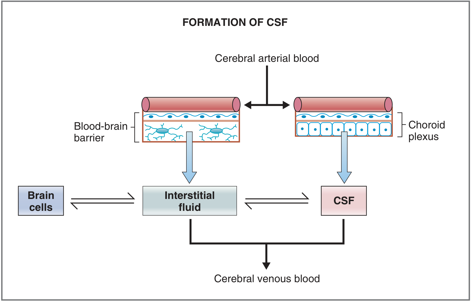 --- ## CIRCULATION OF CSF > **Exam Tip:** Learn this as a one-way flow pathway. This is frequently asked! ``` Lateral Ventricles (choroid plexus) ↓ (via Foramen of Monro / Interventricular foramen) 3rd Ventricle (choroid plexus in roof) ↓ (via Cerebral Aqueduct of Sylvius) 4th Ventricle (choroid plexus in roof) ↓ (via 3 openings) ├── Foramen of Magendie (1, median/midline) └── Foramina of Luschka (2, lateral) ↓ Subarachnoid Space (surrounds brain + spinal cord) ↓ (flows upward over cerebral convexities) Arachnoid Granulations (Pacchionian Granulations) ↓ Superior Sagittal Sinus → Venous Blood ``` 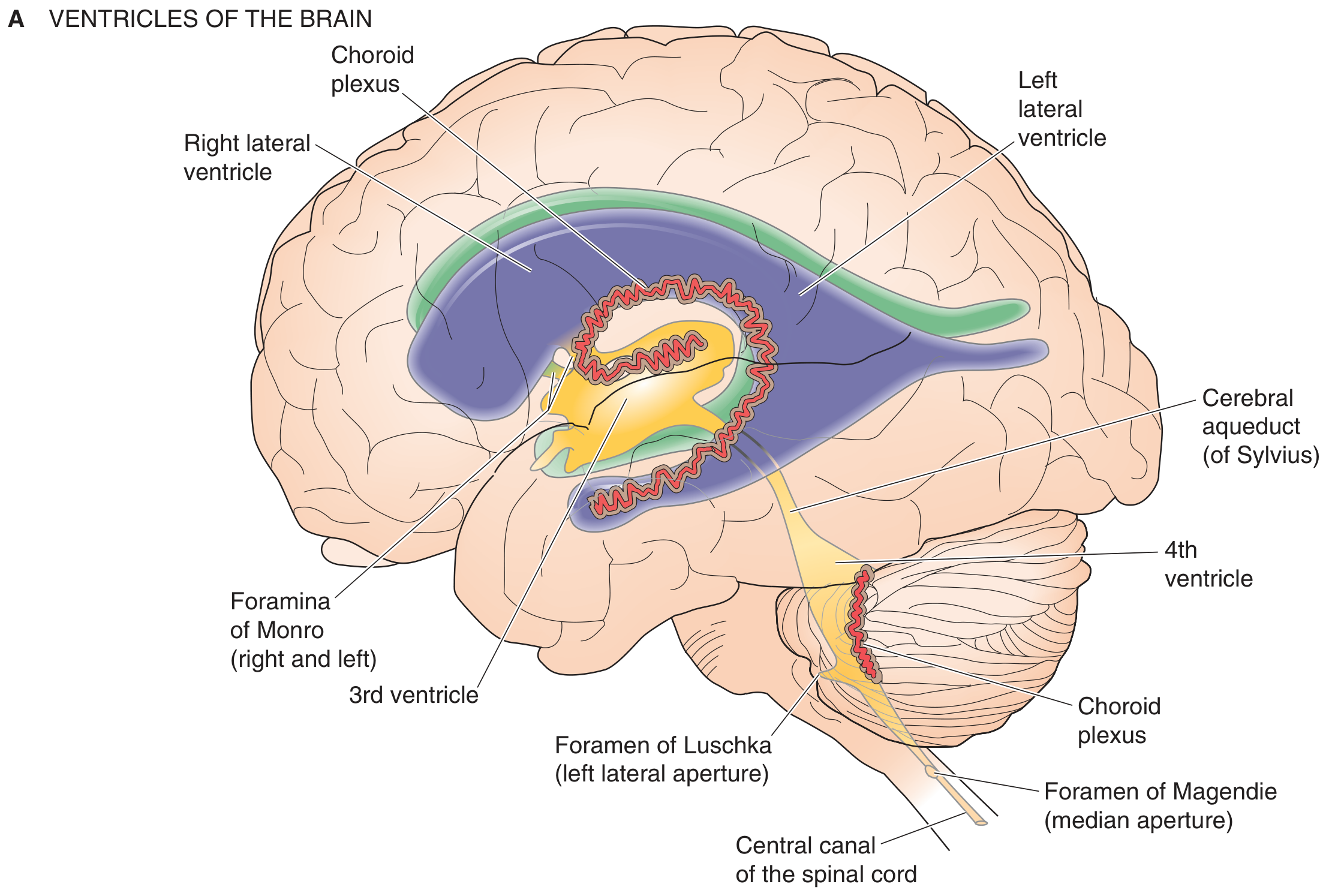 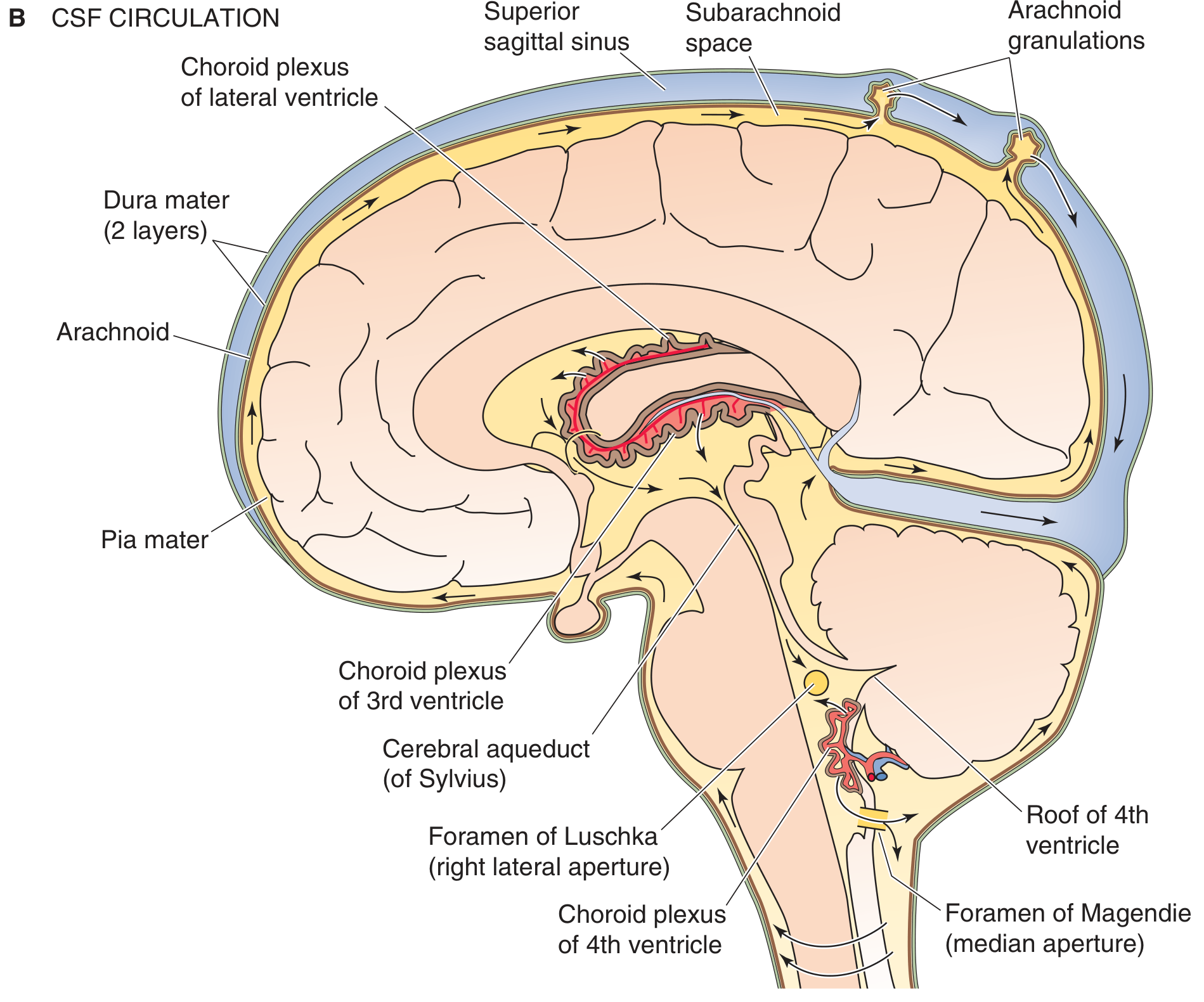 **Key memory tricks:** - **"2L + 1M"** = 2 Luschka (Lateral) + 1 Magendie (Midline) openings of 4th ventricle - **Monro** = connects Lateral to 3rd - **Sylvius** = connects 3rd to 4th --- ## MENINGES (Layers around brain and spinal cord) 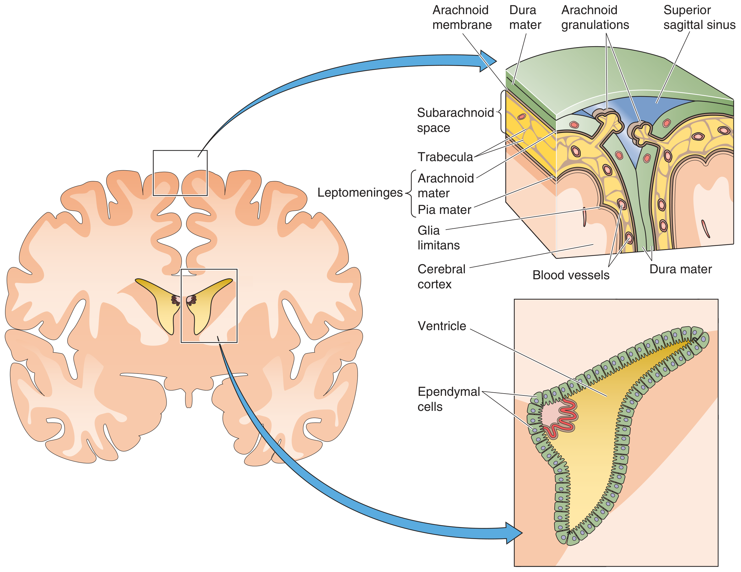 From outside to inside: | Layer | Key Feature | |-------|------------| | **Dura mater** | Outermost, tough; 2 layers intracranially; forms venous sinuses between the layers | | **Arachnoid mater** | Middle; forms **arachnoid granulations** that project into superior sagittal sinus for CSF reabsorption | | **Pia mater** | Innermost; closely stuck to brain surface; follows blood vessels | - **Subarachnoid space** = between arachnoid and pia = filled with CSF - Expanded parts of subarachnoid space = **Cisterns** (e.g., cisterna magna, lumbar cistern) --- ## FUNCTIONS OF CSF 1. **Protection** - cushions brain from trauma (a 1500 g brain weighs only 50 g when floating in CSF!) 2. **Buoyancy** - reduces effective weight of brain 3. **Waste removal** - acts as lymphatics for CNS (brain has no true lymphatics) 4. **Homeostasis** - maintains constant ionic environment for neurons 5. **Transport** - carries hypothalamic hormones to pituitary (median eminence) --- ## APPLIED ANATOMY - LUMBAR PUNCTURE (LP) ### Why do we do LP? To collect CSF for diagnosis of: - Meningitis (bacterial, viral, TB, fungal) - Subarachnoid hemorrhage - Multiple sclerosis - Malignancy (meningeal carcinomatosis) ### Site of LP - **Between L3-L4 or L4-L5** (safest - below conus medullaris which ends at L1-L2) - Patient position: **lateral decubitus (fetal position)** or sitting - to open up the interspaces ### Layers the needle passes through (from outside to inside): > **Mnemonic: "Some Say Liars Love Doing All Procedures"** > **S**kin → **S**ubcutaneous tissue → **S**upraspinous ligament → **I**nterspinous **L**igament → **L**igamentum flavum → **E**pidural space → **D**ura mater → **A**rachnoid mater → **S**ubarachnoid space ✓ 1. Skin 2. Subcutaneous tissue 3. Supraspinous ligament 4. Interspinous ligament 5. Ligamentum flavum 6. Epidural space 7. Dura mater 8. Arachnoid mater 9. **Subarachnoid space** (CSF obtained here) ### Other routes of CSF collection: - **Cisternal puncture** - needle into cisterna magna (between atlas and occipital bone, at back of head) - **Lateral cervical puncture** - **Ventricular tap** - through burr hole (neurosurgical) ### Contraindications to LP: - Raised ICP (risk of **cerebellar tonsillar herniation** - most dangerous complication!) - Infection at LP site (cellulitis, abscess) - Coagulopathy - Spinal cord tumors with neurological deficit ### Complications of LP: - Post-LP headache (most common) - worse on sitting, relieved by lying flat - Cerebellar herniation (most dangerous) - Paresthesia - Infection/meningitis - Hematoma --- ## CSF ANALYSIS ### Collection of tubes: | Tube No. | Goes to | Tests | |----------|---------|-------| | Tube 1 | Chemistry | Glucose, Protein | | Tube 2 | Microbiology | Culture, Gram stain | | Tube 3 | Hematology | Cell count, Differential | | Tube 4 (if needed) | Cytology | Malignant cells | > **Tip:** Tube 3 is used for cell count because it is least contaminated by a traumatic (bloody) tap. --- ## NORMAL CSF VALUES (must memorize!) | Parameter | Normal Value | |-----------|-------------| | **Appearance** | Clear, colorless (like water) | | **Opening pressure** | 90-180 mm H2O (lateral decubitus) | | **WBC** | 0-5 cells/µL (all lymphocytes/monocytes) | | **RBC** | 0 (nil) | | **Protein** | 15-45 mg/dL (some say up to 60) | | **Glucose** | 50-80 mg/dL | | **CSF:Serum glucose ratio** | >0.6 (i.e., CSF glucose = ~2/3 of blood glucose) | | **Chloride** | 115-130 mEq/L | --- ## INTERPRETATION - WHAT ABNORMAL CSF TELLS YOU ### 1. Appearance | CSF Appearance | Cause | |----------------|-------| | Turbid/cloudy | Bacterial meningitis (pus), high WBC | | Xanthochromic (yellow) | Subarachnoid hemorrhage (old blood), high protein, jaundice | | Pink/bloody | SAH or traumatic tap | | Viscous | Cryptococcal meningitis | | Clot forms | Froin syndrome (spinal block), TB meningitis | > **Traumatic tap vs SAH:** In traumatic tap, blood clears from tube 1 to tube 3. In SAH, blood is uniformly present in all tubes + xanthochromia in supernatant after centrifuge. --- ### 2. Pressure | Pressure | Cause | |----------|-------| | High (>180 mm H2O) | Meningitis, brain tumor, cerebral edema, SAH | | Very high (>200 mm H2O) | Remove max 2 mL only | | Low (<80 mm H2O) | CSF leak, dehydration, spinal block above puncture site | --- ### 3. Cells (Pleocytosis) | Cell type increased | Causes | |--------------------|--------| | **Neutrophils (PMNs)** | Bacterial meningitis (early viral/TB too, but shifts to lymphocytes in 2-3 days) | | **Lymphocytes** | Viral meningitis, TB meningitis, fungal, MS, GBS, syphilis | | **Mixed (PMN + lymphocytes)** | TB meningitis, fungal meningitis, Listeria, ruptured brain abscess | | **Eosinophils** | Parasitic infection (most common worldwide), Coccidioides, shunt malfunction | > **High-yield exam fact:** PMN count >1180/µL = 99% predictive of bacterial meningitis --- ### 4. Protein | Protein level | Causes | |--------------|--------| | **Increased** | Bacterial meningitis (highest), TB meningitis, GBS (up to 1000 mg/dL), MS, spinal tumors, acoustic neuroma, DM neuropathy | | **Decreased** | CSF leaks, hyperthyroidism, high ICP | | **Very high (clots)** | Froin syndrome (complete spinal block) | > **GBS (Guillain-Barré):** Classic finding = **Albuminocytologic dissociation** = very high protein + normal/near-normal cell count --- ### 5. Glucose | Glucose | Causes | |---------|--------| | **Low CSF glucose** (ratio <0.4) | Bacterial meningitis, TB meningitis, fungal meningitis, carcinomatous meningitis | | **Normal CSF glucose** | Viral meningitis (KEY feature distinguishing viral from bacterial!) | --- ### 6. Summary Comparison Table (Most Exam-Tested!) | Feature | Normal | Bacterial | Viral | TB/Fungal | |---------|--------|-----------|-------|-----------| | **Appearance** | Clear | Turbid/Purulent | Clear | Slightly turbid | | **Pressure** | 90-180 | High | Normal/slightly high | High | | **WBC** | 0-5 | 100-10,000 (PMN) | 50-1000 (Lymph) | 100-500 (Lymph/Mixed) | | **Protein** | 15-45 | Very high | Mildly high | High | | **Glucose** | 50-80 | Low (<45) | Normal | Low | | **CSF:Serum glucose** | >0.6 | <0.4 | >0.6 | <0.4 | --- ## HYDROCEPHALUS (Applied) | Type | Mechanism | Cause | |------|-----------|-------| | **Non-communicating (Obstructive)** | Block inside ventricular system | Aqueductal stenosis, tumor | | **Communicating** | Block at arachnoid granulations | Post-meningitis, post-SAH | | **Normal Pressure Hydrocephalus (NPH)** | Impaired absorption | Triad: Wet, Wobbly, Wacky (incontinence + gait + dementia) | | **Pseudotumor cerebri (IIH)** | Raised ICP, no structural cause | Obese women, OCP, tetracycline | --- ## QUICK REVISION (Before Exams) **Foramen of Monro** = Lateral → 3rd ventricle **Aqueduct of Sylvius** = 3rd → 4th ventricle **Foramen of Magendie** = 4th ventricle → subarachnoid (midline) **Foramina of Luschka** = 4th ventricle → subarachnoid (lateral, x2) **Arachnoid granulations** = CSF reabsorbed into superior sagittal sinus **LP site** = L3-L4 or L4-L5 (below conus medullaris = L1/L2) **Normal protein** = 15-45 mg/dL **Normal WBC** = 0-5 cells/µL **Normal glucose** = 50-80 mg/dL (2/3 of blood glucose) **Normal pressure** = 90-180 mm H2O **Albuminocytologic dissociation** = GBS (high protein, normal cells) **Low glucose + high PMNs** = Bacterial meningitis **Normal glucose + Lymphocytes** = Viral meningitis **Xanthochromia** = SAH (not traumatic tap)