
Apoptosis
Reading File
Now let me search the medical library for comprehensive content on apoptosis.
Finding Sources
Searching PubMed
apoptosis mechanisms pathways
Good - Robbins and Sleisenger (block1) look most relevant for core pathology content. Let me read those in parallel.
Reading File
Reading File
Excellent content. Let me get the rest of the Robbins apoptosis section and the diagram.
Reading File
Loading Image
Now let me read the core Robbins apoptosis mechanisms section:
Reading File
I have very rich content. Let me also fetch the Robbins mechanisms diagram and read a bit more of the text:
Loading Image
Reading File
I now have comprehensive content. Let me compile the full, detailed answer.
Apoptosis
Apoptosis is a form of programmed cell death in which cells activate intrinsic enzymatic machinery to systematically dismantle themselves. Unlike necrosis, the plasma membrane stays intact throughout the process, and cell contents are packaged into fragments called apoptotic bodies that are rapidly phagocytosed - meaning no inflammatory response is triggered.
The name comes from the Greek for "falling off," as in leaves falling from a tree.
Morphologic Features
- Chromatin condensation and nuclear fragmentation
- Cytoplasmic condensation and cell shrinkage
- Membrane blebbing
- Formation of apoptotic bodies - membrane-bound fragments containing organelles and nuclear debris
- "Eat me" signals (e.g., phosphatidylserine) appear on the outer leaflet of the plasma membrane, directing phagocytes to clear the fragments without triggering inflammation
This stands in sharp contrast to necrosis, which involves cell swelling, membrane rupture, and release of intracellular contents that cause inflammation.
Causes: Physiologic vs. Pathologic
| Condition | Mechanism |
|---|---|
| Embryogenesis (tissue patterning) | Loss of growth factor signaling |
| Turnover of proliferative tissues (intestine, lymphocytes) | Loss of survival signals |
| Involution of hormone-dependent tissues (endometrium) | Decreased hormone levels |
| End of immune responses (leukocyte decline) | Loss of survival signals |
| Elimination of self-reactive lymphocytes | Activation of both pathways by strong self-antigen recognition |
| DNA damage (radiation, cytotoxic drugs) | Activation of proapoptotic BH3-only proteins |
| Misfolded protein accumulation (ER stress) | BH3-only proteins + possible direct caspase activation |
| Viral infections | Viral proteins activate proapoptotic proteins; CTLs activate caspases |
- Robbins & Kumar Basic Pathology, p. 24
Mechanisms: Two Major Pathways
Both pathways converge on the activation of caspases - cysteine proteases that cleave substrates after aspartic acid residues. Caspases exist as inactive zymogens (procaspases) that are activated by cleavage.
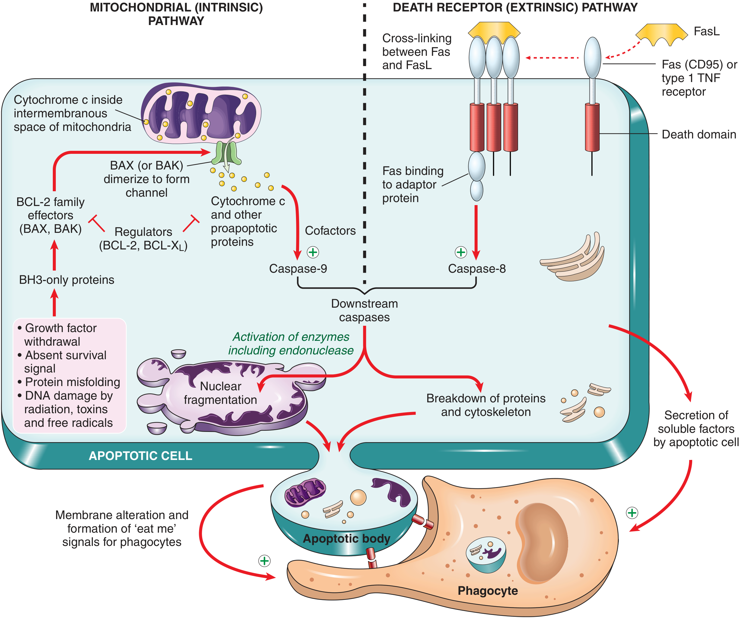
Fig. 1.12 from Robbins & Kumar Basic Pathology - Mitochondrial (intrinsic) and Death Receptor (extrinsic) pathways of apoptosis
1. Mitochondrial (Intrinsic) Pathway
This is the dominant pathway in most physiologic and pathologic apoptosis.
Key trigger: Loss of growth factors/survival signals, DNA damage, protein misfolding, oxidative stress.
The BCL-2 family governs mitochondrial membrane permeability and has three groups:
| Group | Members | Function |
|---|---|---|
| Antiapoptotic | BCL-2, BCL-X | Maintain mitochondrial integrity; oppose BAX/BAK |
| Proapoptotic effectors | BAX, BAK | Dimerize and form pores in the outer mitochondrial membrane |
| BH3-only sensors | BIM, BID, PUMA, NOXA | Sense stress; shift balance toward BAX/BAK |
Sequence of events:
- Stress activates BH3-only proteins, which neutralize BCL-2/BCL-X
L - Freed BAX and BAK dimerize and insert into the outer mitochondrial membrane, forming a channel
- Cytochrome c (and other proteins) leak from the intermembrane space into the cytosol
- Cytochrome c binds Apaf-1, forming the apoptosome complex
- The apoptosome activates initiator caspase-9
- Caspase-9 activates executioner caspases 3 and 7
- Executioner caspases cleave numerous downstream substrates - DNases, cytoskeletal proteins, nuclear lamins - causing the morphologic changes of apoptosis
- Robbins & Kumar Basic Pathology, p. 25
2. Death Receptor (Extrinsic) Pathway
Triggered by extracellular ligands binding death receptors on the cell surface - members of the TNF receptor superfamily that carry a cytoplasmic "death domain."
Key receptors and ligands:
- Fas (CD95) + FasL
- TNFR1 + TNF
Sequence of events:
- Ligand (e.g., FasL) cross-links and clusters the receptor (e.g., Fas)
- The death domain recruits adaptor proteins (e.g., FADD - Fas-associated death domain protein)
- Adaptors recruit and activate initiator caspase-8, forming the DISC (death-inducing signaling complex)
- Caspase-8 directly activates executioner caspases 3 and 7
- (Caspase-8 can also cleave BID, a BH3-only protein, creating tBID - this links the extrinsic pathway to the mitochondrial amplification loop)
This pathway is used by cytotoxic T lymphocytes (CTLs) to kill infected or tumor cells, and eliminates self-reactive lymphocytes.
- Sleisenger and Fordtran's Gastrointestinal and Liver Disease, p. 23
- Robbins & Kumar Basic Pathology, p. 25
Executioner Phase (Common Final Pathway)
Once caspases 3 and 7 are activated:
- CAD (caspase-activated DNase) is released from inhibition, cleaves nuclear DNA into oligonucleosomal fragments (the "DNA ladder" seen on gel electrophoresis)
- Nuclear lamins are cleaved → nuclear fragmentation
- Cytoskeletal proteins are degraded
- Phosphatidylserine flips to the outer leaflet ("eat me" signal)
- Apoptotic bodies form and are phagocytosed by macrophages and neighboring cells
Apoptosis vs. Necrosis
| Feature | Apoptosis | Necrosis |
|---|---|---|
| Trigger | Physiologic or controlled damage | Overwhelming injury |
| Cell size | Shrinkage | Swelling |
| Nucleus | Fragmentation, condensation | Karyolysis/karyorrhexis/pyknosis |
| Plasma membrane | Intact | Disrupted |
| Cell contents | Packaged in apoptotic bodies | Released |
| Inflammation | No | Yes |
| Energy requirement | ATP-dependent | Passive |
Clinical and Pathologic Relevance
Too little apoptosis:
- Cancer - tumor cells evade apoptosis (BCL-2 overexpression in follicular lymphoma via t(14;18), loss of p53)
- Autoimmune disease - failure to delete self-reactive lymphocytes (e.g., mutations in Fas or FasL cause autoimmune lymphoproliferative syndrome, ALPS)
Too much apoptosis:
- Neurodegenerative diseases - excess neuronal death in Alzheimer's, Parkinson's, ALS
- Ischemia-reperfusion injury - mitochondrial pathway activated by oxidative stress
- Viral infections - some viruses (HIV) kill T cells via apoptosis; others (herpesviruses) encode BCL-2 homologs to suppress it
p53 and Apoptosis
p53, the "guardian of the genome," plays a key role: when DNA damage is irreparable, p53 upregulates BH3-only proteins (PUMA, NOXA), shifting the balance toward BAX/BAK and committing the cell to apoptotic death. This is why p53 loss is a hallmark of cancer - damaged cells that should die instead survive and accumulate mutations.
Key Molecules Summary
| Molecule | Role |
|---|---|
| BCL-2, BCL-X | Antiapoptotic; maintain mitochondrial integrity |
| BAX, BAK | Proapoptotic effectors; form the mitochondrial pore |
| BH3-only proteins (BIM, PUMA, NOXA, BID) | Sensors; activate BAX/BAK by neutralizing BCL-2 |
| Cytochrome c | Released from mitochondria; forms apoptosome |
| Apaf-1 | Scaffold for apoptosome assembly |
| Caspase-9 | Initiator caspase (intrinsic pathway) |
| Caspase-8 | Initiator caspase (extrinsic/death receptor pathway) |
| Caspases 3, 7 | Executioner caspases (final common pathway) |
| Fas (CD95), TNFR1 | Death receptors (extrinsic pathway) |
| p53 | Upregulates BH3-only proteins upon DNA damage |
Sources: Robbins & Kumar Basic Pathology, pp. 23-26 | Sleisenger and Fordtran's Gastrointestinal and Liver Disease, p. 23
This is a shared conversation. Sign in to Orris to start your own chat.