
Anatomy and blood supply of brain , circle of Willis, Brain injury forensic medicine, healing and ihc markers , stoke and autopsy findings
Reading File
This is a broad, multi-topic medical query. I'll run parallel searches across all major topics simultaneously.
Finding Sources
Finding Sources
Finding Sources
Finding Sources
Let me try broader single-term searches:
Finding Sources
Finding Sources
Finding Sources
Finding Sources
Good results found. Now let me read the key sections in parallel:
Reading File
Reading File
Reading File
Reading File
Good - I have solid anatomy/circle of Willis and TBI content. Now let me get forensic medicine-specific brain injury, IHC markers for wound age estimation, and stroke/autopsy findings:
Finding Sources
Finding Sources
Finding Sources
I've hit the query_library_db limit. Now let me fetch the Circle of Willis diagram images and read more of the TBI content in parallel, while also doing web searches for the forensic medicine aspects:
Loading Image
Loading Image
Searching the Web
brain injury forensic medicine IHC markers wound age estimation stroke autopsy findings
Excellent! Now let me extract the full content of the PMC article on wound age IHC markers and also fetch the anterior circulation diagram:
Reading Web Page
https://pmc.ncbi.nlm.nih.gov/articles/PMC7241561
Loading Image
Reading File
I now have comprehensive content across all topics. Let me compile the full response.---
Brain: Anatomy, Blood Supply, Circle of Willis, Forensic Injury, Healing, IHC Markers, Stroke & Autopsy
1. Anatomy of the Brain - Blood Supply Overview
The brain receives blood from two paired systems:
Anterior Circulation (Carotid System)
- Originates from the aorta (right side via brachiocephalic artery, left side directly)
- Forms the common carotid arteries, which bifurcate into the internal and external carotid arteries at the carotid bifurcation in the neck
- The internal carotid artery (ICA) has four named segments:
- Cervical segment - in the neck
- Petrous segment - enters carotid canal of temporal bone
- Cavernous segment - S-shaped carotid siphon within cavernous sinus
- Supraclinoid (intracranial) segment - pierces the dura, enters subarachnoid space
Main branches of the supraclinoid ICA - mnemonic OPAAM:
- O - Ophthalmic artery (retinal supply)
- P - Posterior communicating artery (PComm)
- A - Anterior choroidal artery
- A - Anterior cerebral artery (ACA)
- M - Middle cerebral artery (MCA)
Posterior Circulation (Vertebrobasilar System)
- Vertebral arteries arise from the subclavian arteries
- Ascend through foramina transversaria of cervical vertebrae (C6-C1)
- Enter the skull via the foramen magnum
- Unite at the pontomedullary junction to form the basilar artery
- Basilar artery terminates by bifurcating into the two posterior cerebral arteries (PCA)
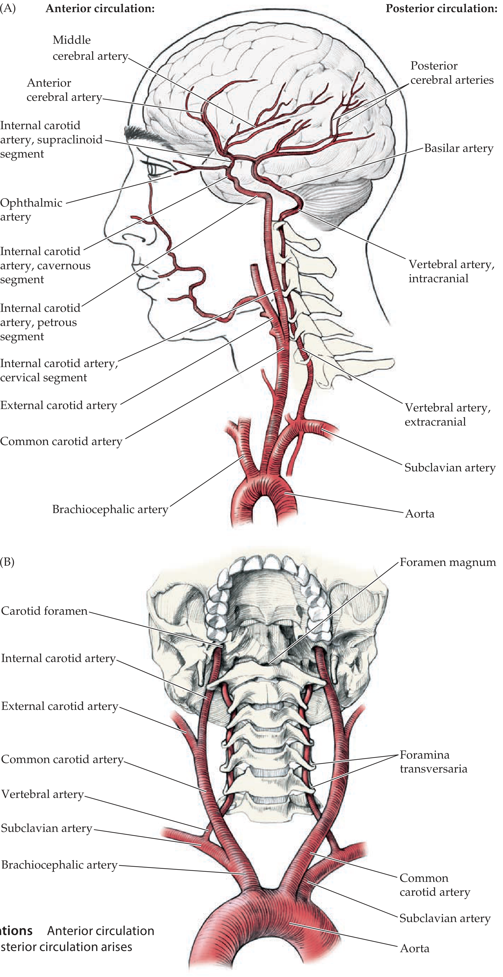
Figure: Anterior circulation (left) arises from the ICA; posterior circulation arises from the vertebral arteries through foramina transversaria - Neuroanatomy through Clinical Cases 3rd Ed.
2. Circle of Willis
The circle of Willis is an anastomotic arterial ring at the base of the brain that connects anterior and posterior circulations, providing collateral flow.
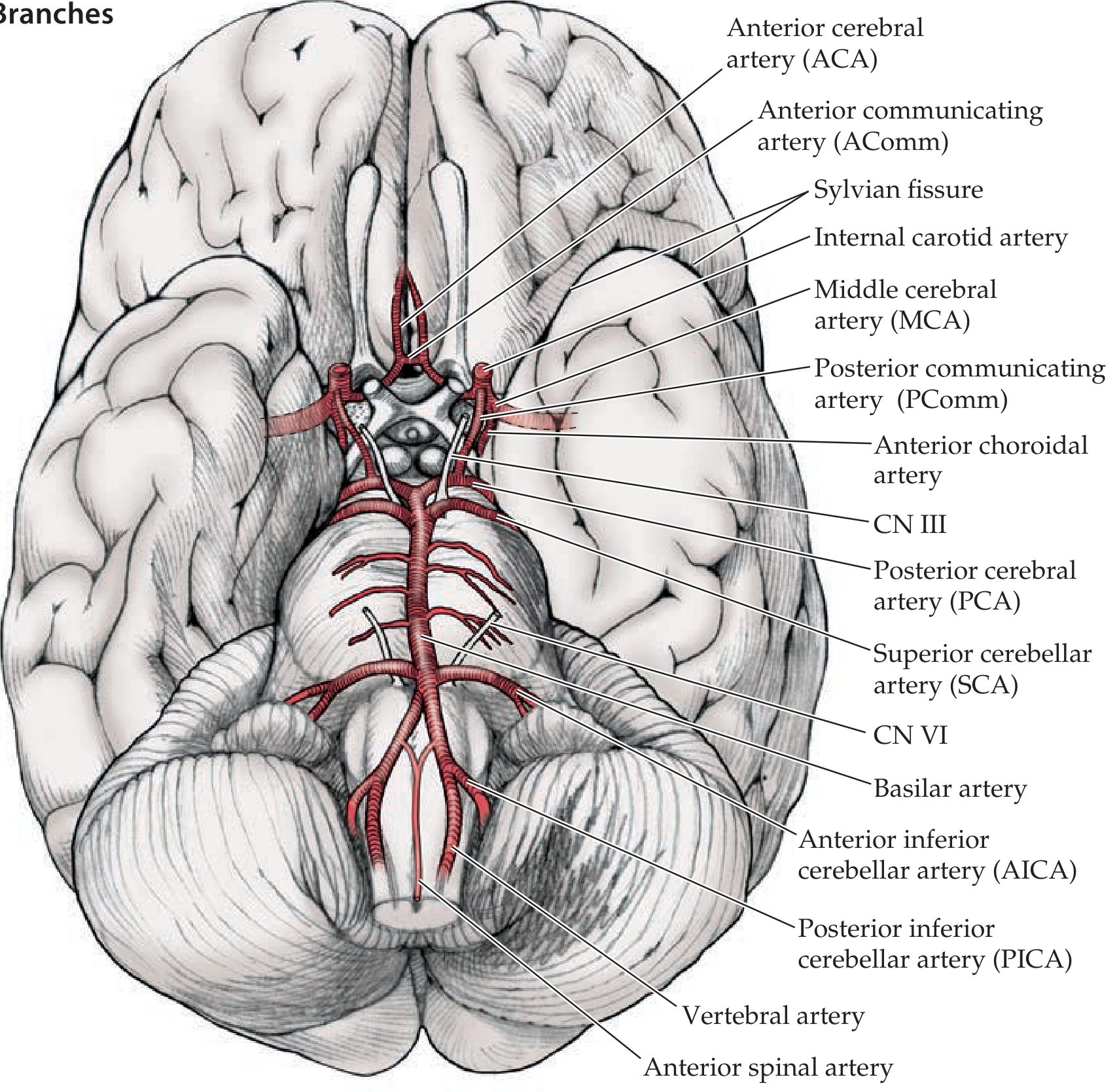
Figure: Circle of Willis - inferior brain view with all named branches - Neuroanatomy through Clinical Cases 3rd Ed.
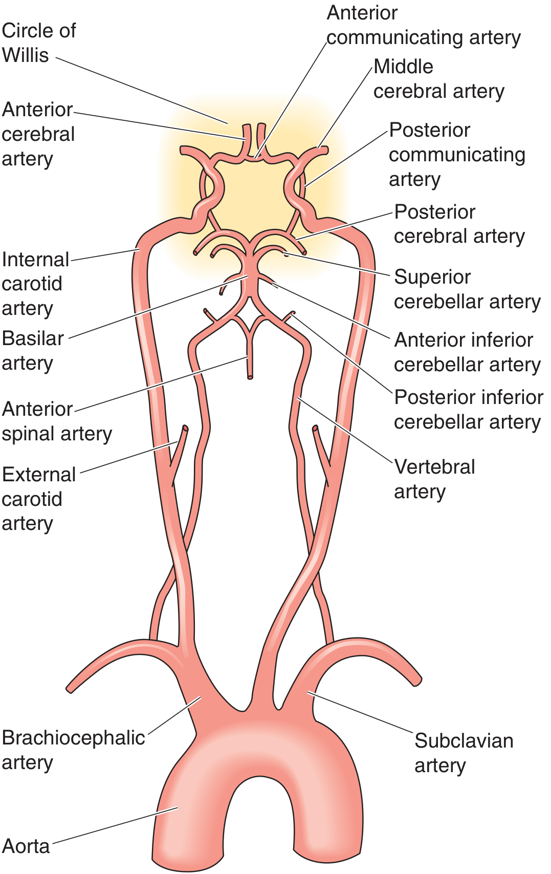
Figure: Schematic showing the full vascular anatomy from aorta through Circle of Willis - Medical Physiology (Boron & Boulpaep)
Components of the Circle of Willis
| Vessel | Connection |
|---|---|
| Anterior communicating artery (AComm) | Links left and right ACA |
| Bilateral ACAs (A1 segments) | From ICA |
| Bilateral ICAs | Entry points |
| Bilateral posterior communicating arteries (PComm) | Link ICA to PCA |
| Bilateral PCAs (P1 segments) | From top of basilar artery |
Key facts:
- A complete, full-caliber ring is present in only ~34% of individuals - anatomical variants are very common
- Provides collateral flow when one vessel is occluded
- Deep penetrating branches from the initial segments of ACA, MCA, PCA near the circle supply the basal ganglia, thalamus, and internal capsule (lenticulostriate arteries from MCA; anterior choroidal artery; thalamoperforators from PCA)
Vascular Territories of the Three Main Cerebral Arteries
| Artery | Superficial Territory | Deep Territory |
|---|---|---|
| ACA | Medial frontal and parietal cortex (medial sensorimotor strip, including leg area) | Anterior limb of internal capsule, head of caudate |
| MCA | Lateral convexity - frontal, parietal, temporal cortex (face and arm sensorimotor, language) | Posterior limb of internal capsule, putamen, globus pallidus (via lenticulostriates) |
| PCA | Occipital and inferior temporal cortex (primary visual cortex) | Thalamus, posterior internal capsule |
The MCA enters the Sylvian fissure and bifurcates into a superior division (frontal/parietal supply) and inferior division (temporal supply). The ACA sweeps over the corpus callosum via the pericallosal and callosomarginal arteries.
3. Brain Injury - Forensic Medicine
Classification of Brain Injuries
Primary injuries (occur at moment of impact):
- Coup injury - directly under point of impact
- Contre-coup injury - opposite side of brain (worse than coup in frontal impacts due to brain movement within CSF)
- Diffuse axonal injury (DAI) - shear-strain injury to axons from angular acceleration; no focal contusion; found in corpus callosum, dorsolateral brainstem, internal capsule
Secondary injuries (develop after the primary event):
- Cerebral edema
- Raised intracranial pressure
- Herniation syndromes (uncal, central, cerebellar tonsillar)
- Ischemia/infarction
- Hydrocephalus
Types of Intracranial Hemorrhage (Forensic Importance)
| Type | Source | Location | Shape | Classic Association |
|---|---|---|---|---|
| Extradural (EDH) | Middle meningeal artery | Between skull and dura | Biconvex/lenticular | Temporal skull fracture, arterial |
| Subdural (SDH) | Bridging veins | Between dura and arachnoid | Crescent-shaped | Shaken baby syndrome, elderly |
| Subarachnoid (SAH) | Ruptured berry aneurysm or trauma | In subarachnoid space | Spreads in CSF cisterns | "Worst headache of life" |
| Intracerebral (ICH) | Small vessels | Brain parenchyma | Round or irregular | HTN, trauma, stroke |
Forensic Assessment at Autopsy
External examination:
- Scalp lacerations, bruising, skull fractures (linear, depressed, basilar)
- Basilar skull fracture signs: Battle's sign (mastoid ecchymosis), raccoon eyes, CSF rhinorrhea/otorrhea, hemotympanum
Internal examination of brain:
- Brain should be fixed in formalin for 2-4 weeks before sectioning (prevents distortion)
- Coronal sections assess contusions, lacerations, hemorrhage, midline shift
- Brain weight (normal adult ~1400g)
- Evidence of raised ICP: Duret hemorrhages in brainstem (central herniation), uncal herniation groove on parahippocampal gyrus
4. Wound Age Estimation - Healing Phases & IHC Markers
Phases of Wound Healing (Chronological)
| Phase | Timeline | Key Events |
|---|---|---|
| Hemostasis | 0-minutes | Platelet plug, fibrin clot, vasoconstriction |
| Inflammatory | Hours-3 days | PMN infiltration (peak 24-48h), then macrophages |
| Proliferative | 3 days-3 weeks | Fibroblasts, angiogenesis, granulation tissue, collagen III |
| Remodeling | Weeks-months | Collagen III → I, scar maturation, myofibroblast apoptosis |
IHC Markers for Brain Wound Age Estimation
The CNS responds to injury with a distinct cellular reaction involving microglia, astrocytes, neurons, and inflammatory cells. These are used in forensic neuropathology to estimate the post-traumatic interval (PTI):
| Biomarker | Method | Peak Expression | Significance |
|---|---|---|---|
| β-APP (β-amyloid precursor protein) | IHC | 1-12 hours | Marker of axonal injury/DAI; accumulates in axons due to impaired axonal transport from 1h onward |
| HIF-1α | IHC | 1h-7 days | Hypoxia marker; peaks at ~1h after injury |
| CD11b (microglia activation) | IHC | 1h-7 days; peak 12h | Activated microglia/macrophage marker |
| IL-6 | IHC | 1h-3 days; peak 12h | Pro-inflammatory cytokine; early marker |
| TNF-α | IHC | 1h-7 days; peak 6h | Inflammatory marker |
| COX-2 | IHC | 1h-5 days; peak 1 day | Cyclooxygenase; inflammation |
| TGF-β1 | IHC | 6h-7 days; peak 3 days | Fibrosis, astrocyte activation |
| HO-1 (heme oxygenase-1) | IHC | 6h-14 days; peak 6-12h | Oxidative stress/hemorrhage marker |
| MMP-9 | IHC | 1h-14 days; peak 5 days | Matrix metalloproteinase; blood-brain barrier disruption |
| Caspase-3 / Caspase-9 | WB/ELISA | 12h-7 days; peak 1-3 days | Apoptosis markers |
| CaMK-II | IHC/WB | 1-7 days; peak 3 days | Neuronal calcium signaling |
| c-Jun / c-Fos | IHC | 1h-7 days | Transcription factors; early response |
| vWF | IHC | 12h-1 day | Neovascularization/endothelial injury |
| MAP-2 | RT-qPCR | 1h-14 days; peak 14 days | Neuronal/dendritic marker |
| RAGE | WB/IHC | 6h-3 days; peak 1 day | Advanced glycation end-product receptor |
| HMGB1 | IHC | 1h-3 days; peak 6h | Damage-associated molecular pattern |
| NFL (neurofilament light) | IHF | 12h-3 days; peak 1 day | Neurofilament injury |
Practical application:
- β-APP is the most widely used forensic IHC marker for DAI - positive axonal staining within 1-6 hours of injury
- CD11b and IL-6 peak at ~12 hours
- TGF-β1 and MMP-9 are useful for injuries surviving >3-5 days
- Intracranial hemorrhage age is assessed with H&E and IHC for hemosiderin (hemosiderin-laden macrophages appear ~3-7 days), fibrin organization, and neovascularization
IHC Markers for Skin Wound Age Estimation (also used in forensic practice)
| Time period | Key markers |
|---|---|
| Minutes-hours | PMN infiltration (MPO+), fibrin |
| 12-24 hours | CD68+ macrophages beginning, PCNA (proliferating cells) |
| 1-3 days | Ki-67 (keratinocyte proliferation), vimentin (fibroblasts) |
| 3-7 days | CD31/Factor VIII (new vessels), α-SMA (myofibroblasts) |
| >7 days | Collagen I deposition, decreased CD68, scar maturation |
5. Stroke
Classification
Ischemic stroke (~80%):
- Thrombotic: in situ atherosclerotic plaque rupture (large vessel)
- Embolic: cardiac source (AF, valvular), arterial-arterial embolism
- Lacunar: small vessel disease (lipohyalinosis) - basal ganglia, internal capsule, thalamus, pons
- Cryptogenic: no identified cause
Hemorrhagic stroke (~20%):
- Intracerebral hemorrhage (ICH): hypertensive - putamen (most common), thalamus, pons, cerebellum
- Subarachnoid hemorrhage (SAH): ruptured berry aneurysm (most often at AComm or MCA bifurcation)
Pathophysiology of Ischemia
- Oxygen/glucose deprivation → ATP failure → Na-K pump failure → cytotoxic edema
- Glutamate excitotoxicity → calcium influx → mitochondrial dysfunction, apoptosis
- Penumbra zone (ischemic but salvageable) surrounds the infarct core
6. Stroke - Autopsy Findings
Gross Findings (Ischemic Infarction)
| Time | Gross Appearance |
|---|---|
| 0-6 hours | Often no visible change; subtle softening |
| 8-24 hours | Pale area, slight swelling, blurred grey-white junction |
| 24-72 hours | Pale/yellow softening, edema, gyral swelling, possible midline shift |
| 3-7 days | Liquefactive necrosis begins; yellow-brown, soft, mushy texture |
| 1-2 weeks | Well-defined liquefaction; yellow gelatinous core |
| Weeks-months | Cavitation (pseudocyst formation); gliosis around margins |
| Old infarct | Fluid-filled cystic cavity with brown hemosiderin staining at edges |
Histological Findings (Ischemic Infarction)
| Time | Histological Changes |
|---|---|
| 0-12 hours | Red (eosinophilic) neurons with pyknotic nuclei ("red dead neurons"), cytoplasmic eosinophilia |
| 12-24 hours | Nuclear pyknosis/karyorrhexis, neuropil pallor |
| 24-48 hours | Neutrophil infiltration (peaks at 48h) |
| 3-5 days | Macrophage infiltration (phagocytosis of debris - "foamy macrophages"), reactive astrocytes |
| 1-2 weeks | Prominent foamy macrophages, neovascularization |
| Weeks-months | Gliosis (GFAP+ reactive astrocytes), cavitation, hemosiderin-laden macrophages |
Hemorrhagic Stroke Autopsy Findings
- ICH: Hematoma in brain parenchyma; surrounding rim of edema and red neurons; hemosiderin-laden macrophages appear at ~7 days; cavity with brown staining in old hematoma
- SAH: Blood in the subarachnoid space, spreading over the convexities and basal cisterns; "Fisher grade" describes extent; blood tracks into ventricles (intraventricular hemorrhage) in severe cases
- EDH: Blood between skull and dura, typically temporal location, biconvex shape; associated with temporal bone (pterion) fracture
- SDH: Crescent-shaped blood over convexity; bridging vein rupture
Specific Forensic Autopsy Considerations for Stroke
- Differentiate primary stroke (natural disease) causing a fall/head injury vs primary trauma causing secondary hemorrhage
- Look for underlying causes: atherosclerosis of carotid/vertebral arteries, cardiac thrombus, coagulopathy, Berry aneurysm
- Neck dissection: carotid artery dissection (trauma or spontaneous)
- Vertebral artery dissection: associated with chiropractic manipulation or blunt neck trauma
- Document brain weight, hemorrhage volume (ml), midline shift, herniation evidence
Summary Table: Forensic Timing of Brain Injury
| Post-Traumatic Interval | Key Histological/IHC Findings |
|---|---|
| 0-1 hour | HIF-1α+, HMGB1+, c-Jun+ (very early); no structural change on H&E |
| 1-6 hours | β-APP+ axons (DAI), CD11b+ microglia, TNF-α+, HIF-1α+ |
| 6-12 hours | IL-6 peak, CD11b peak, HO-1+, COX-2+ |
| 12-24 hours | Neutrophil infiltration, red neurons on H&E, Caspase-9 peak |
| 1-3 days | Macrophage infiltration, Caspase-3 peak, TGF-β1 rising |
| 3-7 days | Foamy macrophages, TGF-β1 peak, MMP-9 peak, early neovascularization (vWF) |
| >7 days | Gliosis (GFAP+), hemosiderin, MAP-2 elevated, scar formation |
| Weeks | Cavitation, GFAP+ reactive astrocyte scar, old hemosiderin |
Sources:
- Neuroanatomy through Clinical Cases 3rd Ed. (Blumenfeld) - Circle of Willis, cerebral vascular territories
- Medical Physiology (Boron & Boulpaep) - Brain vascular anatomy
- Plum and Posner's Diagnosis and Treatment of Stupor and Coma - Traumatic brain injury
- Vitality and wound-age estimation in forensic pathology (PMC7241561) - IHC markers table for brain wound dating
Brain anatomy with diagrams, different part and functioning areas of the brain
Reading File
This is a visual/broad anatomy question - I need to run parallel searches for brain anatomy sections and images simultaneously.
Finding Sources
Finding Sources
Finding Sources
Finding Sources
Finding Sources
Finding Sources
Good results. Now let me read the best sources in parallel - Guyton physiology (functional anatomy), Kaplan psychiatry (cerebral cortex), and Localization in Clinical Neurology (anatomy of cortex):
Reading File
Reading File
Reading File
Excellent content. Now let me fetch the key diagrams and read more about specific lobe functions and brainstem/cerebellum in parallel:
Loading Image
Reading File
Finding Sources
Excellent content and diagram. Let me now get the functional areas (Guyton), the medial view image, and specific lobe function content:
Reading File
Loading Image
Finding Sources
Now let me get the functional areas diagram and read more content about specific regions:
Loading Image
Reading File
Finding Sources
Now fetch the functional map diagram and read limbic/association content in parallel:
Loading Image
Loading Image
Reading File
Reading File
Excellent - I now have rich content with diagrams. Let me get a few more key images - the Broca/Wernicke diagram, limbic system diagram, and cortical layers diagram:
Loading Image
Loading Image
Loading Image
I now have all the diagrams and content needed for a comprehensive answer. Let me compile it.
Brain Anatomy: Parts, Regions & Functional Areas
Overview: Major Divisions of the Brain
The brain is divided into three major parts: the cerebrum (forebrain), brainstem, and cerebellum.
| Division | Embryonic Origin | Key Structures |
|---|---|---|
| Telencephalon | Forebrain | Cerebral hemispheres, basal ganglia, hippocampus, amygdala |
| Diencephalon | Forebrain | Thalamus, hypothalamus, subthalamus, epithalamus |
| Mesencephalon | Midbrain | Midbrain (tectum, tegmentum, cerebral peduncles) |
| Metencephalon | Hindbrain | Pons, cerebellum |
| Myelencephalon | Hindbrain | Medulla oblongata |
1. Cerebral Hemispheres - External Anatomy
The cerebral cortex is a 2-5 mm thick layer of neurons covering the surface of the cerebral hemispheres, containing over 80 billion neurons. It has a total area of ~0.25 m² (folded to fit inside the skull via gyri and sulci).
Lateral Surface - Lobes and Key Landmarks
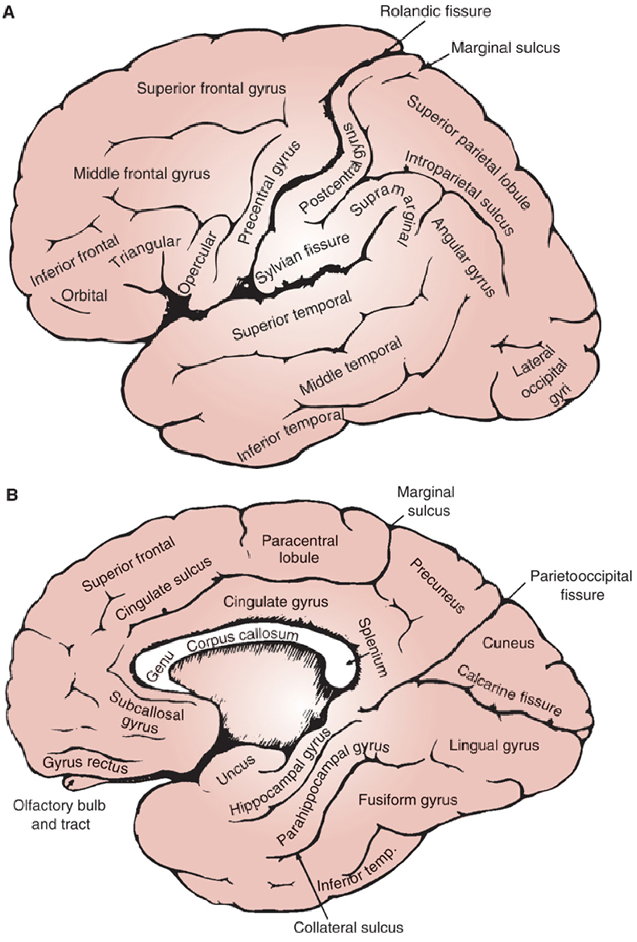
Figure: Lateral surface (A) and medial surface (B) of the cerebral hemisphere - Localization in Clinical Neurology 8e
Key sulci that divide the lobes:
- Rolandic (central) sulcus - separates frontal lobe (anterior) from parietal lobe (posterior)
- Sylvian (lateral) fissure - separates temporal lobe (inferior) from frontal/parietal lobes
- Parietooccipital sulcus - separates parietal from occipital lobe (mainly seen on medial surface)
- Calcarine sulcus - on medial surface of occipital lobe; primary visual cortex lies on its banks
Four lobes on each hemisphere:
| Lobe | Boundaries | Key Gyri |
|---|---|---|
| Frontal | Anterior to central sulcus | Superior, middle, inferior frontal gyri; precentral gyrus |
| Parietal | Behind central sulcus, above Sylvian | Postcentral gyrus, supramarginal, angular gyri |
| Temporal | Below Sylvian fissure | Superior, middle, inferior temporal gyri; Heschl's gyrus (transverse) |
| Occipital | Posterior pole | Lateral occipital gyri, cuneus, lingual gyrus |
Fifth lobe - Insula (Island of Reil): Lies buried deep in the Sylvian fissure; involved in interoception, pain, taste, autonomic regulation, and emotional awareness.
2. Cortical Layers (Neocortex)
The neocortex (>90% of cerebral cortex) has a 6-layer structure:
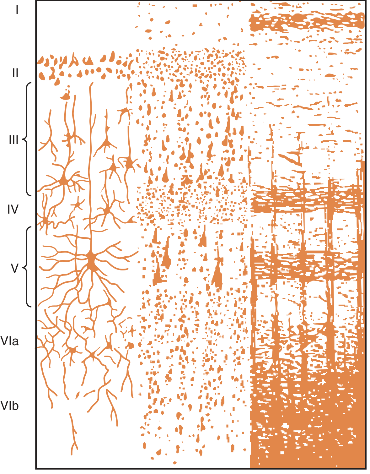
Figure: Cytoarchitecture of the six-layered neocortex (Guyton & Hall Medical Physiology)
| Layer | Name | Contents | Function |
|---|---|---|---|
| I | Molecular layer | Sparse cells, rich in horizontal fibers | Intracortical association |
| II | External granular | Small granule cells | Short-range connections |
| III | External pyramidal | Medium pyramidal cells | Corticocortical connections |
| IV | Internal granular | Small stellate cells + horizontal fibers | Receives thalamic sensory input |
| V | Internal pyramidal (ganglionic) | Large pyramidal cells (Betz cells in motor cortex) | Output to brainstem and spinal cord |
| VI | Multiform (fusiform) | Spindle-shaped cells | Output to thalamus |
Key principle: Sensory signals enter via Layer IV; motor and descending outputs leave via Layers V and VI; intracortical processing occurs mainly in Layers I-III.
3. Functional Areas of the Cerebral Cortex
Primary Functional Map
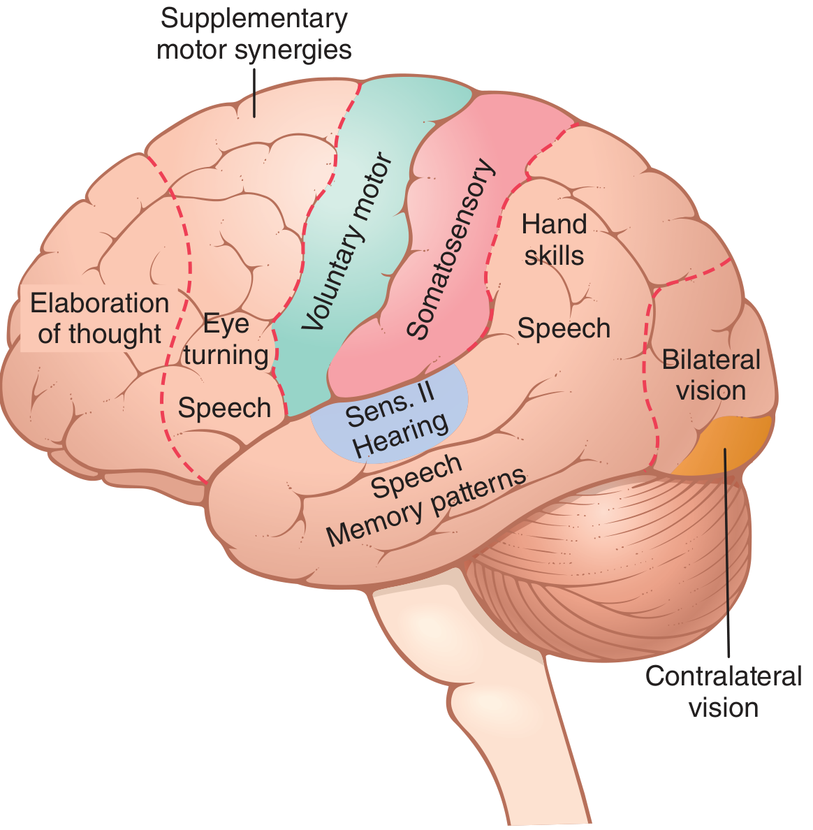
Figure: Functional areas of the cerebral cortex determined by electrical stimulation (Penfield & Rasmussen, from Guyton & Hall)
Detailed Map with Association Areas and Language Zones
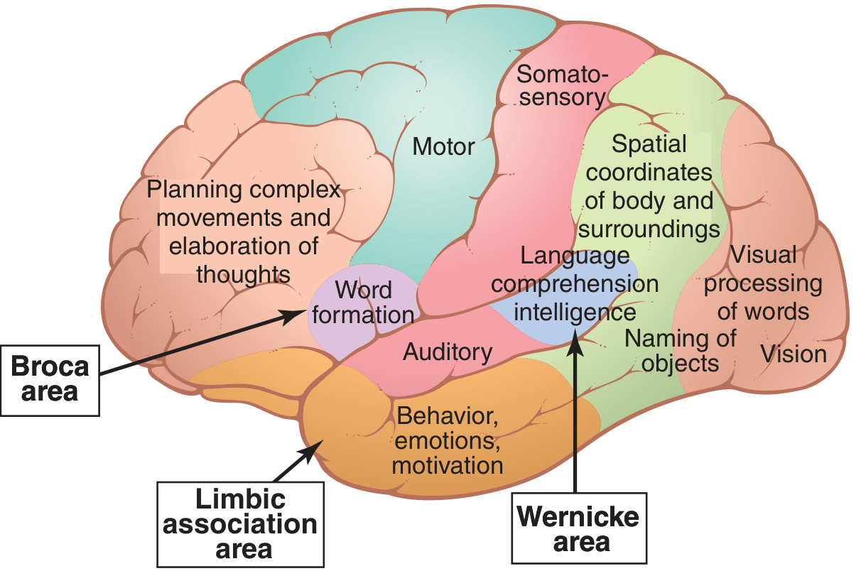
Figure: Specific functional areas including Broca's and Wernicke's areas (Guyton & Hall Medical Physiology)
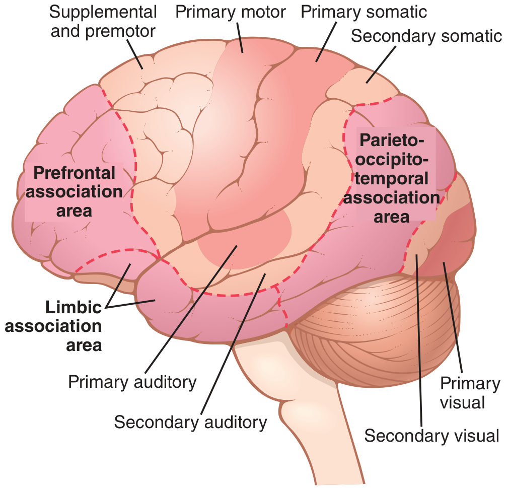
Figure: Major association areas of the cerebral cortex (Guyton & Hall Medical Physiology)
4. Frontal Lobe - Functions
| Area | Location | Function |
|---|---|---|
| Primary motor cortex | Precentral gyrus (area 4) | Voluntary movement of contralateral body (somatotopic - motor homunculus) |
| Premotor cortex | Anterior to precentral gyrus (area 6) | Planning and sequencing movements; patterns of motor activity |
| Supplementary motor area (SMA) | Medial surface, area 6 | Bilateral movement coordination; planning complex motor sequences |
| Frontal eye field | Posterior part of middle frontal gyrus (area 8) | Voluntary conjugate eye movements (contralateral gaze) |
| Broca's area | Posterior inferior frontal gyrus (areas 44, 45) - left hemisphere | Word formation and speech production (expressive language) |
| Prefrontal cortex | Anterior frontal lobe | Working memory, abstract thought, planning, personality, executive function, decision-making |
| Orbitofrontal cortex | Inferior surface of frontal lobe | Emotional regulation, reward processing, social behavior |
Motor Homunculus: The primary motor cortex has a somatotopic map. The leg area lies on the medial surface (supplied by ACA); the hand/face area lies on the lateral convexity (supplied by MCA). The hand and face areas are disproportionately large due to fine motor demands.
5. Parietal Lobe - Functions
| Area | Location | Function |
|---|---|---|
| Primary somatosensory cortex | Postcentral gyrus (areas 3, 1, 2) | Touch, pain, temperature, proprioception from contralateral body (sensory homunculus) |
| Secondary somatosensory cortex | Posterior to postcentral gyrus | Object recognition by touch (stereognosis); texture analysis |
| Superior parietal lobule | Above intraparietal sulcus | Spatial attention, reaching movements, visuospatial processing |
| Inferior parietal lobule | Supramarginal + angular gyri | Spatial coordinates, body image; right side - directed attention; reading/writing/calculation |
| Angular gyrus | At temporoparietal junction | Visual language processing; reading; arithmetic; Gerstmann's syndrome if damaged |
| Supramarginal gyrus | Above Sylvian fissure | Phonological processing; limb apraxia if damaged |
Lesions: Right parietal damage → hemispatial neglect, constructional apraxia. Left parietal damage → Gerstmann's syndrome (agraphia, alexia, acalculia, finger agnosia, left-right disorientation).
6. Temporal Lobe - Functions
| Area | Location | Function |
|---|---|---|
| Primary auditory cortex | Heschl's gyrus (transverse temporal gyrus, area 41) | Detection of sound frequency and intensity |
| Secondary auditory cortex | Superior temporal gyrus (area 42) | Interpretation of sound meaning |
| Wernicke's area | Posterior superior temporal gyrus (area 22) - left hemisphere | Language comprehension (receptive language) |
| Planum temporale | Posterior superior temporal surface | Speech and language processing; larger on left |
| Hippocampus | Medial temporal lobe | Declarative memory formation and consolidation (episodic and semantic) |
| Amygdala | Anterior medial temporal lobe (uncus) | Fear conditioning, emotional memory, threat detection |
| Inferior temporal gyri | Lateral-inferior temporal | Object recognition, face recognition (fusiform face area) |
| Parahippocampal gyrus | Medial temporal | Spatial navigation, context memory; contains entorhinal cortex |
Lesion: Bilateral hippocampal damage → anterograde amnesia (Korsakoff's, Alzheimer's). Dominant temporal lobe damage → Wernicke's aphasia (fluent but incomprehensible speech).
7. Occipital Lobe - Functions
| Area | Location | Function |
|---|---|---|
| Primary visual cortex (V1) | Banks of calcarine sulcus (area 17) | Receives contralateral visual field input from lateral geniculate nucleus |
| Visual association cortex (V2-V5) | Surrounding area 17 | Colour, motion, depth, pattern recognition |
| Dorsal stream ("where" pathway) | Occipital → parietal | Spatial location and visually guided movement |
| Ventral stream ("what" pathway) | Occipital → temporal | Object identity and recognition |
Lesion: Unilateral damage → contralateral homonymous hemianopia. Bilateral damage → cortical blindness (Anton's syndrome - patient unaware of blindness).
8. Brainstem
The brainstem comprises midbrain, pons, and medulla. It contains:
- Nuclei for cranial nerves III-XII
- Reticular activating system (RAS) - consciousness and arousal
- Vital autonomic centers (cardiac, respiratory, vasomotor)
- Major ascending and descending white matter tracts
| Part | Cranial Nerves | Key Functions |
|---|---|---|
| Midbrain | CN III (oculomotor), IV (trochlear) | Superior colliculus (visual reflexes), inferior colliculus (auditory reflexes), red nucleus, substantia nigra, cerebral aqueduct |
| Pons | CN V (trigeminal), VI (abducens), VII (facial), VIII (vestibulocochlear) | Pontine nuclei (relay to cerebellum), respiratory control centers, horizontal gaze center (PPRF) |
| Medulla | CN IX (glossopharyngeal), X (vagus), XI (accessory), XII (hypoglossal) | Cardiac center, respiratory center, vomiting center, swallowing, pyramidal decussation |
9. Cerebellum
- Located in the posterior fossa, connected to brainstem via three cerebellar peduncles (superior, middle, inferior)
- Does not initiate movement - rather coordinates, smooths, and times movement
| Region | Function |
|---|---|
| Vestibulocerebellum (flocculonodular lobe) | Balance and eye movement coordination |
| Spinocerebellum (vermis and intermediate zone) | Coordination of limb and trunk movements; gait |
| Cerebrocerebellum (lateral hemispheres) | Planning and timing of complex skilled movements |
Lesion signs: Ipsilateral ataxia, dysmetria (past-pointing), dysdiadochokinesia, intention tremor, nystagmus, dysarthria (scanning speech).
10. Diencephalon
Thalamus
- The gateway to the cortex - nearly all sensory pathways (except olfaction) relay through thalamic nuclei
- Each thalamic nucleus connects to a specific cortical area (thalamocortical projections)
- Key nuclei: VPL (body sensation), VPM (face sensation), LGN (vision), MGN (hearing), VA/VL (motor), anterior nucleus (memory/limbic), pulvinar (visual association)
Hypothalamus
- Controls the autonomic nervous system and pituitary gland
- Regulates: body temperature, hunger/satiety, thirst, sleep-wake cycles, circadian rhythms, sexual behavior, emotional responses
- Links nervous system to endocrine system via the hypothalamo-pituitary axis
11. Limbic System
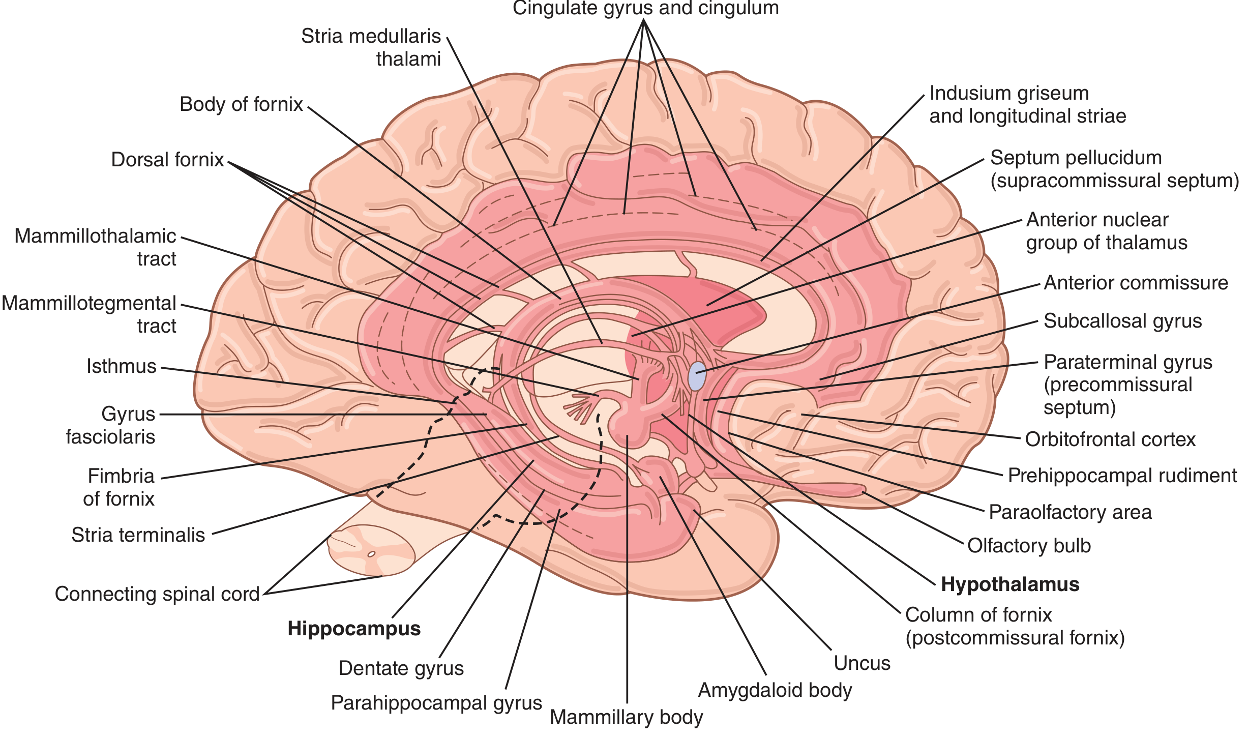
Figure: Anatomy of the limbic system - Guyton & Hall Medical Physiology
The limbic system forms a ring of cortex and subcortical nuclei on the medial surface:
Cortical components: Orbitofrontal cortex → subcallosal gyrus → cingulate gyrus → parahippocampal gyrus → uncus
Subcortical components: Hippocampus, amygdala, hypothalamus, anterior thalamus, mammillary bodies, septal nuclei, olfactory bulbs
Major circuit - Papez Circuit:
Hippocampus → fornix → mammillary bodies → mammillothalamic tract → anterior thalamus → cingulate gyrus → entorhinal cortex → hippocampus
| Structure | Function |
|---|---|
| Hippocampus | Memory formation; spatial navigation |
| Amygdala | Fear, emotion, fight-or-flight responses |
| Cingulate gyrus | Attention, emotional processing, pain modulation, conflict monitoring |
| Hypothalamus | Vegetative functions: hunger, thirst, temperature, hormonal control |
| Mammillary bodies | Memory relay (damaged in Wernicke-Korsakoff syndrome) |
12. Basal Ganglia
A group of subcortical nuclei involved in motor control, habit learning, and reward:
| Nucleus | Notes |
|---|---|
| Caudate nucleus | Head: cognitive and limbic functions |
| Putamen | Motor control; with caudate = striatum |
| Globus pallidus | Internal (GPi) and external (GPe) segments; major output structure |
| Substantia nigra | Dopaminergic input to striatum; loss → Parkinson's disease |
| Subthalamic nucleus | Part of indirect pathway; target for DBS in Parkinson's |
Circuit: Cortex → Striatum → Globus pallidus → Thalamus → Cortex (feedback loop for movement initiation and suppression)
13. Hemispheric Specialization
| Function | Left Hemisphere (dominant in ~95%) | Right Hemisphere |
|---|---|---|
| Language and speech | Broca's area (production) + Wernicke's area (comprehension) | Prosody (emotional tone of speech) |
| Analytical/sequential reasoning | Dominant | Non-dominant |
| Mathematical skills | Dominant | Non-dominant |
| Spatial and constructional skills | Non-dominant | Dominant |
| Directed attention | Non-dominant | Dominant |
| Melody and tone | Non-dominant | Dominant |
| Emotional facial recognition | Non-dominant | Dominant |
| Body image | Non-dominant | Dominant |
Sources:
- Guyton and Hall Textbook of Medical Physiology - Physiological anatomy of the cerebral cortex, functional areas, limbic system
- Localization in Clinical Neurology 8e (Brazis et al.) - Anatomy of the cerebral cortex, gyri, sulci, lobes
- Neuroanatomy through Clinical Cases 3rd Ed. (Blumenfeld) - Limbic system
- Kaplan & Sadock's Comprehensive Textbook of Psychiatry - Neocortex neuron types
This is a shared conversation. Sign in to Orris to start your own chat.