
SAIO me Ryles tube kyu dalte h..medically inka kya benifit h..clinical explanation do
Ryles tube nasogastric tube intestinal obstruction decompression
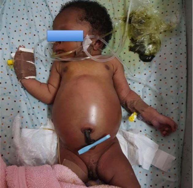
This clinical photograph shows a neonate on the fourth day of life presenting with severe neonatal intestinal obstruction secondary to an anorectal malformation (ARM). The image demonstrates significant, tense abdominal distension with thinned, shiny skin, characteristic of distal bowel obstruction. A nasogastric tube (NG tube) is visible for gastric decompression, and bilateral peripheral intravenous access is established in the upper extremities. In the umbilical region, a blue umbilical cord clamp remains in situ. In the lower abdominal/pelvic area, a surgical stoma is present, draining dark, meconium-like fecal matter into an adjacent collection bag. The image highlights the clinical consequences of delayed presentation in ARM cases, including gross abdominal distension and the necessity for emergency surgical diversion (colostomy). The case is relevant for pediatric surgery and neonatal intensive care, illustrating the typical physical findings and initial stabilization measures—such as nasogastric decompression and intravenous fluid resuscitation—required for neonatal bowel obstruction.
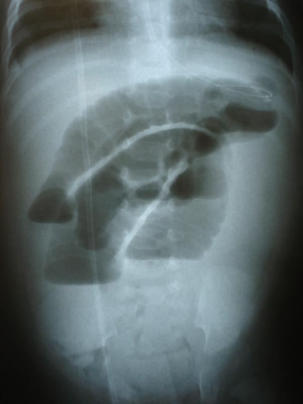
Diagnostic Imaging: Upright plain abdominal radiograph of a pediatric patient. The image demonstrates hallmarks of acute intestinal obstruction, characterized by massively distended intestinal loops localized primarily in the upper abdomen. Key radiographic findings include multiple distinct air-fluid levels and a thickened appearance of the bowel walls, suggesting edema or ischemia. A radiopaque nasogastric tube is visible, descending through the esophagus and terminating within the distended gastrointestinal segment for decompression. The distal abdomen shows a relative paucity of gas, consistent with a high-grade mechanical ileus. This image serves as a classic educational example of neonatal or infant bowel obstruction, emphasizing the importance of recognizing pathological gas patterns and air-fluid levels in the context of clinical signs like bilious vomiting and abdominal distension.
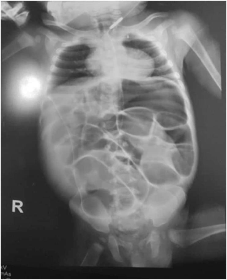
This diagnostic image is an anteroposterior babygram (plain radiograph of the chest and abdomen) of a pediatric patient. The most significant finding is severe, generalized gaseous distention of multiple bowel loops throughout the abdominal cavity, which is characteristic of intestinal obstruction or paralytic ileus. The bowel loops appear stacked and thinned, occupying the majority of the distended abdominal space. A radiopaque nasogastric tube is visible descending into the gastric region for decompression. The thoracic portion demonstrates the cardiac silhouette, rib cage, and pulmonary fields, while the lower portion captures the pelvic girdle and proximal femurs. This visual is highly relevant for pediatric emergency medicine and radiology education, illustrating the radiographic presentation of pediatric bowel distention and the clinical application of decompression tubes in cases of suspected mechanical obstruction or ileus.
SAIO (Small Acute Intestinal Obstruction) mein Ryle's Tube - Clinical Explanation
Ryle's Tube kya hota hai?
SAIO mein Ryle's Tube kyun daala jaata hai? - 5 Key Clinical Reasons
1. 🫀 Gastric & Proximal Bowel DECOMPRESSION
- Ryle's tube stomach aur proximal gut ki collected fluid aur gas ko bahar nikalta hai
- Isse intestinal distension (phoolna) kam hoti hai
- Bowel loops ko thoda "rest" milta hai aur further dilation rok sakta hai
- Bailey & Love Surgery ke anusar: "Nasogastric decompression... facilitating decompression proximal to the obstruction"
2. 🫁 Aspiration ka Risk KHATAM KARNA
- Patient suddenly vomit kare aur stomach contents lungs mein jaayein → Aspiration Pneumonitis/Pneumonia
- Especially during anaesthesia induction ya post-extubation period mein risk bahut zyada hota hai
"They are essential to reducing the risk of subsequent aspiration during induction of anaesthesia and after extubation."
- Bailey & Love's Surgery, 28th Ed.
"Suction with a nasogastric tube empties the stomach, reducing the hazard of pulmonary aspiration of vomitus."
- Sabiston Textbook of Surgery
3. 💧 Further Distension ROKNA (Swallowed Air)
- Yeh extra swallowed air stomach se aspirate hoti rehti hai
- Obstruction further worsen nahin hota
- "Minimizing further intestinal distention from swallowed air" - Sabiston
4. 🧪 Diagnostic Purpose - Monitoring of Output
| Aspirate Nature | Clinical Significance |
|---|---|
| Bilious (green) fluid | Obstruction is below duodenum |
| Large volume (>500 mL/day) | High-grade obstruction |
| Feculent aspirate | Long-standing obstruction, bacterial overgrowth |
| Decreasing output | Obstruction resolving conservatively |
5. 💊 Therapeutic Administration (Contrast Challenge)
- 100 mL water-soluble contrast Ryle's tube se dete hain
- 8 aur 24 hour baad X-ray lete hain
- Agar 24 hours mein contrast colon mein nahin pahuncha → conservative management fail → surgery ki zarurat
- Agar contrast pass ho gaya → obstruction resolve ho sakti hai conservatively
"100 mL of water-soluble contrast given through the nasogastric tube..." - Sabiston Surgery
SAIO Mein Ryle's Tube ka Protocol
| Aspect | Detail |
|---|---|
| Tube type | Non-vented Ryle's tube (ya Salem sump tube) |
| Placement | Free drainage + 4-hourly aspiration |
| Alternative | Continuous ya intermittent suction |
| Monitoring | Daily fluid output measure karo |
| Duration | Till obstruction resolves ya surgery ki jaaye |
Conservative Management mein Ryle's Tube ki Success Rate
- Partial bowel obstruction mein conservative treatment (NG decompression + IV fluids) se 85% tak patients surgery ke bina theek ho jaate hain
- Complete obstruction mein bhi 12-24 hours conservative management safe hai (agar koi fever, tachycardia, tenderness ya leukocytosis naa ho)
- 72 hours tak adhesion-related obstruction mein conservative management continue kar sakte hain
Radiograph Showing NG Tube in Intestinal Obstruction
Summary: SAIO mein Ryle's Tube ke Benefits
| # | Benefit | Mechanism |
|---|---|---|
| 1 | Decompression | Proximal fluid/gas bahar nikalta hai |
| 2 | Aspiration prevention | Stomach empty rakhta hai |
| 3 | No further distension | Swallowed air aspirate hoti hai |
| 4 | Monitoring | Output se obstruction severity assess |
| 5 | Contrast challenge | Gastrografin dene ka route |
| 6 | Peri-op safety | Pre-anaesthesia gastric emptying |
SAIO h ya nhi kaise confirm kre without investigation....conical explanation do
SAIO (Small Acute Intestinal Obstruction) - Bina Investigation ke Clinical Diagnosis
Sabse pehle ek important baat samjho:
"Many patients with acute obstruction can be accurately diagnosed after a thorough history and physical examination is performed before imaging."
- Harrison's Principles of Internal Medicine, 22nd Ed.
SAIO ki Clinical Diagnosis: 4 Cardinal Signs (Tetraad)
1. Colicky Abdominal Pain
2. Vomiting
3. Abdominal Distension
4. Absolute Constipation (Obstipation)
1. COLICKY ABDOMINAL PAIN - "Dard ki Pehchaan"
- Intermittent, crampy, spasmodic dard
- Aata hai - jaata hai - phir aata hai (paroxysmal)
- Har 4-5 minute mein ek episode
- Dard ke saath patient behest hota hai, position badalta rehta hai (vs. peritonitis mein still leta hai)
| Obstruction Level | Pain Location |
|---|---|
| Proximal (High) SBO | Epigastric / Periumbilical |
| Distal (Low) SBO | Periumbilical / Lower abdomen |
2. VOMITING - "Ulti ki Nature se Level Pata chalo"
| Obstruction Level | Vomiting |
|---|---|
| High/Proximal (Duodenum/Jejunum) | Bahut jaldi shuru hoti hai, bilious (green/yellow) hoti hai, profuse hoti hai, pain ke saath ya baad mein aati hai |
| Mid SBO | Thodi der baad, bilious |
| Distal SBO / Late | Feculent vomiting - "Potty jaise smell" - yeh bacterial overgrowth ki wajah se hota hai, late presentation hai |
"As the obstruction becomes more complete with bacterial overgrowth, the vomitus becomes more feculent, indicating a late and established intestinal obstruction."
- Sabiston Textbook of Surgery
3. ABDOMINAL DISTENSION - "Pet Phoolna"
- Obstruction jitna neeche hoga, distension utni zyada hogi (kyunki zyada bowel proximal side mein dilate hota hai)
- Proximal obstruction = minimal/no distension, maximum vomiting
- Distal SBO / Ileum = marked distension, central (periumbilical) predominantly
- Inspection mein visible peristaltic waves dikhti hain - thin patient mein clearly - bowel waves ek side se doosri side move karti hain
- Tympanitic (drum-like) abdomen on percussion - gas-filled loops ki wajah se
4. ABSOLUTE CONSTIPATION (Obstipation) - "Na Potty, Na Gas"
- Na stool pass hoti hai, na flatus (gas) pass hota hai - isko "Absolute Constipation" kehte hain
- Yeh LATE feature hai SBO mein
- Important trap: Early SBO mein patient ek-do baar stool ya loose motion pass kar sakta hai - yeh obstruction ke distal part ki residual content hoti hai - iska matlab obstruction nahi hai yeh sochna GALAT hai!
"A complete bowel obstruction cannot be ruled out on the basis of a history of loose bowel movements."
- Sabiston Textbook of Surgery
Physical Examination - Haath aur Kaan se Diagnose karo
INSPECTION
- Distended abdomen - symmetrical (SBO) vs asymmetrical (volvulus/LBO)
- Visible peristalsis - ladder pattern of bowel loops especially in thin patients
- Previous surgical scars - adhesions ka most common cause
- Groin check - koi hernia toh nahi?
AUSCULTATION - Bowel Sounds ka Character
| Time | Bowel Sound | Meaning |
|---|---|---|
| Early SBO | High-pitched tinkling sounds + borborygmi (rushes/gurgling) | Bowel try kar raha hai obstruction cross karne ki, hyperperistalsis |
| Late SBO | Absent / Hypoactive | Bowel fatigue, impending strangulation |
"Classically, many patients with early small-bowel obstruction will have high-pitched, 'musical' tinkling bowel sounds and peristaltic 'rushes' known as borborygmi."
- Harrison's 22nd Ed.
- Mechanical obstruction (SAIO): Rush of high-pitched tinkling sounds - aate hain, jaate hain (intermittent, with colic)
- Paralytic Ileus: Sounds absent from the start (koi peristalsis nahi)
- Functional obstruction: Metallic tinkling hai but no rushes
PALPATION
| Finding | Significance |
|---|---|
| Mild diffuse tenderness | Simple obstruction |
| Localized tenderness + guarding + rebound | Strangulation / Peritonitis - EMERGENCY |
| Palpable mass | Hernia, tumor, intussusception |
| Hernial orifices tender & irreducible | Strangulated hernia = surgical emergency |
PERCUSSION
- Tympanic (resonant) = gas-filled dilated loops
- Dull = fluid-filled loops ya tumor
History mein kya poochna chahiye - Red Flags
| History Point | Kya batata hai |
|---|---|
| Previous abdominal surgery? | Adhesions - #1 cause of SBO |
| Hernia ka history? | Strangulated hernia |
| Cancer ka history? | Malignant obstruction |
| Crohn's disease / IBD? | Stricture |
| Last stool/flatus kab? | Complete vs partial obstruction |
| Sudden onset severe pain? | Closed loop / strangulation |
| Gradual onset? | Adhesive / partial |
Strangulation ke Warning Signs - Kabhi Miss Mat Karo
⚠ STRANGULATION RED FLAGS:
1. Fever (low-grade bhi)
2. Tachycardia (HR > 100)
3. Localized tenderness
4. Guarding + Rebound (peritonism)
5. Leukocytosis (investigation se)
6. Continuous pain (not colicky anymore - constant pain)
7. Toxemic appearance
"Fever suggests the possibility of strangulation... Localized tenderness, rebound, and guarding suggest peritonitis and the likelihood of strangulation."
- Sabiston Surgery
Proximal vs Distal SBO - Clinical Se Pehchaano
| Feature | Proximal (High) SBO | Distal (Low) SBO |
|---|---|---|
| Pain | Early, severe, epigastric | Later, central/lower |
| Vomiting | Profuse, early, bilious | Late, feculent |
| Distension | Minimal | Marked |
| Obstipation | Late | Earlier |
| Dehydration | Severe (rapid fluid loss) | Slower |
ILEUS vs SAIO - Clinical Differential
| Feature | SAIO (Mechanical) | Paralytic Ileus |
|---|---|---|
| Pain | Colicky, intermittent | Absent / dull |
| Vomiting | Yes | Yes |
| Bowel sounds | High-pitched tinkling | Absent from start |
| History | Surgery/hernia/mass | Post-op, sepsis, drugs |
| Distension | Yes | Yes (more generalized) |
| Flatus | Absent | May pass some |
Summary: Bina Investigation SAIO ka Diagnosis
Continue kro
SAIO Clinical Diagnosis - Continuation (Advanced)
Simple vs Strangulating Obstruction - Clinically Kaise Alag Karo
"No clinical parameters or laboratory measurements can accurately detect or exclude the presence of strangulation in all cases."
- Sabiston Textbook of Surgery
Classic Strangulation Signs (Sabiston + Mulholland):
| Feature | Simple Obstruction | Strangulating Obstruction |
|---|---|---|
| Pain character | Colicky, intermittent | Constant, continuous (colic khatam ho jaati hai) |
| Pain severity | Moderate | "Out of proportion" to findings |
| Fever | Absent | Present (low-grade bhi warning) |
| Pulse | Normal | Tachycardia (>100 bpm) |
| Tenderness | Diffuse, mild | Localized, guarding, rebound |
| Appearance | Ill-looking | Toxic, septic |
| Urine output | Normal initially | Decreasing (oliguria) |
Key Clinical Rule:
Colicky pain → Continuous pain = STRANGULATION tak pahunch rahe ho
Closed-Loop Obstruction - Alag Pattern
- Sudden severe pain, disproportionate to findings (hallmark)
- Pain constant from beginning (colicky phase bahut choti)
- Rapid progression to strangulation
- Asymmetric distension (ek loop phoola hua)
- Surgical emergency - imaging bhi delay mat karo
"Pain out of proportion to physical findings = Closed-loop obstruction excluded karo ya surgical emergency ke roop mein treat karo"
- Mulholland & Greenfield's Surgery
Causes se Clinical Clue Milti Hai - History Ka Jadoo
#1 - Adhesions (60-70% cases)
- Pichle abdominal surgery ka scar dekhna = diagnosis almost confirm
- Any scar: appendectomy, hysterectomy, colectomy, laparotomy
- Lifetime risk: ~4% previous surgery ke baad SBO ka
#2 - Strangulated Hernia (second most common)
| Hernia Site | Kaise Dhundo |
|---|---|
| Inguinal | Groin check - medial aur lateral inguinal ring |
| Femoral | Femoral triangle - below inguinal ligament, medial side |
| Umbilical | Navel ke around |
| Incisional | Purani surgery scar ke upar |
| Obturator | Inner thigh - rare but miss hoti hai! |
- Previously reducible hernia → aaj tender aur irreducible ho gayi
- Overlying skin red/erythematous
- Nahi hoti reduce by gentle pressure
- Bowel obstruction ke signs + local hernia tenderness
"Hernia is an important cause of obstruction that is not infrequently missed on clinical examination."
- Mulholland & Greenfield's Surgery
#3 - Malignancy (third cause)
- Weight loss + anorexia + fatigue + change in bowel habits
- Older patient
- Blood in stool ya anemia
#4 - Crohn's Disease / IBD
- Young patient + chronic recurrent episodes
- Diarrhea history bhi ho sakta hai between episodes
Inspection Mein "Ladder Pattern" - S Das ka Concept
"The characteristic 'ladder pattern' peristalsis may be found in small bowel obstruction. Watch for a while patiently to detect visible peristalsis."
- S. Das Manual on Clinical Surgery, 13th Ed.
- Dilated small bowel loops ek ke baad ek horizontally soothe hoti hain
- Peristaltic wave ek loop se doosre loop ki taraf jaati hai
- Dekh ke lagta hai seedi (ladder) ki rungs move ho rahi hain
- Thin patient mein clearly dikh jaata hai
- Yeh pathognomonic hai mechanical SBO ka
Partial vs Complete Obstruction - Clinically Samjho
| Feature | Partial (Incomplete) | Complete |
|---|---|---|
| Flatus | Pass hota rehta hai | Completely absent |
| Stool | Kuch pass hota hai | Absent |
| Distension | Moderate | Marked |
| Vomiting | Present | Present (more severe) |
| Conservative success | Up to 85% recover bina surgery | Usually surgery lagti hai |
Ileus vs SAIO - Advanced Clinical Distinction
| Feature | SAIO (Mechanical) | Paralytic Ileus | Pseudo-obstruction |
|---|---|---|---|
| Pain | Colicky, severe | Mild/absent, dull ache | Mild discomfort |
| Bowel sounds | High-pitched tinkling → absent (late) | Absent from start | Absent/hypoactive |
| History | Surgery scar/hernia/tumor | Post-op, pancreatitis, drugs, sepsis | Elderly, bed-ridden, opioids |
| Vomiting | Yes | Yes | Sometimes |
| Flatus | Absent | May pass some | Often passes some |
| Distension | Central (SBO) | Generalized | Colonic (massive) |
| Onset | Sudden | Gradual post-op | Gradual |
"Colicky abdominal pain is typically absent in ileus, and patients may not have nausea or emesis. Ongoing, regular discharge of stool or flatus can sometimes help distinguish patients with ileus from those with complete mechanical bowel obstruction."
- Harrison's 22nd Ed.
Dehydration Assessment - Clinically Gauge Karo
| Sign | Assessment |
|---|---|
| Tachycardia | Early sign - HR >100 |
| Hypotension | Late / severe |
| Dry mucous membranes | Tongue check karo |
| Decreased skin turgor | Skin pinch test |
| Oliguria | Urine output <0.5 mL/kg/hr |
| Sunken eyes | Severe dehydration |
| Hemoconcentration | (investigation) elevated Hct |
Differential Diagnosis Jo Confuse Karti Hai
| Condition | Confusing feature | Distinguishing point |
|---|---|---|
| Appendicitis | Abdominal pain + vomiting | RIF tenderness, Rovsing's sign, no bowel distension initially |
| Diverticulitis | Pain + fever | LIF tenderness, older patient, no classic obstruction pattern |
| Perforated PU | Sudden pain + rigidity | Board-like rigidity, free air, no colicky character |
| Mesenteric ischemia | Pain out of proportion | AF, atherosclerosis history, no bowel sounds, continuous pain from start |
| Pancreatitis | Upper abdominal pain + vomiting | Epigastric radiation to back, no distension initially |
| Gastroenteritis | Vomiting + pain | Diarrhea prominent, no obstipation, bowel sounds high but no obstruction pattern |
Rectal Examination - Kabhi Mat Bhulo
| Finding on PR | Meaning |
|---|---|
| Empty rectum | Proximal SBO / Complete obstruction |
| Ballooned empty rectum | Large bowel obstruction (LBO) |
| Mass palpable | Rectal carcinoma causing obstruction |
| Blood on finger | Malignancy, intussusception, infarction |
| Fecal loading | Constipation / partial obstruction distal |
Clinical Algorithm - Bedside Decision Making
Patient aaya abdominal pain + vomiting ke saath
            ↓
HISTORY: Colicky? + Previous surgery? + Last flatus?
            ↓
INSPECTION: Distension? Scars? Ladder pattern? Hernias?
            ↓
AUSCULTATION: Tinkling bowel sounds present?
            ↓
PALPATION: Diffuse mild tenderness OR localized + guarding?
            ↓
PERCUSSION: Tympanic?
            ↓
PR EXAM: Empty rectum? Blood?
            ↓
    ┌─────────────────────────────┐
    ↓                             ↓
SIMPLE SAIO likely          STRANGULATION / EMERGENCY
(Diffuse tenderness,         (Constant pain, fever,
tinkling BS, colicky         localized guarding,
pain, distension)            tachycardia, toxic)
    ↓                             ↓
Conservative management      URGENT SURGERY
(Ryle's tube + IVF)         Do NOT delay
Key Clinical Pearls - Yaad Rakhne wali Baatein
- "Sun should not rise and set" on unrelieved obstruction - classic surgical dictum
- Diarrhea ka matlab obstruction nahi hai - bowel ka distal content flush hota hai
- Hernia orifices check karna MANDATORY hai - miss hone wala common cause
- Colicky → Constant pain = strangulation ka signal
- High-pitched tinkling = mechanical, Absent = ileus ya late SBO
- Virgin abdomen (koi surgery ka scar nahi) mein SBO = malignancy / hernia ka zyada chance
- PR examination kabhi mat chodo
- Strangulation ko koi bhi test 100% exclude nahin kar sakta - clinical judgment final