
I'm getting swelling on my eyes many times what could be the problem
causes of recurrent periorbital eyelid swelling medical
periorbital edema swollen eyes causes
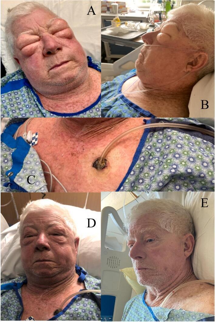
This clinical photographic series documents the presentation and treatment of severe Grade IV subcutaneous emphysema (SSE) in an adult patient. Images A and B show massive facial and cervical swelling on hospital day 5, with significant periorbital edema resulting in the eyes being swollen shut and loss of normal facial contours. Image C illustrates the procedural intervention: incisional negative pressure wound therapy (iNPWT) applied to the left anterior chest wall. A small incision is visible with a suction tube and occlusive dressing, designed to facilitate the evacuation of trapped subcutaneous air. Images D and E show a chronological timeline of clinical improvement. At 24 hours post-intervention (D), there is a noticeable reduction in facial puffiness, allowing the patient to partially open their eyes. By 48 hours (E), there is near-total resolution of the SSE, with facial features returning to baseline and the disappearance of the previously taut, distended skin. This series demonstrates the efficacy of iNPWT in managing rapidly progressive subcutaneous air accumulation.
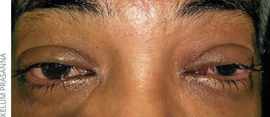
This clinical photograph shows a close-up frontal view of a patient's eyes exhibiting hallmark signs of viral conjunctivitis. Bilateral conjunctival injection is evident, characterized by diffuse redness and prominent vascularization across the bulbar and palpebral sclera, particularly concentrated in the inferior regions. There is visible eyelid edema (chemosis) resulting in a slightly swollen or puffy appearance of the periorbital tissues. The eyes demonstrate increased lacrimation or watery discharge, giving the lid margins a glistening, moist texture. The periocular skin appears hyperpigmented and somewhat shiny compared to the forehead, likely secondary to chronic irritation or fluid accumulation. The primary clinical focus is on identifying acute inflammatory changes of the ocular surface, including hyperemia and epiphora, which are typical manifestations of a viral infection such as adenovirus. This image serves as an educational reference for distinguishing viral etiologies from bacterial or allergic conjunctivitis based on the nature of the discharge and the pattern of inflammation.
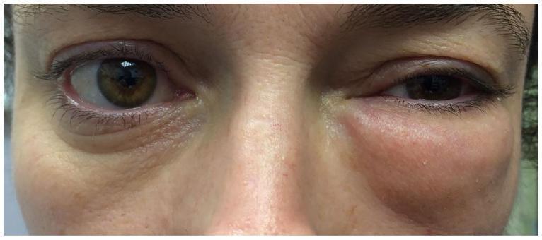
Clinical photograph of a patient's mid-face showing significant, asymmetrical left periorbital swelling. The left orbital region exhibits diffuse edema extending from the upper eyelid to the malar area, causing mechanical ptosis and narrowing of the palpebral fissure. The skin over the swollen area appears mildly erythematous and tense, distorting the natural facial contours. In contrast, the right eye and periorbital tissues appear normal with preserved lid margins and orbital architecture. This visual finding is characteristic of conditions leading to sudden volume increase in the subcutaneous periorbital space, such as orbital emphysema (often following trauma or nose blowing in the setting of an orbital floor fracture) or inflammatory processes. The presentation highlights the clinical signs of unilateral periorbital edema and its impact on ocular adnexa.
Recurrent Swelling Around the Eyes - Possible Causes
1. Allergies (Most Common)
- Allergic rhinitis or hay fever - pollen, dust mites, pet dander, mold
- Contact allergy - eye makeup, face creams, contact lens solution, soap
- Food allergies - especially nuts, shellfish, dairy, eggs
- Symptoms: itching, redness, watery eyes alongside swelling
- Usually affects both eyes
2. Lifestyle & Dietary Factors
- High salt intake - causes the body to retain fluid, pooling in loose tissue around the eyes
- Alcohol consumption - causes dehydration and fluid imbalance
- Sleep deprivation or poor sleep position - fluid pools when you sleep flat
- Crying - tear irritation causes temporary swelling
- Smoking - causes vascular imbalances
3. Kidney Problems (Nephrotic Syndrome)
- One of the classic signs - morning periorbital puffiness that improves during the day
- The kidneys lose protein through urine, dropping blood protein levels, so fluid leaks into tissues
- Often accompanied by: puffy face, legs, frothy urine, reduced urine output
- This textbook finding is specifically noted in Comprehensive Clinical Nephrology (7th Ed.) - morning periorbital edema in nephrotic patients that resolves with gravity during the day
4. Thyroid Disease
- Hypothyroidism (underactive thyroid): causes a particular type of puffiness called myxedema - non-pitting swelling around the eyes
- Hyperthyroidism / Graves' disease (overactive thyroid): can cause thyroid eye disease - periorbital edema, eye protrusion (proptosis), redness, double vision
- Other symptoms: weight changes, fatigue, hair loss, heart palpitations
5. Eye Infections
- Conjunctivitis (pink eye) - viral or bacterial - causes lid swelling + redness + discharge
- Blepharitis - inflammation of the eyelid margins, often recurrent
- Stye (hordeolum) - a blocked oil gland or hair follicle on the eyelid
- Chalazion - a painless lump/swelling from a blocked meibomian gland
6. Periorbital Cellulitis (Important - Seek Care Urgently)
- A skin infection of the eyelid/skin around the eye
- Looks like: red, warm, painful, swollen eyelid, sometimes with fever
- This requires urgent antibiotic treatment - can become dangerous if untreated
7. Angioedema
- Sudden, deeper swelling triggered by allergies, medications (especially ACE inhibitors like ramipril/lisinopril), or hereditary conditions
- Recurrent episodes without clear cause = see a specialist
8. Blepharochalasis
- A rare condition with recurrent, painless episodes of periorbital swelling starting in adolescence
- Thought to be a variant of angioedema; episodes may be triggered by stress
- Over time leads to sagging eyelid skin
9. Sinusitis
- Infection or inflammation of the sinuses near the eyes can cause swelling of the surrounding tissues
10. Heart or Liver Disease (Less Common)
- Advanced heart failure or liver cirrhosis can cause generalized fluid retention, including around the eyes
When to See a Doctor Promptly
- Swelling is painful, red, or hot (infection)
- You have fever with eye swelling
- Your vision is blurred or eye movement is painful
- Swelling is accompanied by difficulty breathing or throat tightening (anaphylaxis - emergency)
- Swelling keeps returning with no obvious cause
- You also have frothy urine, leg swelling, or extreme fatigue (kidney/systemic disease)
- You have symptoms of thyroid disease (weight changes, hair loss, palpitations)
Practical Next Steps
- Keep a diary - note when the swelling happens, how long it lasts, what you ate, products you used
- Reduce salt intake and drink more water
- Check your cosmetics/skin products for contact allergens
- See a doctor for basic blood tests: kidney function, thyroid function (TSH, T3, T4), urine protein test, and possibly allergy testing
Disclaimer: This is general health information, not a substitute for a personal medical evaluation. Please consult a doctor for a proper diagnosis.
Not around the eyes on the eyelids
recurrent eyelid swelling causes stye chalazion blepharitis treatment
stye chalazion eyelid swelling comparison
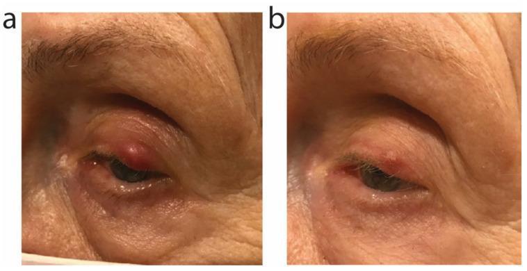
Clinical photograph comparison of the left eye in an adult patient, demonstrating the progression and treatment of an external hordeolum (stye) associated with ocular rosacea. Image (a) shows the baseline presentation: a prominent, well-circumscribed, erythematous, and edematous nodule located on the margin of the left upper eyelid. There is visible peri-orbital erythema and inflammatory swelling of the surrounding skin. Image (b) shows clinical improvement 15 days later, following a 9-day course of oral sarecycline. The lesion exhibits a significant reduction in volume, height, and vascular congestion. The surrounding skin shows resolution of the inflammatory edema and a return to a more natural texture and color. This comparison illustrates the efficacy of tetracycline-class antibiotics in managing painful inflammatory eyelid lesions by reducing both bacterial load and localized inflammation. Educational focus includes ophthalmology, dermatology, and the clinical response of staphylococcal infections or ocular rosacea to systemic antibiotic therapy.
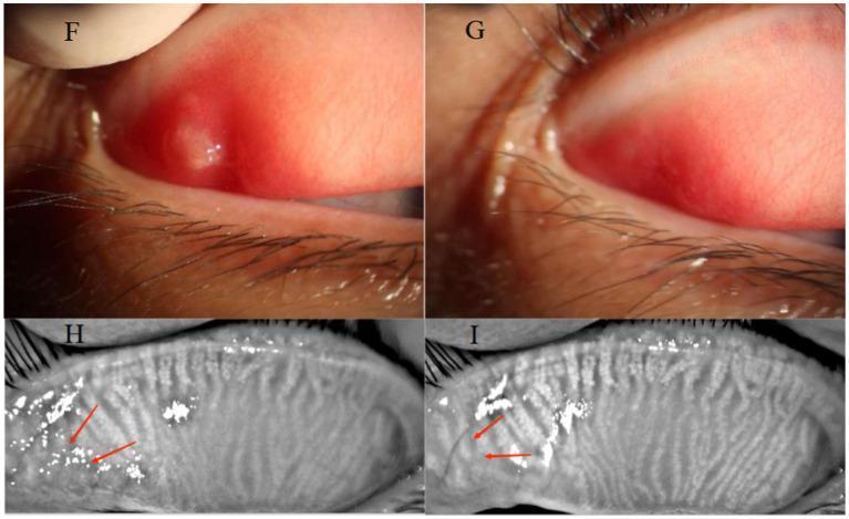
A multi-panel medical comparison showing a 32-year-old female patient with an upper eyelid chalazion before and after Optimal Pulse Technology (OPT) treatment. Panels F and G are clinical photographs of the everted upper palpebral conjunctiva. Panel F (baseline) demonstrates a raised, circumscribed chalazion with significant surrounding palpebral conjunctival hyperemia. Panel G (post-treatment) shows complete regression of the cyst and resolution of the hyperemia. Panels H and I provide infrared meibography of the same eyelid. Panel H shows significant meibomian gland dropout and architectural distortion at the site of the original chalazion (marked with red arrows). Panel I (post-treatment) displays a comparative increase in meibomian gland density and improved definition of glandular structures in the previously affected area. This comparison illustrates the efficacy of non-invasive light therapy in reducing eyelid inflammation and promoting meibomian gland recovery.
Swelling Specifically on the Eyelids - What's Causing It?
1. Stye (Hordeolum) - Most Common
- Looks like: a red, painful lump on the edge of the eyelid, like a pimple
- Usually caused by Staphylococcus bacteria
- Can cause swelling of the whole eyelid
- Tends to come to a head and drain on its own
2. Chalazion - Very Common, Especially Recurrent Cases
- Looks like: a firm, pea-like lump under the eyelid skin
- Usually not painful (unlike a stye)
- Can cause the entire eyelid to swell
- If large, can press on the eye and blur vision
- Tends to recur if the root cause (see below) is not treated
3. Blepharitis - The #1 Root Cause of Recurrence
- Eyelid margins look red, crusty, or scaly
- You may notice flaking at the base of the lashes (like dandruff on the lids)
- Caused by bacterial overgrowth and/or meibomian gland dysfunction (MGD)
- Strongly associated with: rosacea, seborrheic dermatitis (dandruff), oily skin
- Without treating blepharitis, styes and chalazia keep coming back
4. Meibomian Gland Dysfunction (MGD)
5. Demodex Mite Infestation
6. Allergic Eyelid Swelling
Important Warning - When to See a Doctor Urgently
- The swelling is very painful, red, and warm (could be preseptal/orbital cellulitis - a serious infection)
- You have fever with eyelid swelling
- Your vision is affected
- The lump keeps coming back in the exact same spot - this can occasionally be a sign of a sebaceous gland carcinoma (rare, mostly in older people), and a biopsy may be needed
What You Can Do Now
| Step | Action |
|---|---|
| Warm compress | Apply a warm, damp cloth to the eyelid for 10-15 minutes, 3-4 times a day - helps unblock glands |
| Lid hygiene | Gently clean the eyelid margins daily with diluted baby shampoo or lid-scrub wipes |
| Avoid touching/squeezing | Squeezing a stye can spread infection |
| See an eye doctor | If it keeps recurring, an ophthalmologist can treat the root cause (blepharitis, MGD, Demodex) properly |
Disclaimer: This is general health information and not a substitute for a personal medical examination. Please see an eye doctor for proper diagnosis and treatment.
What are the best at-home remedies for an active stye or chalazion?
at home remedies stye chalazion warm compress evidence based treatment
stye chalazion home treatment warm compress tea tree oil lid scrub evidence
At-Home Remedies for a Stye or Chalazion
#1 - Warm Compresses (THE Most Important Step)
- Wash your hands thoroughly first
- Soak a clean cloth in comfortably hot (not scalding) water and wring it out - or microwave a damp cloth for 20-30 seconds
- Hold it against the closed eyelid for 10 minutes
- Repeat 4 times a day (every few hours)
- After each compress, use a clean fingertip to gently massage the lump in a circular motion - this active compression is much more effective than just resting the cloth on the eye
- For a stye: heat softens the pus and helps it come to a head and drain on its own
- For a chalazion: heat melts the thick, solidified oil clogging the meibomian gland, allowing it to drain naturally
Styes and chalazia often start shrinking within a few days of consistent compresses. Allow it to open and drain on its own - do not squeeze it.
#2 - Eyelid Margin Cleaning (Lid Scrubs)
- Pre-made lid scrub wipes (e.g., Ocusoft Lid Scrub pads) - the easiest option
- Diluted baby shampoo - put a tiny drop on a damp cotton pad or your clean fingertip, gently scrub along the lash line, then rinse off
- Do this once or twice a day
#3 - OTC Stye Ointments
#4 - Stop Wearing Makeup and Contact Lenses
- Remove all eye makeup until fully healed
- Stop wearing contact lenses while the stye/chalazion is active - both can introduce bacteria and worsen inflammation
- Throw away any eye makeup that may have been used while the stye was present (mascara, eyeliner) as it can be contaminated
What NOT to Do (Common Mistakes)
| Avoid | Why |
|---|---|
| Squeezing or popping it | Spreads infection, can push bacteria deeper, may cause scarring |
| Neat tea tree oil directly on the lid | Toxic to the eye surface - can cause chemical burns to the cornea |
| Touching the eye repeatedly | Introduces more bacteria |
| Wearing eye makeup | Clogs glands further and risks contamination |
| Stopping compresses early | A chalazion can take 3-4 weeks to fully resolve - consistency matters |
Stye vs Chalazion - Small Difference in Approach
| Stye | Chalazion | |
|---|---|---|
| Pain | Painful, tender | Usually painless |
| Goal of compress | Help it drain/rupture | Melt blocked oil |
| Typical timeline | Resolves in 1-2 weeks | May take 3-6 weeks |
| Massage | Gentle | Gentle circular massage especially helpful |
When to Stop Home Treatment and See a Doctor
- No improvement after 3-4 weeks of consistent warm compresses
- The swelling is getting larger or more painful
- You develop fever, redness spreading beyond the eyelid, or pain with eye movement (signs of spreading infection - preseptal cellulitis)
- Your vision becomes blurred (large chalazion pressing on the eye)
- The lump keeps returning in the exact same spot - this needs a biopsy to rule out rare eyelid tumors
Disclaimer: This is general health guidance and not a substitute for personal medical advice. See an eye doctor if symptoms worsen or persist.
How can I prevent styes and chalazion from coming back?
prevent recurrent stye chalazion blepharitis meibomian gland dysfunction long term prevention
https://wexnermedical.osu.edu/eye-care-ophthalmology/conditi…
How to Prevent Styes and Chalazia from Coming Back
Daily Lid Hygiene Routine (Most Important)
- Use a pre-made lid scrub pad (Ocusoft, Blephaclean), or
- Put a tiny drop of diluted baby shampoo on a damp cotton pad
- Gently scrub along the base of the lashes on both upper and lower lids
- This removes bacterial buildup, debris, and stale oil that clog the glands
Omega-3 Fatty Acids (Fish Oil)
- They improve the quality and fluidity of meibomian gland oil, making it less likely to solidify and block the glands
- Dose: typically 1,000-2,000 mg of EPA/DHA per day from fish oil - ask your doctor
- Effects build up over weeks to months, so be patient
Makeup and Cosmetic Habits
- Remove all makeup thoroughly every night - never sleep with mascara or eyeliner on
- Residue left overnight accumulates at gland openings and directly causes blockages
- Replace mascara every 3 months and eyeliner regularly - old products harbor bacteria
- Avoid applying eyeliner to the waterline (inside the lash line) - this directly blocks meibomian gland openings
- Use clean brushes and applicators - contaminated tools reintroduce bacteria
- Consider switching to lighter, mineral-based formulas if you are prone to recurrence
Hands and Hygiene
- Don't touch or rub your eyes - hands carry Staphylococcus bacteria which is the main bug that infects the eyelid glands
- Wash hands before any eye contact (removing contacts, applying drops, etc.)
- Don't share towels, pillowcases, or washcloths - bacteria transfer easily
- Change your pillowcase at least once a week
Address Underlying Skin Conditions
| Condition | How it contributes | What helps |
|---|---|---|
| Rosacea | Causes chronic lid inflammation and poor oil quality | Treat with low-dose doxycycline, IPL therapy, prescribed skin care |
| Seborrheic dermatitis (dandruff) | Flaking skin clogs lid margins | Medicated shampoo (selenium sulfide, ketoconazole), regular lid scrubs |
| Demodex mites | Microscopic mites on lashes block glands | Tea tree oil-based lid scrubs (diluted/in wipes - never neat), or prescription XDEMVY eye drops for confirmed cases |
| Hormonal fluctuations | Affect gland secretion quality | Discuss with your doctor if symptoms track with menstrual cycle or menopause |
Screen Use and Blinking
- Heavy screen use reduces your blink rate (from ~15 blinks/min to ~5), causing meibomian gland oil to stagnate and thicken
- Follow the 20-20-20 rule: every 20 minutes, look at something 20 feet away for 20 seconds, and blink deliberately
- Consider lubricating eye drops if you have dry eyes - dry eye and MGD are closely linked
Diet
- Eat more leafy greens and oily fish (salmon, sardines, mackerel) - supports gland health
- Stay well hydrated
- Some evidence that a diet high in saturated fats worsens oil gland secretions
When You Need a Doctor for Prevention
- Low-dose oral doxycycline (20-50 mg daily) - the Wills Eye Manual specifically recommends this for multiple or recurrent chalazia, particularly with rosacea. It works through anti-inflammatory rather than antibiotic action and is taken long-term
- Intense Pulsed Light (IPL) therapy - a clinic treatment that uses controlled light pulses to liquefy blocked gland secretions and reduce lid inflammation; effective for rosacea-related and MGD-related recurrence
- Meibomian gland probing - a procedure that physically opens blocked gland ducts, used when conservative measures fail
- Demodex testing and treatment - a slit-lamp exam can confirm mite infestation; prescription drops (XDEMVY/lotilaner) can eradicate them
Quick Daily Prevention Checklist
- Warm compress for 2-3 minutes each morning
- Lid scrub along the lash line
- Remove all eye makeup before bed
- Don't touch or rub your eyes
- Take daily omega-3 supplement
- Blink consciously during screen time
- Change pillowcase weekly
Disclaimer: This is general health guidance. See an eye doctor (ophthalmologist or optometrist) for a personalised plan, especially if flare-ups are frequent or severe.
What are the clinical treatment for blepharitis or meibomian gland dysfunction
clinical treatment blepharitis meibomian gland dysfunction guidelines 2024 2025
https://www.reviewofophthalmology.com/article/unraveling-ble…
https://www.college-optometrists.org/clinical-guidance/clini…
"blepharitis" AND "meibomian gland dysfunction" AND "treatment"
"meibomian gland dysfunction"[MeSH Terms] AND "treatment"
PMID: 38350160
Clinical Treatments for Blepharitis & Meibomian Gland Dysfunction
| Type | Location | Main Cause | Primary Treatment Focus |
|---|---|---|---|
| Anterior blepharitis | Outer lash margin | Staph bacteria, Demodex mites, seborrheic dermatitis | Antibiotics, lid hygiene, anti-mite agents |
| Posterior blepharitis / MGD | Inner lid margin, meibomian glands | Blocked/dysfunctional oil glands | Heat therapy, gland expression, anti-inflammatory agents |
| Mixed | Both | Both | Combined approach |
Step 1 - Lid Hygiene (Foundation for Both Types)
- Warm compresses 2-4 times daily to soften meibum and scales
- Eyelid scrubs along the lash margin (commercial wipes, diluted baby shampoo)
- Vertical lid massage after compresses to physically express thickened meibomian secretions
- This is continuous, long-term management - not a short-term fix
Step 2 - Topical Medications (Prescribed by a Doctor)
For Anterior Blepharitis (Staph / Seborrheic)
| Medication | What It Does | Typical Use |
|---|---|---|
| Topical antibiotics (azithromycin eye drops, bacitracin/erythromycin ointment, tobramycin drops) | Reduce Staphylococcus bacterial load on the lid margin | Short course, applied to lid margin |
| Antibiotic-steroid combination (neomycin/polymyxin B/dexamethasone, tobramycin/dexamethasone) | Treats both infection and inflammation together | Short course only - steroids carry risk of glaucoma and cataracts with prolonged use |
| Topical cyclosporine (Restasis, Cequa) | Immunomodulator - reduces lid and ocular surface inflammation | Long-term use; particularly useful when dry eye coexists |
| Lifitegrast (Xiidra) | Lymphocyte integrin inhibitor - reduces T-cell driven inflammation | Newer agent; used for inflammatory dry eye/blepharitis overlap |
For Posterior Blepharitis / MGD
| Medication | What It Does |
|---|---|
| Topical azithromycin (AzaSite) | Has both antibacterial and anti-inflammatory properties; penetrates eyelid tissue well |
| Antibiotic-steroid ointments | Reduce lid margin inflammation |
Step 3 - Oral Medications (For Moderate-Severe or Rosacea-Associated Cases)
Tetracycline class (first-line oral treatment)
- Doxycycline 20-100 mg once or twice daily - the most widely used oral agent
- Minocycline - similar class, alternative option
- Low-dose doxycycline (20-50 mg) is used specifically for its anti-inflammatory (not antibiotic) effect - at this dose it improves meibum quality without the antibiotic risks of resistance or gut flora disruption
- Duration: weeks to months, tapered based on response; can be restarted during flares
- Especially effective when rosacea is the underlying trigger
Macrolides
- Azithromycin oral - used when tetracyclines are not tolerated (e.g., children, pregnancy)
- Erythromycin - alternative option
Step 4 - Demodex-Specific Treatments
| Treatment | Details |
|---|---|
| Tea tree oil-based lid scrubs (50% dilution in clinic, 5-10% for home use) | First-line; disrupts mite life cycle at lash roots |
| XDEMVY (lotilaner 0.25% ophthalmic solution) | FDA-approved 2023 - first prescription drop specifically for Demodex blepharitis; applied twice daily for 6 weeks; paralyzes and eliminates mites |
Step 5 - In-Office Procedures (For MGD Not Responding to Medications)
Thermal Pulsation (Heat + Pressure)
- LipiFlow (Johnson & Johnson) - applies controlled heat (42.5°C) to the inner lid surface and simultaneous pulsatile pressure from outside, liquefying and expressing blocked meibum from all glands in one 12-minute session
- Systane iLux2 (Alcon) - similar concept, handheld device
- A single treatment has shown symptom improvement lasting up to 1 year in RCTs
- Best results when glands have not yet atrophied
Intense Pulsed Light (IPL)
- Broad-spectrum light pulses applied to the skin around the eyes
- Reduces inflammation in lid blood vessels, melts meibum, and kills Demodex
- Particularly effective for rosacea-associated MGD
- Typically 3-4 sessions, 2-4 weeks apart, then maintenance sessions
- Evidence building since 2002; now widely used in dry eye/blepharitis clinics
Automated Eyelid Debridement
- BlephEx - a motorized micro-sponge tip that spins along the lash line, physically removing biofilm, bacterial debris, and Demodex from the lid margin in clinic
- Performed every 4-6 months as maintenance
- Immediately improves symptoms in many patients
Intraductal Meibomian Gland Probing (MGP)
- A fine probe is inserted directly into blocked meibomian gland orifices to mechanically open them
- Used for severe obstructive MGD when heat therapy has failed
- Can be uncomfortable but provides rapid and lasting relief in case series data
Summary Treatment Ladder
STEP 1 (Everyone) → Daily lid hygiene: warm compress + lid scrubs + massage

STEP 2 (Mild-Moderate) → Add topical antibiotics ± steroid-antibiotic combo
                       → Omega-3 supplements

STEP 3 (Moderate-Severe or Rosacea) → Oral doxycycline low-dose long-term
                                     → Topical cyclosporine / lifitegrast

STEP 4 (Demodex confirmed) → Tea tree oil scrubs + XDEMVY drops (6-week course)

STEP 5 (Not responding to meds) → In-office: LipiFlow / IPL / BlephEx / MGP

STEP 6 (Recurrent chalazia, atypical) → Pathology biopsy to rule out sebaceous carcinoma
Important Caveats
- Blepharitis has no definitive cure - it is a chronic condition managed, not cured, as confirmed by the 2024 clinical review
- Topical steroids must be used short-term only (risk of raised intraocular pressure and cataracts with long-term use)
- Treatment is tailored to the type (anterior vs posterior vs mixed) and severity
- In older patients with recurrent or atypical chalazia that don't respond, a sebaceous gland carcinoma biopsy should be considered
Disclaimer: These treatments require proper diagnosis and prescription by an eye doctor (ophthalmologist or optometrist). Do not self-prescribe antibiotics or steroids for the eye. See a specialist for a tailored treatment plan.
I'm getting alternative on both eyes in a month gap. My doctor adviced me to drain and clean. But he said it costs 3500. What should I do now is there any alternative
Your Situation
- A new or early chalazion (less than 4-6 weeks old)
- A lesion that hasn't been tried on conservative treatment yet
Alternatives to Surgical Drainage
Option 1 - Aggressive Warm Compress Therapy (Try This First)
- 10 minutes, 4 times a day with active massage after each session
- Use a heated eye mask (reusable ones cost ₹200-500 at pharmacies) rather than a wet cloth - they maintain the right temperature longer and work better
- Give it a full 4 weeks of strict daily compliance before deciding on surgery
- Many chalazia that look like they need draining resolve completely with this alone
Option 2 - Steroid Injection (Less Expensive Than Surgery)
- Shrinks the lump from the inside without cutting
- Is less expensive than I&D in most clinics
- Takes only a few minutes
- Works in roughly 60-80% of chalazia
- Ask your doctor specifically about this option - "Can we try an intralesional steroid injection instead of cutting?"
Option 3 - Topical Antibiotic-Steroid Ointment (For Early/Inflamed Lesions)
Option 4 - Oral Doxycycline (Treats the Root Cause)
- Low-dose doxycycline 20-50 mg daily (very affordable, widely available) treats the underlying blepharitis/MGD
- It reduces meibomian gland inflammation, improves oil quality, and breaks the monthly recurrence cycle
- Costs a few hundred rupees per month at most
- Ask your doctor to prescribe this alongside or instead of repeated drainage
What I Would Suggest Doing
-
Start warm compresses immediately - 4 times a day, 10 minutes each, with massage. Use a proper warm eye mask if possible.
-
Go back to your doctor and have an honest conversation:
- "The current one is [X] weeks old - can we try compresses + a steroid injection first?"
- "Can you prescribe low-dose doxycycline to stop them coming back every month?"
- "Is the steroid injection option available, and what does it cost?"
-
If the chalazion is already hard and more than 6-8 weeks old, conservative treatment is less likely to work and drainage may genuinely be the most practical solution. At that point, ₹3,500 for a minor procedure done properly is reasonable.
-
After this episode resolves - commit to the daily lid hygiene routine (warm compress + lid scrub every morning) to prevent the next one. This is free and is the most powerful long-term fix.
When Drainage IS Necessary
- The chalazion is large, hard, and has been present for more than 6-8 weeks
- It is pressing on your eye and blurring your vision
- It keeps refilling after a steroid injection
- It is growing despite conservative treatment
Bottom Line
Disclaimer: This is general health guidance. Your doctor has examined you in person and their advice is based on the specific state of your lesion - please discuss these options with them before deciding.
When it came last time it only lasted for a week
What This Means
- It was likely a stye (hordeolum) rather than a true chalazion - styes are acute infections that often drain and resolve quickly on their own
- Your body is actively clearing them - your immune response is working
- The lesion was not severely blocked or hardened - which is a good sign
- Surgical drainage is very unlikely to have been needed for that one
This Changes the Picture Significantly
- Start warm compresses right away - 10 minutes, 4 times a day with gentle massage
- Let it come to a head and drain naturally - do not squeeze it
- Keep the lid clean with gentle scrubs at the lash line
What to Ask Your Doctor
"The last one resolved completely on its own in one week without any procedure. This current one has been present for [X days/weeks]. Can we try warm compresses first before considering drainage?"
The Bigger Issue - Why Do They Keep Returning?
Practical Summary
| Question | Answer based on your history |
|---|---|
| Will this one resolve on its own? | Possibly yes - the last one did in 1 week |
| Should you rush to get drainage? | No - try warm compresses for 2-3 weeks first |
| What should you actually spend money on? | A consultation to get low-dose doxycycline + a reusable warm eye mask (₹200-400) |
| What stops them coming back monthly? | Daily lid hygiene + doxycycline to treat the root cause |
Disclaimer: This is general guidance based on what you've described. Your doctor has examined you directly - please keep them informed about your decision and seek their input before declining any recommended treatment.
The swelling dose not have any heaf
Stye vs Chalazion - Key Difference
| Feature | Stye (Hordeolum) | Chalazion (what you have) |
|---|---|---|
| Pus head | Yes - red, tender, comes to a point | No - smooth lump under the skin |
| Pain | Painful, tender | Usually painless or mildly sore |
| Cause | Bacterial infection | Blocked oil gland - no infection |
| Feels like | A pimple on the lid | A firm pea under the lid skin |
| Resolves how | Drains and pops on its own | Needs the blocked oil to be reabsorbed or drained |
Why This Matters for You
- The last one that resolved in a week was likely a stye that drained spontaneously
- This current one is different - it is a chalazion (blocked gland, no infection), which behaves differently and takes longer to resolve
- Chalazia do not "pop" - the blocked thickened oil has to either be reabsorbed by the body or drained by a doctor
What This Means for Treatment
Warm Compresses - Still the First Step
- 10 minutes, 4 times a day, with firm circular massage after each session
- This is slow - a chalazion can take 4-8 weeks to fully resolve with compresses alone
- Consistency every single day is what makes the difference
Realistic Expectations
- Small or recent chalazia (a few weeks old) - good chance compresses resolve it
- Large or older chalazia (6+ weeks, hard lump) - less likely to fully resolve without a procedure
- How long has this current one been there? That is the key question
The Steroid Injection Option
- A triamcinolone steroid injection directly into the lump is an excellent option
- It shrinks the granulomatous inflammation from inside
- Less invasive and often cheaper than I&D
- Works in about 60-80% of chalazia
- Ask your doctor: "Since there's no infection, can we try a steroid injection first instead of drainage?"
Bottom Line
| What you have | Chalazion - blocked oil gland, no pus, no infection |
|---|---|
| Will it drain naturally? | No - chalazia don't drain like styes |
| Best home approach | Warm compresses 4x daily for 4-6 weeks |
| Best clinic alternative to surgery | Steroid injection into the lump |
| When is drainage necessary? | If it's large, old, or not responding to compresses/injection after 6-8 weeks |
Disclaimer: Please keep your doctor informed and follow their direct clinical advice, as they have examined you in person.
It is on the upper eyelid the new one is 3 days old and keeps growing daily
What's Happening Right Now
What You Should Do Right Now (Today)
1. Start Warm Compresses Immediately - This Is Urgent Now
- Heat a clean cloth or use a warm eye mask to 40-45°C (warm but not burning)
- Hold against the closed upper eyelid for 10-15 minutes
- After each compress, place a clean finger on the outer surface of the upper lid and massage downward toward the lash line - this pushes blocked oil toward the gland opening
- Do this every 3-4 hours - at least 4 times today and every day going forward
2. Do NOT Rub or Press the Lump Randomly
3. Keep the Lid Clean
The "Growing Daily" Part - When to Act Fast
| Warning Sign | What It May Mean |
|---|---|
| Swelling spreading beyond the eyelid onto the surrounding skin | Early preseptal cellulitis (infection spreading) |
| Skin becoming red, warm, and painful to touch | Infected chalazion / cellulitis |
| Fever developing | Spreading bacterial infection |
| Vision becoming blurred | Lump pressing on the cornea |
| Eye becoming difficult to open | Significant size - needs prompt treatment |
| Swelling crossing to the nose or cheek | Requires urgent medical attention |
Your Specific Situation - Honest Assessment
| Factor | What It Means |
|---|---|
| Upper eyelid location | More noticeable, can affect vision if large - worth monitoring closely |
| Only 3 days old | Ideal time to start compresses - early intervention works best |
| Still growing | Expected at this stage - watch if it plateaus or keeps growing past day 7 |
| No pus head | Chalazion - won't drain naturally, needs reabsorption |
| Recurring monthly | Underlying MGD driving it - root cause untreated |
Realistic Timeline From Here
- Days 1-7: Inflammation phase - may continue to grow slightly, then plateau
- Week 2-3: With consistent warm compresses, most early chalazia begin to soften and shrink
- Week 4-6: Full resolution possible in early/small ones
- If still growing at day 10-14 with no sign of slowing - see your doctor again and ask specifically about a steroid injection before agreeing to surgical drainage
Most Important Action Right Now
Disclaimer: You have an active, growing eyelid lesion. Please monitor it closely and do not delay seeking care if any warning signs develop. This guidance does not replace in-person medical evaluation.
Find recent clinical guidelines on early chalazion management and natural resolution rates
"chalazion" AND "natural resolution" OR "conservative treatment" OR "warm compress"
chalazion natural resolution rate early management clinical guidelines 2023 2024 2025
PMID: 37937798
https://pmc.ncbi.nlm.nih.gov/articles/PMC12178684
"chalazion"[MeSH Terms] AND "steroid injection" AND "incision curettage"
https://emedicine.medscape.com/article/1212709-treatment
"chalazion"[MeSH Terms] AND "triamcinolone" AND "incision"
Recent Clinical Guidelines & Evidence on Early Chalazion Management
Sources Used
- Tashbayev et al., Current Eye Research 2024 - Chalazion Treatment: A Concise Review of Clinical Trials (PMID 37937798)
- Shin et al., Korean Journal of Ophthalmology 2025 - Management Practice for Hordeolum and Chalazion: A Survey of KSOPRS Members (PMID 40267992)
- Medscape Clinical Treatment Guidelines - Chalazion, Updated 2024
- Perth Children's Hospital Clinical Guidelines, March 2024
- College of Optometrists Clinical Management Guideline - Chalazion, Updated January 2025
- Wills Eye Manual (medical library)
1. Natural Resolution Rate - What the Evidence Says
"Studies have found resolution rates of 25% to 50% with conservative management alone"
- Korean Journal of Ophthalmology survey of ophthalmologists, 2025
"Most chalazions and styes do not require treatment. The natural history is initial expansion followed by spontaneous rupture before healing."
- Roughly 1 in 3 to 1 in 2 chalazia resolve on their own with warm compresses and lid hygiene alone
- Your chalazion is only 3 days old - you are in the best possible window for conservative treatment to work
- The other 50-75% that do not resolve need either a steroid injection or surgical drainage
2. How Long Do Doctors Wait Before Escalating?
- For chalazion: most doctors attempt conservative treatment for 1-4 weeks before moving to procedures
- Medscape guidelines: "If the chalazion does not resolve within 1 month, the patient should be referred to an ophthalmologist for definitive treatment"
- ~57% of ophthalmologists surveyed consider warm compresses and lid hygiene to be effective or somewhat effective as first-line treatment
3. Treatment Options Ranked by Evidence (Least to Most Invasive)
| Treatment | Evidence / Resolution Rate | When Used |
|---|---|---|
| Warm compress + lid massage | 25-50% resolution | First-line, always tried first |
| Topical antibiotic-steroid ointment | Modest additional benefit | Added to compresses for inflamed lesions |
| Intralesional steroid injection (triamcinolone) | ~80-85% resolution rate | After compresses fail, or as first-line for persistent/large lesions |
| Incision & curettage (I&D) | ~85-90% resolution | When compresses + injection fail, or large/old lesions |
| Steroid injection + I&D combined | ~95% resolution | Recurrent/stubborn cases |
4. Is There a "No Treatment Needed" Option?
"Small, inconspicuous, asymptomatic chalazia may be ignored."
5. What the Guidelines Say About Antibiotics
- 51.3% of ophthalmologists consider topical antibiotics ineffective for chalazion (Korean survey 2025)
- Antibiotics are only useful if there is a secondary infection present
- Chalazia are non-infectious - they are a blocked oil gland reaction, not a bacterial infection
- Guidelines: "Topical or systemic antibiotics are not usually required" for chalazia (Perth PCH Guidelines 2024)
What This Evidence Means Specifically for You
| Your Situation | What the Evidence Says |
|---|---|
| 3-day-old chalazion, upper lid | Way too early for surgical drainage per all guidelines |
| No infection/pus head | Conservative management appropriate |
| Doctor recommending immediate I&D | More aggressive than guideline-recommended approach |
| Recurring monthly | Needs long-term doxycycline/root cause treatment, not just drainage |
| Asking about alternatives | Steroid injection is evidence-based, equally effective, less invasive, and likely cheaper |
Your Clearest Path Forward (Evidence-Based)
- Start warm compresses today - 4 times daily, 10-15 minutes each - give it 3-4 weeks
- Ask your doctor for a steroid injection as the next step if compresses don't work - it has ~80-85% success and avoids cutting
- Ask for low-dose doxycycline to treat the root cause and stop monthly recurrence - this is the most clinically important step
- Surgical drainage (I&D) is valid but should come after a trial of conservative management and/or steroid injection per current evidence
Disclaimer: Clinical guidelines are general frameworks. Your doctor's recommendation is based on their direct examination. The evidence above supports a trial of conservative management first for a 3-day-old chalazion, but discuss this with your doctor before changing your treatment plan.