


Give this point wis e So I can remember all of this
Nictitating Membrane - Surgical Anatomy
- Roughly triangular fold of mucosa located at the medial canthus
- Base of the triangle = its free/leading margin
- Bulbar (posterior) and palpebral (anterior) surfaces are confluent with conjunctival mucosa
- A T-shaped cartilage lies within the membrane
- The "arms" of the T run along the leading margin
- Functions: supports the membrane, supports the corneal contour, and protects it
- Surrounds the base of the cartilage
- Produces seromucoid tears
- Excretory ducts emerge at the middle section of the bulbar mucosal surface
- Can be the major or minor producer of the aqueous tear film layer
- Found primarily on the bulbar surface
- Appear as raised translucent spots
- Comes from branches of the internal maxillary artery
- Protects the cornea
- Spreads the tear film
- Contributes the essential aqueous layer to the preocular tear film

Which one is free or leading margin
Simple way to remember:
Medial canthus        Center of eye
(where it's fixed) → [MEMBRANE] → FREE/LEADING MARGIN
                                        ↑
                              This edge "leads" as the
                              membrane sweeps across the eye
- Base of the triangle = the free/leading margin - this is the outermost/exposed edge that you can see in the diagram, the border of the nictitating membrane labeled at the bottom of the eye
- It's called "leading" because when the membrane moves across the cornea (like a windshield wiper), this edge leads the movement
- It's called "free" because it is not attached to anything - it floats freely
Memory tip:
"The BASE is FREE" - the base of the triangle is the free margin, and it's the edge closest to the cornea/center of the eye, NOT the part tucked in the corner.
Give me diagram
Veterinary anatomy diagram of the nictitating membrane (third eyelid) of a dog's eye, labeled cross-section and front view. Show: the triangular fold of mucosa at the medial canthus (nasal corner), the T-shaped cartilage inside with arms along the leading/free margin, the nictitans gland surrounding the base of the cartilage, the bulbar (posterior) surface and palpebral (anterior) surface, the free/leading margin (base of triangle) pointing toward the center of the eye, excretory ducts emerging at the bulbar mucosal surface, and the medial canthus attachment point. Use clear labels and arrows, educational medical illustration style, clean white background.
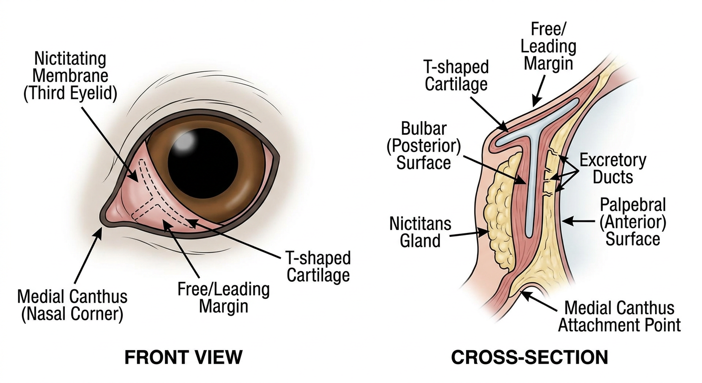
| Structure | Key Point |
|---|---|
| Medial canthus | Where the membrane is anchored (nasal corner) |
| Free/Leading margin | The BASE of the triangle - exposed edge facing the cornea |
| T-shaped cartilage | Arms run ALONG the leading margin |
| Nictitans gland | Wraps around the BASE of the cartilage |
| Bulbar surface | Posterior face - where ducts open |
| Palpebral surface | Anterior face |