
Gastric outlet obstruction full topic gen surg theory and clinical
Gastric outlet obstruction full topic gen surg theory and clinical
gastric outlet obstruction pyloric stenosis succussion splash radiology
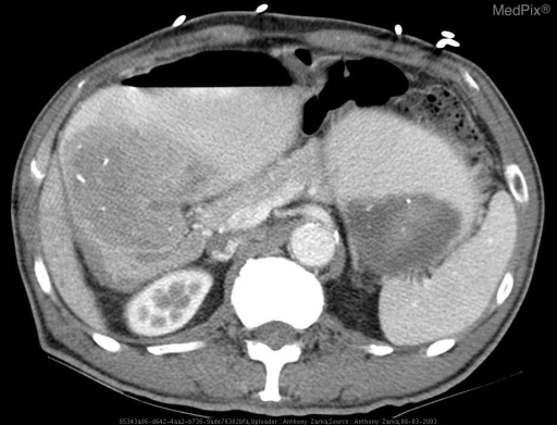
**Imaging Modality:** Contrast-enhanced axial Computed Tomography (CT) of the upper abdomen. **Anatomical Region:** Upper abdomen, specifically visualizing the stomach, liver, spleen, pancreas, and left kidney. **Observed Pathology:** Significant Gastric Outlet Obstruction (GOO). The image reveals marked distention of the stomach, which is filled with heterogeneous material (likely a combination of ingested food, secretions, and air), characteristic of a "succussion splash" appearance on imaging. **Characteristic Visual Features:** - **Gastric Dilation:** Severe gastric expansion occupying a large portion of the upper abdominal cavity. - **Transition Zone:** The obstruction appears localized to the distal antrum or pyloric region, consistent with cicatricial narrowing from chronic peptic ulcer disease. - **Associated Findings:** Presence of an air-fluid level within the stomach and compression of adjacent viscera. Small metallic surgical clips are visible on the anterior abdominal wall. **Clinical Context & Diagnosis:** Findings are highly suggestive of Gastric Outlet Obstruction secondary to peptic ulcer disease (PUD), leading to chronic pyloric or duodenal stenosis. **Differentiating Features:** The degree of gastric distention without an obvious intraluminal mass suggests a benign, chronic inflammatory or fibrotic etiology rather than acute malignancy.
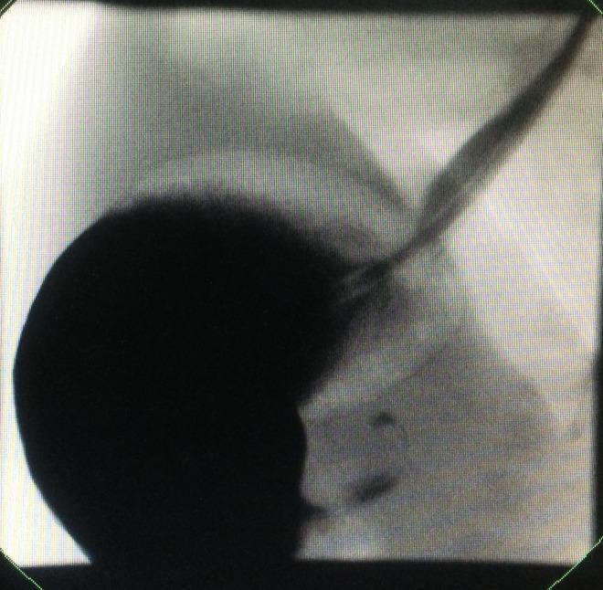
This diagnostic image is an upper gastrointestinal contrast study (fluoroscopy/X-ray) demonstrating gastric outlet obstruction. The image reveals a significantly dilated, globular stomach filled with radiopaque contrast material, appearing as a dense dark mass. There is a marked delay in gastric emptying, with only a thin, elongated stream of contrast (the 'string sign') visualized passing through a narrowed, hypertrophied pylorus into the distal duodenum. The clinical significance of these findings is highly suggestive of Infantile Hypertrophic Pyloric Stenosis (IHPS). The study serves as an educational example for pediatric gastroenterology and radiology, highlighting the visual contrast between the massive proximal gastric distension and the restricted flow into the small intestine caused by pyloric hypertrophy.
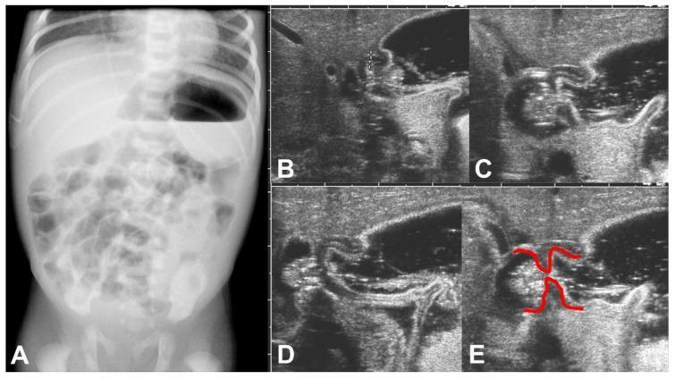
This composite figure presents multimodality imaging of a neonate with hypertrophic pyloric stenosis (HPS). Panel A shows an erect anteroposterior plain abdominal X-ray, demonstrating a significantly dilated stomach with a prominent air-fluid level, indicative of gastric outlet obstruction. Small bowel gas distribution appears distal but sparse. Panels B-E display a temporal sequence of abdominal ultrasonography focused on the pyloric region. The grayscale images show a thickened, elongated pyloric canal with failed passage of gastric contents during active peristaltic waves. In Panel E, red annotations highlight the 'shouldering' sign, where the thickened pyloric muscle bulges into the gastric antrum. Educational features include the hallmark radiographic finding of gastric distension and the sonographic characteristics of a non-relaxing, hypertrophic pylorus, which are critical for the diagnosis of HPS in pediatric radiology.
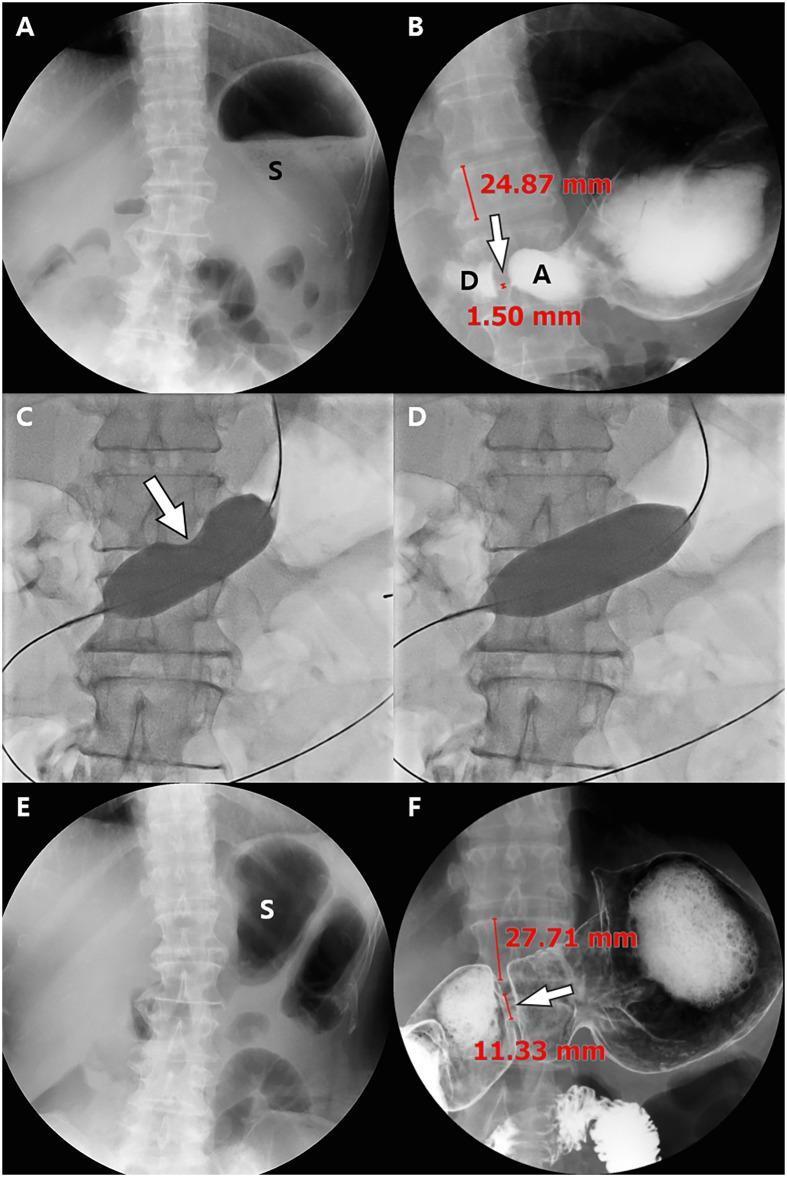
This composite of abdominal radiographic and fluoroscopic images demonstrates the management of impaired pyloric function following a pylorus-preserving gastrectomy (PPG). Panel A (scout) and B (Upper GI Series - UGIS) show pre-procedural state, highlighting significant gastric stasis with residual food (S) and severe narrowing of the pyloric canal (1.50 mm) between the antrum (A) and duodenum (D). Panels C and D provide intra-procedural fluoroscopic visualization of balloon dilatation. Panel C shows an 'hourglass deformity' (arrow) of the balloon at the point of stenosis, which resolves in Panel D upon full inflation, indicating successful dilatation. Panels E and F show the post-procedural follow-up at 11 months. The scout image (E) shows a marked reduction in residual gastric food material. The follow-up UGIS (F) demonstrates a patent, dilated pyloric canal measuring 11.33 mm. Quantitative assessment is provided by comparing the pyloric width to the height of the adjacent vertebral body (24.87 mm vs 27.71 mm). The series illustrates the clinical utility of interventional radiology in treating post-operative gastric outlet obstruction via balloon dilatation.
"gastric outlet obstruction"[MeSH Terms] AND management
Gastric Outlet Obstruction (GOO) - Full Topic: General Surgery Theory & Clinical
1. DEFINITION
2. AETIOLOGY / CAUSES
Benign Causes
| Category | Specific Causes |
|---|---|
| Peptic Ulcer Disease (most common benign) | Pyloric/prepyloric/duodenal bulb ulcer - oedema (acute/reversible) or fibrosis/scarring (chronic/fixed) |
| Inflammatory | Crohn's disease involving the duodenum, eosinophilic gastroenteritis |
| Congenital | Hypertrophic pyloric stenosis (neonates; also rarely adults), pyloric web/diaphragm |
| Extrinsic compression | Annular pancreas, superior mesenteric artery syndrome |
| Caustic ingestion | Acid/alkali ingestion causing pyloric stricture |
| Tuberculosis | Hypertrophic gastroduodenal TB |
| Bezoar | Phytobezoar, trichobezoar obstructing the pylorus |
| Gastric volvulus / hernias | Paraesophageal hernia, post-traumatic diaphragmatic hernia |
Malignant Causes (now the most common overall)
- Carcinoma of the antrum/pylorus - most common malignant cause
- Carcinoma of the head of pancreas / periampullary tumours
- Duodenal carcinoma
- Lymphoma
- Metastatic deposits (e.g., peritoneal carcinomatosis involving the duodenum)
Key Point (Bailey & Love 28e): With the decreasing incidence of PUD, GOO should be considered malignant until proven otherwise.
3. PATHOPHYSIOLOGY
Mechanism of obstruction
- Acute/functional: Oedema around an active peptic ulcer causing reversible outlet narrowing
- Chronic/mechanical: Progressive fibrosis and scarring of the pyloric canal from repeated ulceration/healing cycles - produces fixed, non-reversible stenosis
Metabolic Consequences - HYPOCHLORAEMIC HYPOKALAEMIC METABOLIC ALKALOSIS
- Repeated vomiting leads to loss of HCl (gastric acid) → primary hypochloraemia and alkalosis
- Early: Urine has low Cl⁻, high HCO₃⁻ (renal compensation for alkalosis)
- Progressive dehydration → sodium and water conservation by kidneys → aldosterone activated
- Aldosterone promotes Na⁺ retention with K⁺ and H⁺ excretion in distal tubule
- Result: Paradoxical aciduria (urine becomes acidic despite systemic alkalosis) + hypokalaemia
- Alkalosis → decreased ionised calcium → tetany (Trousseau's/Chvostek's signs)
4. CLINICAL FEATURES
Symptoms
- Vomiting - the cardinal symptom:
- Projectile, large volume
- Non-bilious (obstruction proximal to duodenal entry of bile duct)
- Contains undigested food eaten hours or even days earlier
- Characteristically occurs several hours after meals (often in the evening)
- Vomiting may bring temporary relief of upper abdominal discomfort
- Epigastric fullness and distension
- Early satiety
- Progressive weight loss
- Anorexia
- Long history of peptic ulcer disease (in benign cases) OR short history with no prior PUD (malignant)
Signs
- Dehydration, cachexia, sunken eyes
- Signs of malnutrition
- Visible gastric peristalsis - left-to-right peristaltic waves visible in epigastrium (more common in thin patients with longstanding obstruction)
- Distended stomach visible as epigastric fullness
- Succussion splash - audible splashing sound heard with stethoscope over epigastrium when patient is gently rocked side-to-side (or by placing the ear near the epigastrium) - indicates retained gastric fluid more than 4 hours after a meal/drink - highly characteristic
- Epigastric tenderness on palpation
- Palpable epigastric mass - if underlying carcinoma
- Hard irregular epigastric mass
- Virchow's node (left supraclavicular lymphadenopathy - Troisier's sign)
- Hepatomegaly (liver metastases)
- Umbilical nodule (Sister Mary Joseph's nodule)
- Krukenberg tumour on pelvic exam
- Ascites (shifting dullness, fluid thrill)
Benign vs Malignant GOO - Clinical Distinction
| Feature | Benign (PUD) | Malignant (Carcinoma) |
|---|---|---|
| Duration of history | Long (months-years) | Short (weeks-months) |
| Previous PUD history | Usually present | Usually absent |
| Vomitus | Food, no bile, no blood | May have "coffee ground" altered blood |
| Anorexia | Variable | Constant/severe |
| Weight loss | Moderate | Marked |
| Palpable mass | Absent | May be present |
| Metabolic alkalosis | Severe | Mild (hypochlorhydria) |
5. INVESTIGATIONS
Bloods
- FBC - anaemia (chronic disease/blood loss)
- U&E - hypokalaemia, hyponatraemia (late)
- Serum Cl⁻ - low
- ABG - metabolic alkalosis (pH ↑, HCO₃⁻ ↑, BE +ve)
- LFTs - liver metastases
- Serum albumin - nutritional status
- Tumour markers - CA 19-9, CEA (if malignancy suspected)
Endoscopy (OGD) - Most Important Investigation
- Confirms diagnosis, localises obstruction
- Biopsy is mandatory to exclude malignancy (endoscopic biopsy of the area around the pylorus is essential)
- Assesses degree of narrowing
- Can be therapeutic (balloon dilatation)
Imaging
- Plain AXR - massively dilated stomach, often filling most of the upper abdomen; air-fluid level
- Erect CXR - large gastric air bubble
- Barium meal (upper GI series) - shows dilated stomach, narrowed pyloric canal ("rat-tail" appearance in carcinoma, smooth tapered narrowing in benign disease), delayed gastric emptying; the "string sign" in pyloric stenosis
- CT abdomen/pelvis with contrast - investigation of choice for malignant GOO; shows:
- Gastric distension
- Site and nature of obstruction
- Lymphadenopathy
- Liver metastases
- Peritoneal disease
- Ultrasound - for pyloric stenosis in neonates (muscle thickness >4mm, canal length >16mm)
6. MANAGEMENT
Step 1 - Resuscitation and Correction of Metabolic Abnormalities (Preoperative)
- Nil by mouth - stop all oral intake
- Wide-bore nasogastric tube - gastric lavage and decompression
- May need orogastric tube if NGT cannot cope with contents
- Lavage until stomach is completely empty (allows subsequent investigation)
- IV fluid resuscitation - Isotonic (0.9%) saline with potassium supplementation
- This replaces the lost NaCl and water, allowing the kidney to independently correct the acid-base abnormality
- Do NOT use dextrose alone or Hartmann's solution as primary fluid
- Monitor - strict fluid balance, daily U&E, urine output
- Nutritional support - NG/NJ feeding or TPN if prolonged period before surgery
- Acid suppression - IV PPI (omeprazole/pantoprazole)
- H. pylori eradication - if confirmed positive
Step 2 - Diagnosis (while resuscitating)
- OGD + biopsy, CT scan, barium meal
Step 3 - Definitive Treatment
A. BENIGN GOO (PUD)
- Aggressive PPI therapy + H. pylori eradication
- May resolve oedema-related obstruction in early/acute cases
- Medical therapy can delay surgery for 1-2 years in approximately 50% of patients
- Endoscopic balloon dilatation (EBD) - most useful in early/acute benign obstruction
- Usually requires multiple sessions
- Durable response in ~50% of cases
- Preferred in high surgical-risk patients
| Procedure | Description | Notes |
|---|---|---|
| Vagotomy + Antrectomy (V/A) | Gold standard for obstructing DU | Lower recurrence; higher mortality (2%); can confirm benign disease |
| Vagotomy + Gastrojejunostomy (V/GJ) | Posterior retrocolic GJ with truncal vagotomy | Easier laparoscopically; lower mortality; risk of missing malignancy |
| Vagotomy + Pyloroplasty | Heineke-Mikulicz or Finney pyloroplasty | Used for less severe cases |
| Highly selective vagotomy (HSV) + dilatation | Preserves pyloric function | Less common |
- Peritoneum over abdominal oesophagus is incised
- Gastrohepatic ligament opened above hepatic vagal branches
- Anterior vagal trunk identified, clipped, severed - segment sent to histology
- Phrenoesophageal ligament opened along right crus
- Posterior vagus located in retroesophageal space, clipped, severed - sent to histology
- Retrocolic posterior GJ - preferred; posterior wall of distal stomach anastomosed to proximal jejunum, side-to-side, ~50cm from Treitz
- Can be done open or laparoscopically
- Laparoscopic approach allows faster return to systemic oncologic therapy
B. MALIGNANT GOO
- First-line for advanced/unresectable malignancy
- Can be placed as outpatient procedure
- ~90% of patients can tolerate soft/regular diet post-stenting
- Re-intervention possible (covered SEMS within existing uncovered stent)
- If jaundice present: biliary stenting FIRST before duodenal stenting (reverse order makes biliary access very difficult)
- Recent meta-analysis (2026) in Surg Endosc confirms EUS-guided gastroenterostomy is now competitive with stenting for malignant GOO
- Open or laparoscopic retrocolic GJ
- Better long-term patency than SEMS
- Preferred if:
- Expected survival >3-6 months
- Patient fit for surgery
- Neoadjuvant therapy planned (resectable disease)
- Laparoscopic GJ allows earlier start of chemotherapy
- If resectable gastric antral carcinoma - subtotal gastrectomy (Billroth II / Roux-en-Y reconstruction)
- Pancreaticoduodenectomy for resectable periampullary/pancreatic head carcinoma
- 5-year survival in resectable cases: 15-25%
Malignant GOO
  ├── Resectable → Surgical resection (± neoadjuvant)
  ├── Locally advanced, planned neoadjuvant → Laparoscopic GJ + biliary drainage
  ├── Advanced/metastatic, good PS → Duodenal SEMS
  └── Advanced/metastatic, poor PS → Venting gastrostomy / conservative
- Creating a gastrojejunal anastomosis under EUS guidance using a lumen-apposing metal stent (LAMS)
- Systematic review and meta-analysis (Canakis et al, 2026 J Clin Gastroenterol, PMID 39761155) supports its use for both benign and malignant GOO
- Lower re-intervention rates compared to conventional SEMS for malignant GOO
7. COMPLICATIONS OF UNTREATED GOO
- Severe dehydration and electrolyte imbalance
- Malnutrition and cachexia
- Aspiration pneumonia (from vomiting)
- Tetany (from alkalosis-induced hypocalcaemia)
- Renal failure
- Death (in neglected/elderly patients)
8. COMPLICATIONS OF SURGERY
| Complication | Associated Procedure |
|---|---|
| Duodenal stump leak ("blow-out") | Antrectomy with difficult duodenal stump |
| Dumping syndrome (early/late) | Any gastric resection or bypass |
| Marginal ulceration | Gastrojejunostomy |
| Afferent loop syndrome | Billroth II reconstruction |
| Bile reflux gastritis | Gastrojejunostomy |
| Diarrhoea | Post-vagotomy diarrhoea |
| Retained antrum syndrome | Incomplete antrectomy |
| Anastomotic leak | Any anastomosis |
9. SPECIAL SCENARIO - INFANTILE HYPERTROPHIC PYLORIC STENOSIS (IHPS)
- Presents at 2-4 weeks of age (male:female = 4:1)
- Projectile non-bilious vomiting immediately after feeding
- "Olive-shaped" pyloric mass palpable in right epigastrium
- Visible left-to-right gastric peristalsis
- Same metabolic derangement: hypochloraemic hypokalaemic metabolic alkalosis
- Diagnosis: Ultrasound (muscle thickness >4mm, canal length >16mm)
- Treatment: Medical resuscitation FIRST, then Ramstedt's pyloromyotomy (open or laparoscopic)
10. SUMMARY BOX (Exam Rapid Revision)
| Feature | Key Point |
|---|---|
| Most common cause overall | Malignancy (gastric/pancreatic cancer) |
| Most common benign cause | PUD (duodenal ulcer scarring) |
| Cardinal symptom | Large-volume, non-bilious, projectile vomiting hours after meal |
| Pathognomonic sign | Succussion splash |
| Metabolic abnormality | Hypochloraemic hypokalaemic metabolic alkalosis |
| Paradoxical finding | Acidic urine despite systemic alkalosis (paradoxical aciduria) |
| IV fluid of choice | 0.9% normal saline + KCl |
| Mandatory investigation | OGD + biopsy (exclude malignancy) |
| Gold standard surgery (benign) | Vagotomy + Antrectomy |
| Palliation (malignant) | Duodenal SEMS or gastrojejunostomy |
| IHPS treatment | Ramstedt's pyloromyotomy (after metabolic correction) |