
Hi
Hi there! How can I help you today?
Hyperbilirubinemia
Asking for Preferences
What aspect of hyperbilirubinemia are you interested in?
Hyperbilirubinemia is a condition where bilirubin levels in the blood exceed the normal range (>1 mg/dL total, or >0.3 mg/dL direct). It presents clinically as jaundice when levels exceed ~2.5 mg/dL. Let me pull detailed information from the medical library.
Searching sources...
Reading File
Finding Sources
Finding Sources
I found excellent sources. Let me read the most comprehensive ones - Harrison's (pathophysiology/types), Robbins (hereditary), Harper's Biochemistry (metabolism), and Tintinalli's (overview).
Reading File
Reading File
Reading File
Reading File
I now have rich, comprehensive content from multiple authoritative textbooks. Let me compile the full answer.
Hyperbilirubinemia - General Overview
Definition
Hyperbilirubinemia is a blood bilirubin level exceeding 1 mg/dL (17 µmol/L). When it reaches 2-2.5 mg/dL, bilirubin diffuses into tissues producing the yellow discoloration known as jaundice (icterus). Scleral icterus typically appears at serum bilirubin >5 mg/dL. - Harper's Illustrated Biochemistry, 32nd Ed
Bilirubin Metabolism (Quick Review)
Bilirubin is a breakdown product of hemoglobin. The sequence:
- Production: Heme from RBC breakdown is converted to unconjugated (indirect) bilirubin
- Transport: Unconjugated bilirubin travels bound to albumin in blood
- Conjugation: In hepatocytes, UDP-glucuronosyltransferase 1A1 (UGT1A1) conjugates bilirubin with glucuronic acid → water-soluble conjugated (direct) bilirubin
- Excretion: Conjugated bilirubin is secreted into bile via MRP2 (canalicular transporter) → enters GI tract
- Enterohepatic circulation: Gut bacteria convert bilirubin to urobilinogen (excreted in urine) or passed in feces (gives stool its yellow-brown color)
Classification: Two Major Types
1. Unconjugated (Indirect) Hyperbilirubinemia
Also called retention hyperbilirubinemia - due to overproduction or impaired conjugation.
Key features:
- Bilirubin is hydrophobic → cannot appear in urine → acholuric jaundice
- Can cross the blood-brain barrier → risk of kernicterus (bilirubin encephalopathy)
Causes:
| Category | Examples |
|---|---|
| Overproduction | Hemolytic anemias (ABO/Rh incompatibility, G6PD deficiency, sickle cell), ineffective erythropoiesis |
| Impaired hepatic uptake | Gilbert syndrome, some drugs |
| Impaired conjugation | Crigler-Najjar types I & II, Gilbert syndrome, physiologic neonatal jaundice, breast milk jaundice, drugs (novobiocin, chloramphenicol, atazanavir) |
Note: Even with extensive hemolysis, bilirubinemia is usually modest (<4 mg/dL) because a healthy liver has high capacity to metabolize bilirubin. - Harper's Illustrated Biochemistry
2. Conjugated (Direct) Hyperbilirubinemia
Also called regurgitation hyperbilirubinemia - due to reflux into the bloodstream from biliary obstruction or hepatocellular failure.
Key features:
- Conjugated bilirubin is water-soluble → appears in urine → choluric jaundice (dark urine)
- Does NOT cross the blood-brain barrier (no kernicterus risk in adults)
- Associated with pale/acholic stools when biliary obstruction is present
Causes:
| Category | Examples |
|---|---|
| Hepatocellular disease | Viral hepatitis, alcoholic hepatitis, cirrhosis, drug-induced liver injury |
| Biliary obstruction (intrahepatic) | Primary biliary cholangitis, primary sclerosing cholangitis, intrahepatic cholestasis of pregnancy |
| Biliary obstruction (extrahepatic) | Choledocholithiasis, pancreatic head cancer, cholangiocarcinoma, biliary strictures |
| Hereditary | Dubin-Johnson syndrome, Rotor syndrome |
Hereditary Hyperbilirubinemia Syndromes
(Source: Robbins Pathologic Basis of Disease; Harrison's Principles)
| Syndrome | Type | Defect | Bilirubin Level | Clinical Features |
|---|---|---|---|---|
| Gilbert Syndrome | Unconjugated | Reduced UGT1A1 activity (~30% of normal); impaired hepatic uptake. Affects ~5% of Caucasians | Mild, fluctuates 1-3 mg/dL | Benign; exacerbated by fasting, infection, stress; no treatment needed |
| Crigler-Najjar Type I | Unconjugated | Complete absence of UGT1A1; autosomal recessive | Very high: 20-45 mg/dL | Fatal without treatment (kernicterus); phototherapy 10-12 hrs/day; liver transplant curative |
| Crigler-Najjar Type II | Unconjugated | Residual UGT1A1 activity; autosomal recessive | Moderate: 7-20 mg/dL | Milder; responds to phenobarbital (induces UGT1A1) |
| Dubin-Johnson Syndrome | Conjugated | MRP2 gene mutation → impaired canalicular excretion of conjugated bilirubin; autosomal recessive | Mild-moderate | Benign; striking black liver due to melanin-like pigment deposition |
| Rotor Syndrome | Conjugated | Defective hepatic storage/re-uptake of conjugated bilirubin | Mild-moderate | Benign; clinically innocuous; no pigment in liver |
Pathophysiologic Classification at a Glance
PRE-HEPATIC          HEPATIC              POST-HEPATIC
(overproduction)   (hepatocellular)      (obstructive)
     ↓                   ↓                    ↓
Hemolysis         Hepatitis/Cirrhosis    Bilestones
G6PD deficiency   Gilbert/Crigler-Najjar  Pancreatic cancer
Ineffective       Alcoholic liver         Cholangiocarcinoma
erythropoiesis    disease                 Biliary stricture
      ↓                  ↓                    ↓
UNCONJUGATED     MIXED/UNCONJUGATED    CONJUGATED
Key Distinguishing Points
| Feature | Unconjugated | Conjugated |
|---|---|---|
| Urine bilirubin | Absent (acholuric) | Present (dark urine) |
| Urine urobilinogen | Increased (hemolysis) | Decreased/absent (obstruction) |
| Stool color | Normal/dark | Pale/acholic (if obstruction) |
| Kernicterus risk | Yes | No |
| Crosses BBB | Yes (lipid-soluble) | No (water-soluble) |
| Albumin binding | Yes | No |
Clinical Progression / Complications
- Kernicterus: Unconjugated bilirubin deposits in basal ganglia and brainstem nuclei → irreversible neurological damage (choreoathetosis, sensorineural deafness, intellectual disability). Primarily a neonatal risk.
- Biliary cirrhosis: Persistent cholestatic obstruction → portal fibrosis → cirrhosis
- Ascending cholangitis: Bacterial infection of biliary tree secondary to obstruction - presents with Charcot's triad (fever, right upper quadrant pain, jaundice)
- Gallstone formation: Chronic hemolysis → pigment (bilirubin) gallstones
Sources: Harrison's Principles of Internal Medicine 22E | Harper's Illustrated Biochemistry 32nd Ed | Robbins & Cotran Pathologic Basis of Disease | Tintinalli's Emergency Medicine
Saltatory conduction Physiology in flow chart
Saltatory conduction is the mechanism by which action potentials "jump" between Nodes of Ranvier in myelinated nerve fibers, greatly increasing conduction velocity. Let me pull the detailed physiology from the medical library.
Searching sources...
Finding Sources
Finding Sources
Excellent sources found. Let me read Guyton & Hall, Neuroscience (Exploring the Brain), and Medical Physiology simultaneously.
Reading File
Reading File
Reading File
I also spotted textbook diagram images - let me fetch those, and also read the continuation of the Guyton/Neuroscience texts for the full picture.
Reading File
Reading File
Loading Image
I now have everything needed for a comprehensive flowchart-style physiology answer. Here it is:
Saltatory Conduction - Physiology in Flowchart
Structural Basis First
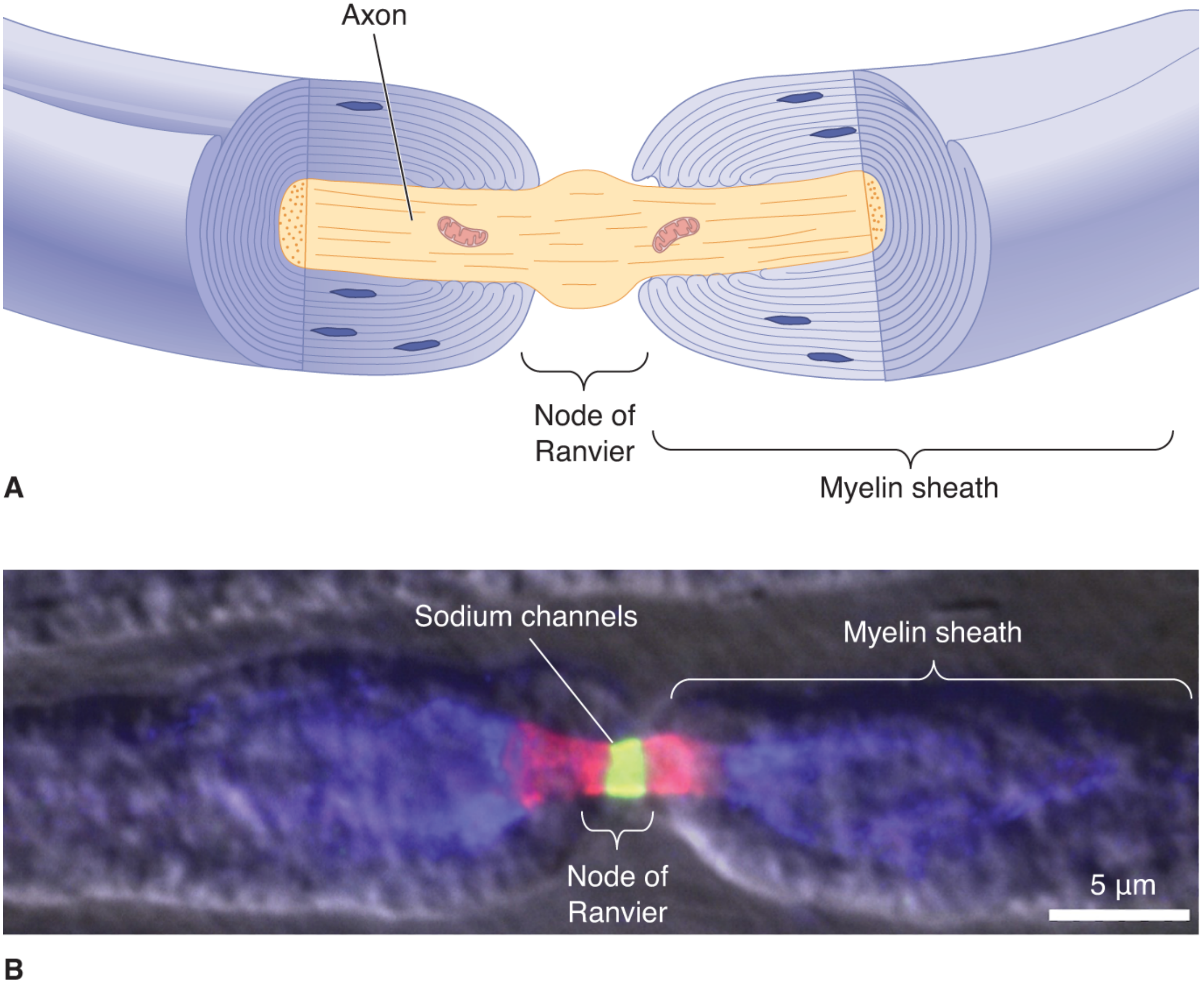
Fig: Node of Ranvier. (A) Axon wrapped in myelin with exposed node. (B) Voltage-gated Na⁺ channels (green/red) densely concentrated at the node. - Neuroscience: Exploring the Brain, 5th Ed
Physiology Flowchart
PREREQUISITE ANATOMY
─────────────────────────────────────────────────────────────
Schwann cell (PNS) / Oligodendrocyte (CNS)
        │
        ▼
Wraps axon in multiple lipid bilayer layers
        │
        ▼
Forms MYELIN SHEATH (sphingomyelin-rich)
  • Electrically insulating
  • Reduces ion flow ~5,000-fold through membrane
  • Decreases membrane capacitance ~50-fold
        │
        ▼
Gaps between adjacent Schwann cells = NODES OF RANVIER
  • Only 1-2 μm long
  • Spaced every 0.2 - 2.0 mm (internodal distance)
  • Dense concentration of voltage-gated Na⁺ channels
  • K⁺ channels also present at/near nodes
  • Ion flow occurs ONLY here


STEP 1 — STIMULUS ARRIVES AT NODE 0
─────────────────────────────────────────────────────────────
Action potential initiated at one node (Node 0)
        │
        ▼
Depolarization: Node 0 reaches threshold (~-55 mV)
        │
        ▼
Voltage-gated Na⁺ channels OPEN at Node 0
        │
        ▼
Na⁺ rushes IN → membrane depolarizes to ~+30 mV
        │
        ▼
Local current generated INSIDE axoplasm (intracellular)
AND OUTSIDE in extracellular fluid simultaneously


STEP 2 — CURRENT JUMPS ACROSS THE INTERNODE
─────────────────────────────────────────────────────────────
Current flows from Node 0 → toward Node 1
  • Myelin = high membrane resistance → prevents ion leak
  • Forces current to travel longitudinally down axoplasm
  • Current arrives at Node 1 with minimal loss
        │
        ▼
Node 1 is depolarized by this arriving current
        │
        ▼
Threshold reached at Node 1


STEP 3 — ACTION POTENTIAL REGENERATED AT NODE 1
─────────────────────────────────────────────────────────────
Voltage-gated Na⁺ channels open at Node 1
        │
        ▼
New action potential fires at Node 1
        │
        ▼
Process REPEATS: Node 1 → Node 2 → Node 3 → ...
        │
        ▼
Impulse "LEAPS" (Latin: saltare) from node to node
= SALTATORY CONDUCTION


STEP 4 — REPOLARIZATION & UNIDIRECTIONALITY
─────────────────────────────────────────────────────────────
At each node after firing:
        │
        ▼
Voltage-gated Na⁺ channels INACTIVATE (absolute refractory)
        │
        ▼
K⁺ channels OPEN → K⁺ flows OUT → membrane repolarizes
        │
        ▼
Refractory period at prior node prevents backward propagation
        │
        ▼
Signal travels UNIDIRECTIONALLY forward only


FINAL RESULT
─────────────────────────────────────────────────────────────
Fast, efficient, energy-saving nerve impulse conduction
Why Saltatory Conduction is Advantageous
| Advantage | Mechanism | Magnitude |
|---|---|---|
| Speed | AP jumps long internodal distances instead of creeping step-by-step | Up to 120 m/s in myelinated vs ~0.5-2 m/s in unmyelinated |
| Energy conservation | Only nodes depolarize; far fewer ions cross membrane | ~100x less ion movement needed |
| Space efficiency | A thin myelinated axon (~20 µm) conducts as fast as an unmyelinated squid giant axon (1000 µm) | Same velocity in 1/50th the volume |
| Less metabolic cost | Fewer ions displaced = less Na⁺/K⁺-ATPase pump activity required to restore gradients | Conserves ATP |
Source: Guyton & Hall Textbook of Medical Physiology; Medical Physiology (Boron & Boulpaep)
Conduction Velocity by Fiber Type
| Fiber Type | Myelination | Diameter | Velocity | Function |
|---|---|---|---|---|
| Aα | Thick myelin | 12-20 µm | 70-120 m/s | Motor, proprioception |
| Aβ | Thick myelin | 5-12 µm | 30-70 m/s | Touch, pressure |
| Aδ | Thin myelin | 2-5 µm | 5-30 m/s | Fast pain, temperature |
| C | Unmyelinated | 0.2-1.5 µm | 0.5-2 m/s | Slow pain, postganglionic autonomic |
What Happens When Myelin is Lost?
DEMYELINATION (e.g., Multiple Sclerosis, Guillain-Barré)
        │
        ▼
Myelin sheath degraded or absent between nodes
        │
        ▼
Current leaks through internode → cannot jump to next node
        │
        ▼
Saltatory conduction FAILS
        │
        ▼
Either:
  ├─ Slowed conduction (partial demyelination)
  └─ Complete conduction block (severe demyelination)
        │
        ▼
Clinical: Weakness, sensory loss, visual disturbance,
          lack of coordination
Key Concept Summary:
Myelin acts like insulating tape on a leaky hose - it forces current to travel fast and far down the axoplasm, regenerating the action potential only at exposed nodes, achieving high-speed conduction at a fraction of the metabolic cost.
Sources: Guyton & Hall Textbook of Medical Physiology | Neuroscience: Exploring the Brain, 5th Ed | Medical Physiology (Boron & Boulpaep)
This is a shared conversation. Sign in to Orris to start your own chat.