
Sensory supply of face
The face is supplied by the trigeminal nerve (CN V), which has three divisions. Here's a quick overview:
- V1 (Ophthalmic) - forehead, scalp (anterior), upper eyelid, nose (dorsum)
- V2 (Maxillary) - cheek, lower eyelid, upper lip, lateral nose
- V3 (Mandibular) - lower lip, chin, jaw, temporal region
Searching sources...
Reading File
Finding Sources
Searching Images
sensory supply of face trigeminal nerve divisions dermatomes
Finding Sources
Finding Sources
There's a great image in Andrews' Diseases of the Skin. Let me fetch that and read the key textbook sections in parallel.
Loading Image
Reading File
Reading File
Excellent - I have rich, detailed content from two authoritative dermatology textbooks plus a clear anatomical diagram. Here is the complete answer:
Sensory Supply of the Face
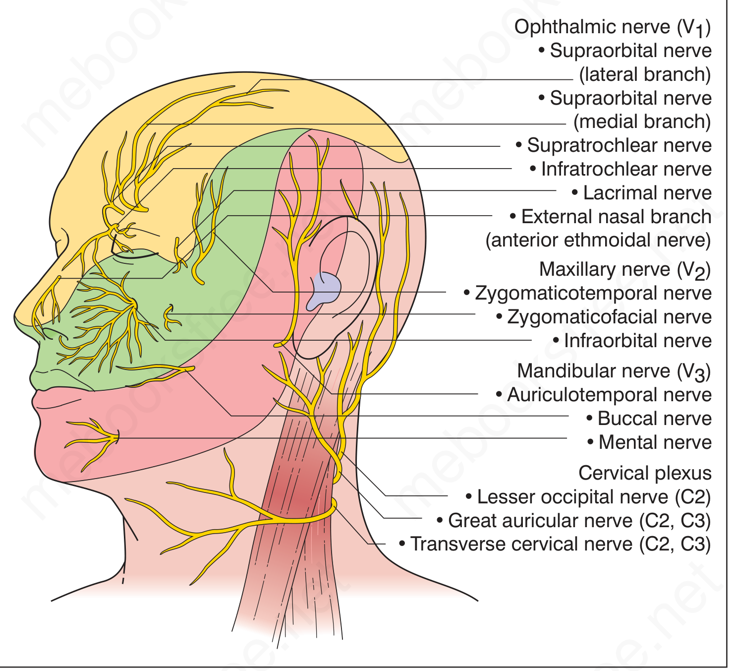
The face is supplied primarily by the trigeminal nerve (CN V), the largest cranial nerve, which divides into three branches at the trigeminal (Gasserian) ganglion. A small contribution also comes from the cervical plexus (C2, C3) and from CN VII, IX, and X to the ear.
CN V - Trigeminal Nerve
The three divisions exit the skull through foramina arranged along the mid-pupillary line: supraorbital, infraorbital, and mental foramina.
V1 - Ophthalmic Division (smallest, uppermost)
Gives off three branches before exiting the orbit: nasociliary, frontal, and lacrimal nerves.
| Branch | Area Supplied |
|---|---|
| Supraorbital nerve (lateral) | Forehead and anterior scalp |
| Supratrochlear nerve (medial) | Medial forehead, medial upper eyelid, frontal scalp |
| Infratrochlear nerve (from nasociliary) | Glabella, nasal root, medial canthus, bridge of nose |
| External nasal nerve (anterior ethmoidal) | Nasal dorsum, tip, supratip, columella |
| Lacrimal nerve | Upper eyelid (lateral), lacrimal gland area |
Clinical pearl: The nasociliary branch also supplies the corneal surface via ciliary nerves. Herpes zoster involving the nasal tip (Hutchinson's sign) implies corneal involvement - urgent ophthalmology referral is needed.
V2 - Maxillary Division
Exits via the infraorbital foramen.
| Branch | Area Supplied |
|---|---|
| Infraorbital nerve | Lower eyelid, superior cheek, upper lip, lateral nose, part of nasal ala |
| Zygomaticofacial nerve | Prominence of the cheek |
| Zygomaticotemporal nerve | Anterior temporal region |
V3 - Mandibular Division
The only division with both sensory and motor components (motor: muscles of mastication). Exits via the foramen ovale.
| Branch | Area Supplied |
|---|---|
| Auriculotemporal nerve | Temple, most of the ear (auricle), external auditory canal, scalp |
| Buccal nerve | Cheek mucosa and skin, buccal gingiva |
| Mental nerve | Lower lip, chin, lower teeth |
Cervical Plexus Contributions
The upper cervical nerves (C2, C3) contribute to the posterior and inferior areas:
| Nerve | Area Supplied |
|---|---|
| Lesser occipital nerve (C2) | Upper posterior neck, back of ear |
| Great auricular nerve (C2, C3) | Lower face/jaw angle, lower ear, parotid region |
| Transverse cervical nerve (C2, C3) | Anterior neck |
The Ear - Mixed Supply
The auricle and external auditory canal receive a unique mixed innervation:
- Auriculotemporal nerve (V3) - most of the auricle
- Great auricular nerve (C2, C3) - lower auricle and skin over parotid
- Lesser occipital (C2) - upper posterior ear
- Facial nerve (CN VII), glossopharyngeal (CN IX), vagus (CN X) - concha and external auditory canal (variable)
Key Anatomical Points
-
Sensory nerves run superficially - between subcutaneous fat and the SMAS, often in neurovascular bundles with arteries and veins. This makes them vulnerable during facial surgery.
-
Three foramina along mid-pupillary line - supraorbital, infraorbital, and mental - are the key exit points used for regional nerve blocks.
-
Collateral re-innervation is possible after unilateral trigeminal root injury due to contralateral sprouting.
-
Scalp above a line from glabella to external occipital protuberance - supplied by trigeminal anteriorly and cervical nerves posteriorly.
Sources: Andrews' Diseases of the Skin, p. 37.3; Fitzpatrick's Dermatology, p. 3732; Dermatology 2-Volume Set 5e, p. 2929
This is a shared conversation. Sign in to Orris to start your own chat.