
Iris antomy
I'll look up iris anatomy from the medical library right away.
Reading File
Now let me search the library for iris anatomy content and images in parallel.
Finding Sources
Finding Sources
Finding Sources
Finding Sources
Excellent - multiple great sources found. Let me read the most detailed ones in parallel - Junqueira's Histology, Gray's Anatomy, and the Histology Atlas.
Reading File
Reading File
Reading File
Excellent content. Now let me fetch the key anatomical diagrams in parallel.
Loading Image
Loading Image
Loading Image
I have rich content and three excellent images. Here is a comprehensive answer on iris anatomy.
Iris Anatomy
The iris is the most anterior part of the uveal (vascular) layer of the eye. It is a circular, pigmented diaphragm that projects inward from the ciliary body and contains a central aperture - the pupil - through which light enters the eye.
Position and Relations
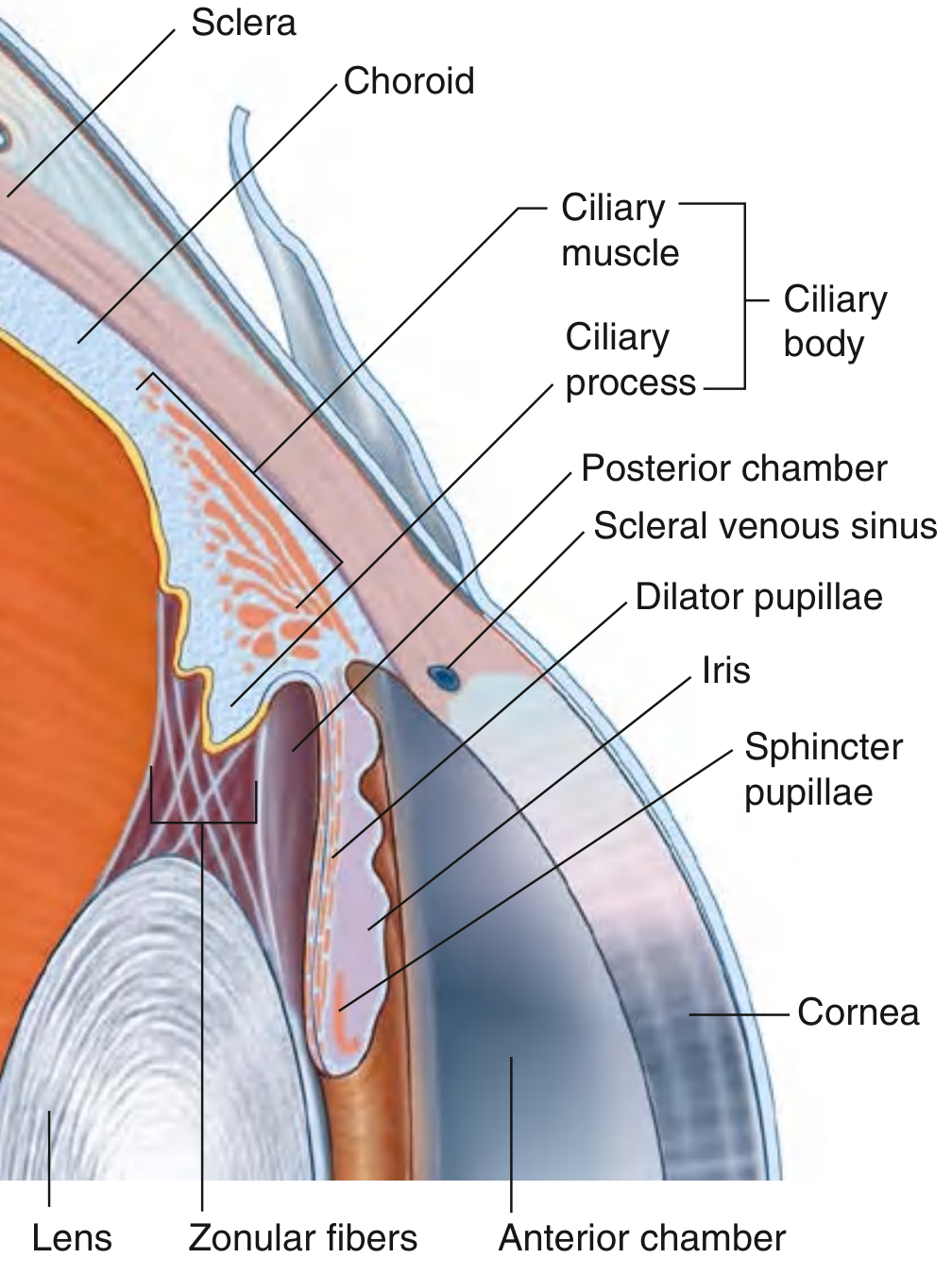
The iris sits between the cornea anteriorly and the lens posteriorly, dividing the anterior segment of the eye into:
- Anterior chamber - between cornea and iris, filled with aqueous humor
- Posterior chamber - between iris and lens, also filled with aqueous humor
Its peripheral root attaches to the ciliary body, while its free inner edge forms the pupillary margin.
Gross Anatomy Diagram
Layers of the Iris (Anterior to Posterior)
1. Anterior Surface (No Epithelium)
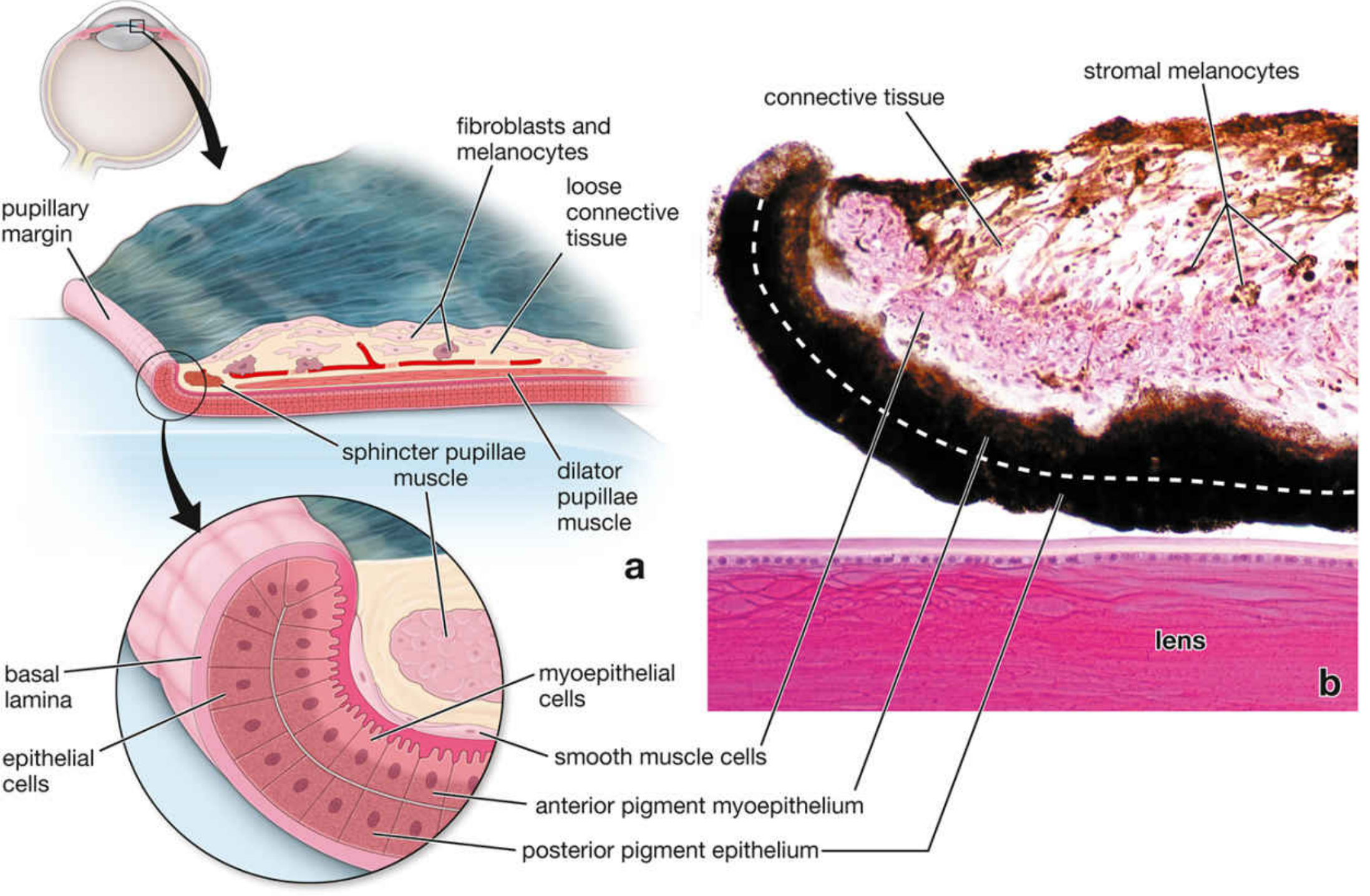
Unlike virtually every other surface in the body, the anterior face of the iris has no epithelial covering. Instead, it consists of a dense, matted layer of interdigitating fibroblasts and melanocytes exposed directly to the aqueous humor of the anterior chamber. Scanning electron microscopy reveals numerous ridges and crypts (Fuchs' crypts) on this surface.
2. Stroma
The bulk of the iris consists of loose connective tissue stroma containing:
- Melanocytes - variable in number; their density determines eye color (see below)
- Sparse collagen fibers
- Blood vessels (sparse microvasculature)
- Nerve fibers
- Sphincter pupillae muscle - located in the stroma near the pupillary margin
3. Posterior Epithelium - Bilaminar (Two Cell Layers)
The posterior surface carries two layers of pigmented epithelium, continuous with the epithelium of the ciliary body:
| Layer | Cell Type | Pigmentation | Special Feature |
|---|---|---|---|
| Posterior pigment epithelium (innermost) | Columnar epithelial cells | Very heavily pigmented - nuclei obscured | Basal lamina faces posterior chamber |
| Anterior pigment myoepithelium | Myoepithelial cells | Apical portions heavily pigmented | Basal portions extend radially as the dilator pupillae muscle |
Intrinsic Muscles of the Iris
| Muscle | Arrangement | Cell Type | Innervation | Action |
|---|---|---|---|---|
| Sphincter pupillae | Circular fibers near pupillary margin, within stroma | Smooth muscle | Parasympathetic (CN III via ciliary ganglion) | Constricts pupil (miosis) |
| Dilator pupillae | Radial processes extending from myoepithelial cells of anterior pigment layer | Myoepithelial (unique - neuroectodermal origin) | Sympathetic (superior cervical ganglion, T1) | Dilates pupil (mydriasis) |
Key point: the dilator pupillae is not a classic smooth muscle - it is formed by contractile processes of myoepithelial cells derived from the anterior pigment epithelium layer.
Histological Detail
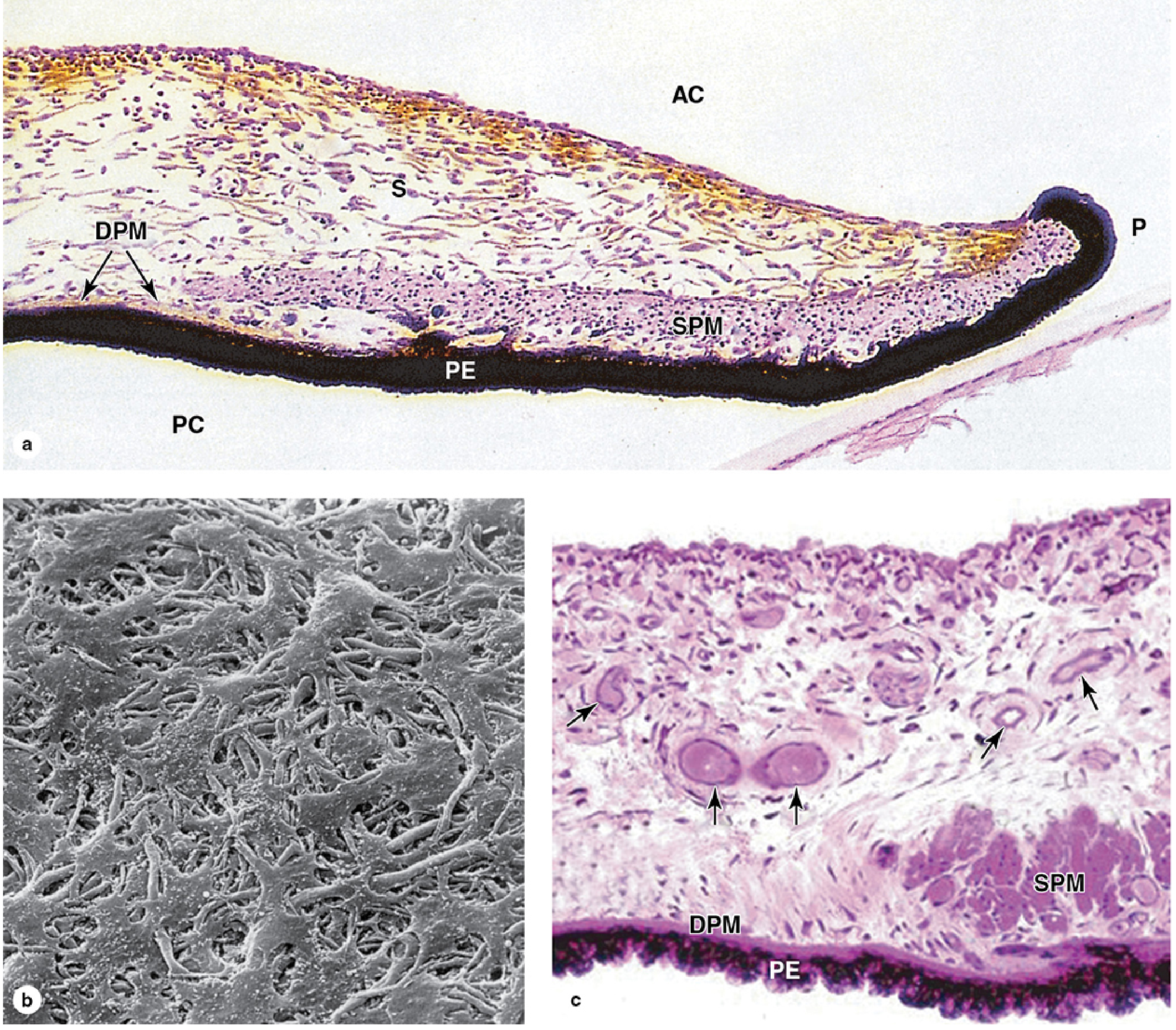
- AC = anterior chamber, PC = posterior chamber, S = stroma, PE = posterior pigment epithelium, DPM = dilator pupillae muscle, SPM = sphincter pupillae muscle, P = pupil
Eye Color
Eye color is determined by the number and melanin content of stromal melanocytes:
- Few, lightly pigmented stromal melanocytes → light reflected from the densely pigmented posterior epithelium → blue iris
- Increasing stromal melanocytes → progressively absorb and reflect longer wavelengths → green → gray → hazel → brown
- The posterior pigment epithelium is densely pigmented in all individuals regardless of eye color.
Blood Supply and Innervation
- Arterial supply: The major arterial circle of the iris (circulus arteriosus major) sits at the ciliary body root, formed by long posterior ciliary arteries and anterior ciliary arteries. From it, radial branches run toward the pupil and form the minor arterial circle (circulus arteriosus minor) near the pupillary margin.
- Innervation: Sympathetic fibers (via the nasociliary branch of CN V1, traveling with the long ciliary nerves) innervate the dilator. Parasympathetic fibers (from CN III via the short ciliary nerves from the ciliary ganglion) innervate the sphincter.
Summary Table
| Feature | Detail |
|---|---|
| Part of | Uvea (middle vascular layer) |
| Anterior surface | No epithelium; fibroblasts + melanocytes |
| Stroma | Loose connective tissue, melanocytes, blood vessels |
| Posterior surface | Bilaminar pigmented epithelium (myoepithelial + pigment layers) |
| Pupil constriction | Sphincter pupillae - circular smooth muscle - parasympathetic (CN III) |
| Pupil dilation | Dilator pupillae - radial myoepithelium - sympathetic (T1) |
| Eye color | Determined by stromal melanocyte density |
Sources:
- Junqueira's Basic Histology, 17e, pp. 1189-1190
- Histology: A Text and Atlas, pp. 2392-2393
- Gray's Anatomy for Students, p. 1090
This is a shared conversation. Sign in to Orris to start your own chat.